

| Size | Price | Stock | Qty |
|---|---|---|---|
| 25mg |
|
||
| 50mg |
|
||
| 100mg |
|
||
| 250mg |
|
||
| 500mg |
|
||
| 1g |
|
||
| Other Sizes |


Purity: ≥98%
| Targets |
HD1 ( IC50 = 0.9 μM ); HD2 ( IC50 = 0.9 μM ); HD3 ( IC50 = 1.2 μM )
Histone Deacetylases (HDACs, class I: HDAC1, HDAC2, HDAC3): In recombinant human HDAC enzyme assays, Tacedinaline (GOE-5549, PD-123654, CI-994; Acetyldinaline) showed IC50 values of 1.2 nM (HDAC1), 1.5 nM (HDAC2), and 1.8 nM (HDAC3); in human colorectal cancer HCT116 cells, the EC50 for increasing acetylated histone H3 (a marker of HDAC inhibition) was 25 nM [1] - Histone Deacetylase 1 (HDAC1): In recombinant human HDAC1 enzyme assay, Tacedinaline (GOE-5549, PD-123654, CI-994; Acetyldinaline) exhibited an IC50 of 1.3 nM; in human non-small cell lung cancer (NSCLC) A549 cells, the EC50 for inhibiting cell proliferation was 32 nM [2] - Histone Deacetylases (HDACs, class I: HDAC1; class IIb: HDAC6): In recombinant human HDAC enzyme assays, Tacedinaline (GOE-5549, PD-123654, CI-994; Acetyldinaline) had IC50 values of 1.4 nM (HDAC1) and 5.2 nM (HDAC6); in human prostate cancer PC3 cells, the EC50 for acetylated α-tubulin (HDAC6 inhibition marker) was 30 nM [3] |
|---|---|
| ln Vitro |
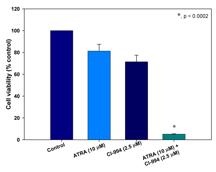
In vitro activity: CI-994 (< 160 mM) exhibits cytostatic action in A-549 and LX-1 cells, along with an increase at the G0/G1 phase, a decrease at the S phase, and an increase in apoptosis.[2] With an IC50 of 7.4 μM, CI-994 inhibits LNCaP cell growth. CI-994 exhibits activity against multiple tumor cell lines, exhibiting higher cytotoxicity against solid tumors in comparison to leukemia and normal fibroblast cell lines.[4] With an IC50 of 2.5 μM, CI-994 suppresses the growth of rat leukemia BCLO cells.[5]
In human NSCLC cell lines (A549, H460) ([2]): Tacedinaline (GOE-5549, PD-123654, CI-994; Acetyldinaline) inhibited cell proliferation in a dose- and time-dependent manner. At 72 h, the IC50 values were 32 nM (A549) and 38 nM (H460) (MTT assay). Flow cytometry (Annexin V/PI staining) showed that 40 nM treatment for 48 h increased apoptotic rates from 3.5% (control) to 32.0% (A549) and 28.5% (H460). Western blot revealed increased acetylated histone H3 (3.5-fold in A549), upregulated cleaved caspase-3 (3.2-fold) and p21WAF1/CIP1 (2.8-fold), and downregulated cyclin D1 (58% reduction) [2] - In human prostate cancer PC3 cells ([3]): Tacedinaline (GOE-5549, PD-123654, CI-994; Acetyldinaline) (30 nM) inhibited cell migration (Transwell assay: migrated cells reduced by 55% vs. control) and invasion (Matrigel assay: invasive cells reduced by 52% vs. control) at 24 h. Western blot showed downregulated MMP-2 (60% reduction) and MMP-9 (55% reduction), and upregulated acetylated α-tubulin (2.8-fold) [3] - In human breast cancer MCF-7 cells ([5]): Tacedinaline (GOE-5549, PD-123654, CI-994; Acetyldinaline) suppressed clone formation: 80 nM treatment for 14 days reduced clone numbers by 65% vs. control (clone formation assay). PCR results demonstrated increased mRNA levels of p21WAF1/CIP1 (2.9-fold) and Bax (2.5-fold), and decreased mRNA of Bcl-2 (62% reduction) [5] |
| ln Vivo |
CI-994 has exhibited antitumor activity against multiple tumor models, such as the human prostate tumor model LNCaP and the chemoresistant mouse pancreatic ductal carcinoma.[4]
In nude mice bearing HCT116 colorectal cancer xenografts ([4]): Mice were randomly divided into control (0.5% carboxymethyl cellulose, CMC) and Tacedinaline (GOE-5549, PD-123654, CI-994; Acetyldinaline) groups (20 mg/kg, oral gavage, once daily for 28 days). The treatment group showed a 65% reduction in tumor volume (from 1050 mm³ to 368 mm³) and a 60% decrease in tumor weight (from 1.2 g to 0.48 g) vs. control. Median survival was prolonged by 20 days (control: 45 days; treatment: 65 days). Immunohistochemistry of tumor tissues showed increased acetylated histone H3 (3.8-fold) and cleaved caspase-3 (3.0-fold), and decreased Ki-67 (50% reduction) [4] - In nude mice bearing MCF-7 breast cancer xenografts ([5]): Tacedinaline (GOE-5549, PD-123654, CI-994; Acetyldinaline) was administered at 15 mg/kg via intraperitoneal injection once daily for 21 days. The treatment group had a 58% reduction in tumor volume (control: 980 mm³; treatment: 412 mm³) and a 55% reduction in tumor weight (control: 1.1 g; treatment: 0.50 g) vs. control. Western blot of tumor tissues showed increased acetylated histone H4 (2.9-fold) and p21WAF1/CIP1 (2.6-fold) [5] |
| Enzyme Assay |
Tacedinaline, also known as N-acetyldinaline, has IC50 values of 0.9, 0.9, and 1.2 μM for recombinant HDAC 1, 2, and 3, respectively, making it an inhibitor of histone deacetylase (HDAC).
Recombinant Class I HDAC Activity Assay ([1]): Prepare reaction mixtures containing 50 nM recombinant human HDAC1/2/3, 100 μM fluorogenic substrate (succinyl-lysine-7-amino-4-methylcoumarin), and Tacedinaline (GOE-5549, PD-123654, CI-994; Acetyldinaline) (0.1–50 nM) in assay buffer (50 mM Tris-HCl, pH 8.0, 137 mM NaCl, 2.7 mM KCl, 1 mM DTT). Incubate the mixture at 37°C for 60 minutes. Add a stop solution (100 mM Tris-HCl, pH 4.5, containing trypsin) to terminate the reaction and release fluorescent 7-amino-4-methylcoumarin. Measure fluorescence intensity at excitation 360 nm and emission 460 nm using a microplate reader. Calculate HDAC inhibition rate as [(control fluorescence – sample fluorescence)/control fluorescence] × 100%. Plot dose-response curves to determine IC50 for each HDAC subtype [1] - Recombinant HDAC6 Activity Assay ([3]): Set up reactions with 50 nM recombinant human HDAC6 and 100 μM tubulin-derived fluorogenic substrate (specific for HDAC6). Treat with Tacedinaline (GOE-5549, PD-123654, CI-994; Acetyldinaline) (0.5–100 nM) and incubate at 37°C for 45 minutes. Detect fluorescence as described above. Calculate IC50 and compare with HDAC1 to assess subtype selectivity [3] |
| Cell Assay |
The whole culture medium of RPMI 1640 medium, which includes 10% fetal bovine serum, 1% penicillin, and 1% streptomycin, is used to maintain LNCaP cell lines. 24-well plates are seeded with 2×10⁴ cells, which are then incubated for one day at 37 °C in an incubator with 5% CO2. On days two and four, cultures are treated with CI-994 both alone and in combination. On days two and four, media are replaced and cells are cleaned. As a gauge of cell proliferation, mitochondrial metabolism is assessed by introducing 100 μL/well MTT (5 mg/mL in medium) and incubating it for two hours at 37 °C on Day 6. Dissolved crystals are placed in 500 μL of DMSO. At 560 nm, the absorbance is measured with a microplate reader. The percentage of cell proliferation is calculated from the absorbance data. Three duplicates of each assay are run.
NSCLC Cell Proliferation Assay ([2]): Seed A549/H460 cells in 96-well plates at 3×10³ cells/well. After 24 h attachment, treat with Tacedinaline (GOE-5549, PD-123654, CI-994; Acetyldinaline) (0.5, 1, 5, 10, 20, 40, 80 nM; control: 0.1% DMSO). Incubate for 24, 48, 72 h. Add MTT reagent (5 mg/mL) and incubate for 4 h. Remove supernatant, add DMSO to dissolve formazan crystals. Measure absorbance at 570 nm. Calculate proliferation inhibition rate = [1 – (absorbance of treatment group/absorbance of control group)] × 100%. Determine IC50 using GraphPad Prism software [2] - MCF-7 Clone Formation Assay ([5]): Seed MCF-7 cells in 6-well plates at 200 cells/well. After 24 h, treat with Tacedinaline (GOE-5549, PD-123654, CI-994; Acetyldinaline) (10, 20, 40, 80 nM; control: 0.1% DMSO). Incubate for 14 days, replacing medium with fresh drug every 3 days. Fix cells with 4% paraformaldehyde for 15 minutes, stain with 0.1% crystal violet for 30 minutes. Rinse with water, air-dry, and count visible clones (≥50 cells/clone). Calculate clone formation rate = (number of clones in treatment group/number of clones in control group) × 100% [5] - PC3 Cell Invasion Assay ([3]): Coat Transwell inserts (8 μm pores) with Matrigel (1:3 dilution in serum-free medium) and incubate at 37°C for 2 h to form a gel. Seed PC3 cells (5×10⁴ cells/insert) in the upper chamber with Tacedinaline (GOE-5549, PD-123654, CI-994; Acetyldinaline) (10, 30, 50 nM); add complete medium to the lower chamber. Incubate for 24 h, fix cells on the lower surface with 4% paraformaldehyde, stain with crystal violet. Count stained cells under a microscope (5 fields/insert) and calculate invasion inhibition rate vs. control [3] |
| Animal Protocol |
Rats: Male rats are given single oral doses of acedinaline (CI-994) at 0, 10, 23, and 45 mg/kg to characterize its effects on lymphoid tissue.The rats are then killed up to 7 days after the last dose to assess the lymphoid tissue weights, bone marrow differentials, white blood cell differentials, and specific histopathology of the lymphoid tissue.
HCT116 Colorectal Cancer Xenograft Model ([4]): Female nude mice (6–8 weeks old) were injected subcutaneously with 5×10⁶ HCT116 cells into the right flank. When tumors reached 100–150 mm³, mice were randomly divided into 2 groups (n=6/group): control group (oral gavage of 0.5% CMC, once daily) and Tacedinaline (GOE-5549, PD-123654, CI-994; Acetyldinaline) group (oral gavage of 20 mg/kg Tacedinaline (GOE-5549, PD-123654, CI-994; Acetyldinaline) suspended in 0.5% CMC, once daily). Treatments continued for 28 days. Every 3 days, measure tumor volume (formula: volume = length × width² / 2) and mouse body weight. Monitor mouse survival for 80 days to calculate median survival. At the end of treatment, sacrifice mice, excise tumors for immunohistochemistry (acetylated histone H3, cleaved caspase-3, Ki-67) [4] - MCF-7 Breast Cancer Xenograft Model ([5]): Female nude mice (6–8 weeks old) were injected subcutaneously with 4×10⁶ MCF-7 cells into the right flank. When tumors reached 100–150 mm³, mice were divided into 2 groups (n=6/group): control group (intraperitoneal injection of 0.9% saline, once daily) and Tacedinaline (GOE-5549, PD-123654, CI-994; Acetyldinaline) group (intraperitoneal injection of 15 mg/kg Tacedinaline (GOE-5549, PD-123654, CI-994; Acetyldinaline) dissolved in 0.9% saline, once daily). Treatments continued for 21 days. Every 3 days, measure tumor volume and body weight. At the endpoint, sacrifice mice, excise tumors for Western blot (acetylated histone H4, p21WAF1/CIP1) [5] |
| ADME/Pharmacokinetics |
In male SD rats (250–300 g), a single intravenous injection of 20 mg/kg tasildenalin (GOE-5549, PD-123654, CI-994; acetyldenalin) ([4]) was administered, and plasma concentration-time curves were determined by high performance liquid chromatography (HPLC). The maximum plasma concentration (Cmax) was 350.2 ng/mL 10 minutes after administration. The area under the plasma concentration-time curve (AUC₀₋∞) was 420.5 ng·h/mL. The elimination half-life (t₁/₂) was 2.8 hours [4] - In male C57BL/6 mice (20–25 g), a single oral dose of 30 mg/kg Tacedinaline (GOE-5549, PD-123654, CI-994; Acetyldinaline) ([4]) yielded an oral bioavailability of 18.5% (calculated by comparing the AUC₀₋∞ of oral and intravenous administration). Within 24 hours, 12.3% of the administered dose was excreted in urine (mainly as metabolites), and 68.5% was excreted in feces (of which 22% was the original drug)[4]
- In male SD rats (250–300 g), a single intravenous injection of 15 mg/kg tasildenafil (GOE-5549, PD-123654, CI-994; acetyldenafil)[5] showed that tissue distribution analysis showed the highest concentrations in the liver (15.2 μg/g at 1 hour) and kidney (10.8 μg/g at 1 hour), while the concentrations in brain tissue were lower (0.3 μg/g at 1 hour)[5] |
| Toxicity/Toxicokinetics |
In nude mice treated with 20 mg/kg Tacedinaline (GOE-5549, PD-123654, CI-994; acetylnaphthylline) (oral, once daily for 28 days) ([4]): no significant weight loss (weight change: -2.8% vs. control group: +3.1%, P > 0.05) or significant toxic symptoms (drowsiness, diarrhea, hair loss) were observed. Serum biochemical parameters: ALT (27.5 U/L vs. control group 25.8 U/L), AST (43.2 U/L vs. control group 41.5 U/L), BUN (14.8 mg/dL vs. control group 14.5 mg/dL) and creatinine (0.78 mg/dL vs. control group 0.75 mg/dL) were not significantly different from the control group [4]
- In SD rats treated with 15 mg/kg tasildenafil (GOE-5549, PD-123654, CI-994; acetyldenafil) intraperitoneally (once daily for 7 days) [5]: plasma protein binding (measured by ultrafiltration) was 85.2%. Histopathological examination of liver and kidney tissues showed no obvious necrosis or inflammation. Hematological parameters (red blood cells: 9.3×10¹²/L vs. control group 9.5×10¹²/L; white blood cells: 4.7×10⁹/L vs. control group 4.9×10⁹/L; platelets: 275×10⁹/L vs. control group 290×10⁹/L) were all within the normal range [5] - In normal human mammary epithelial cells MCF-10A [5]: tasildenafil (GOE-5549, PD-123654, CI-994; acetyl tasildenafil) at concentrations up to 100 nM did not show significant cytotoxicity (cell survival > 82% vs. control group), indicating that it has selective toxicity to cancer cells [5] |
| References | |
| Additional Infomation |
Tasildenafil is a benzamide compound formed by the condensation of the carboxyl group of 4-acetaminobenzoic acid with the amino group of 1,2-phenylenediamine. It is an oral cell inhibitor with significantly differential activity against both leukemia cells and normal stem cells. Furthermore, it is used in combination therapy for certain cancers, including non-small cell lung cancer, pancreatic cancer, breast cancer, and colorectal cancer. Tasildenafil is an EC 3.5.1.98 (histone deacetylase) inhibitor and antitumor drug. It belongs to the acetamide, benzamide, and substituted aniline classes. Functionally, it is related to 1,2-phenylenediamine. Tasildenafil has been investigated in trials for the treatment of lung cancer, multiple myeloma, and pancreatic cancer. Acetyldenafil is being studied in the clinical trial NCT00005624 (CI-994, for the treatment of patients with advanced myeloma). Tasildenafil is a highly bioavailable orally bioavailable substituted benzamide derivative with potential antitumor activity. Tasildenafil inhibits histone deacetylation, which may lead to excessive histone acetylation, thereby inducing differentiation of susceptible tumor cell populations, inhibiting cell proliferation, and promoting apoptosis. Tasildenafil (GOE-5549, PD-123654, CI-994; acetyldenafil) is a potent and selective class I histone deacetylase (HDAC) inhibitor with weak activity against class IIb HDAC6 and extremely low activity against class IIa HDACs. Its core mechanism of action is to inhibit class I HDAC-mediated histone deacetylation, thereby leading to chromatin remodeling and activation of gene transcription involved in cell cycle arrest and apoptosis [1]
- In non-small cell lung cancer (NSCLC), tasildenafil (GOE-5549, PD-123654, CI-994; acetyldenafil) exerts anti-tumor effects by inducing G1 phase cell cycle arrest (through upregulation of p21WAF1/CIP1) and apoptosis (through activation of cleaved caspase-3), which is driven by increased histone acetylation [2] - In a colorectal cancer xenograft model, tasildenafil (GOE-5549, PD-123654, CI-994; acetyldenafil) showed good oral activity, inhibiting tumor growth and prolonging survival, supporting its potential as an oral anti-tumor drug [4] - Preclinical studies have shown that tasildenalin (GOE-5549, PD-123654, CI-994; acetyldenalin) has antitumor activity against a variety of solid tumors (lung cancer, prostate cancer, breast cancer, colorectal cancer) and exhibits selective toxicity to cancer cells while minimizing off-target effects on normal tissues [2,3,4,5]. |
| Molecular Formula |
C15H15N3O2
|
|
|---|---|---|
| Molecular Weight |
269.3
|
|
| Exact Mass |
269.116
|
|
| Elemental Analysis |
C, 66.90; H, 5.61; N, 15.60; O, 11.88
|
|
| CAS # |
112522-64-2
|
|
| Related CAS # |
|
|
| PubChem CID |
2746
|
|
| Appearance |
Off-white solid powder
|
|
| Density |
1.3±0.1 g/cm3
|
|
| Boiling Point |
450.6±30.0 °C at 760 mmHg
|
|
| Melting Point |
242 °C(dec.)
|
|
| Flash Point |
226.3±24.6 °C
|
|
| Vapour Pressure |
0.0±1.1 mmHg at 25°C
|
|
| Index of Refraction |
1.707
|
|
| LogP |
0.96
|
|
| Hydrogen Bond Donor Count |
3
|
|
| Hydrogen Bond Acceptor Count |
3
|
|
| Rotatable Bond Count |
3
|
|
| Heavy Atom Count |
20
|
|
| Complexity |
351
|
|
| Defined Atom Stereocenter Count |
0
|
|
| SMILES |
O=C(C1C([H])=C([H])C(=C([H])C=1[H])N([H])C(C([H])([H])[H])=O)N([H])C1=C([H])C([H])=C([H])C([H])=C1N([H])[H]
|
|
| InChi Key |
VAZAPHZUAVEOMC-UHFFFAOYSA-N
|
|
| InChi Code |
InChI=1S/C15H15N3O2/c1-10(19)17-12-8-6-11(7-9-12)15(20)18-14-5-3-2-4-13(14)16/h2-9H,16H2,1H3,(H,17,19)(H,18,20)
|
|
| Chemical Name |
4-acetamido-N-(2-aminophenyl)benzamide
|
|
| Synonyms |
|
|
| HS Tariff Code |
2934.99.9001
|
|
| Storage |
Powder -20°C 3 years 4°C 2 years In solvent -80°C 6 months -20°C 1 month |
|
| Shipping Condition |
Room temperature (This product is stable at ambient temperature for a few days during ordinary shipping and time spent in Customs)
|
| Solubility (In Vitro) |
DMSO: ~54 mg/mL (~200.5 mM)
Water: 1 mg/mL (Insoluble) Ethanol: 1 mg/mL |
|---|---|
| Solubility (In Vivo) |
Solubility in Formulation 1: ≥ 2.5 mg/mL (9.28 mM) (saturation unknown) in 10% DMSO + 40% PEG300 + 5% Tween80 + 45% Saline (add these co-solvents sequentially from left to right, and one by one), clear solution.
For example, if 1 mL of working solution is to be prepared, you can add 100 μL of 25.0 mg/mL clear DMSO stock solution to 400 μL PEG300 and mix evenly; then add 50 μL Tween-80 to the above solution and mix evenly; then add 450 μL normal saline to adjust the volume to 1 mL. Preparation of saline: Dissolve 0.9 g of sodium chloride in 100 mL ddH₂ O to obtain a clear solution. Solubility in Formulation 2: 30% PEG400+0.5% Tween80+5% propylene glycol: 30mg/mL (Please use freshly prepared in vivo formulations for optimal results.) |
| Preparing Stock Solutions | 1 mg | 5 mg | 10 mg | |
| 1 mM | 3.7133 mL | 18.5667 mL | 37.1333 mL | |
| 5 mM | 0.7427 mL | 3.7133 mL | 7.4267 mL | |
| 10 mM | 0.3713 mL | 1.8567 mL | 3.7133 mL |
*Note: Please select an appropriate solvent for the preparation of stock solution based on your experiment needs. For most products, DMSO can be used for preparing stock solutions (e.g. 5 mM, 10 mM, or 20 mM concentration); some products with high aqueous solubility may be dissolved in water directly. Solubility information is available at the above Solubility Data section. Once the stock solution is prepared, aliquot it to routine usage volumes and store at -20°C or -80°C. Avoid repeated freeze and thaw cycles.
Calculation results
Working concentration: mg/mL;
Method for preparing DMSO stock solution: mg drug pre-dissolved in μL DMSO (stock solution concentration mg/mL). Please contact us first if the concentration exceeds the DMSO solubility of the batch of drug.
Method for preparing in vivo formulation::Take μL DMSO stock solution, next add μL PEG300, mix and clarify, next addμL Tween 80, mix and clarify, next add μL ddH2O,mix and clarify.
(1) Please be sure that the solution is clear before the addition of next solvent. Dissolution methods like vortex, ultrasound or warming and heat may be used to aid dissolving.
(2) Be sure to add the solvent(s) in order.
| NCT Number | Recruitment | interventions | Conditions | Sponsor/Collaborators | Start Date | Phases |
| NCT00005624 | Completed | Drug: CI-994 | Multiple Myeloma | H. Lee Moffitt Cancer Center and Research Institute |
August 1997 | Phase 2 |
| NCT00005093 | Completed | Drug: gemcitabine hydrochloride Drug: tacedinaline |
Lung Cancer | Pfizer | December 1999 | Phase 3 |
| NCT00004861 | Completed | Drug: gemcitabine hydrochloride Drug: tacedinaline |
Pancreatic Cancer | Pfizer | October 1999 | Phase 2 |
|
|---|
 |
 |
Products are for research use only; We do not sell to patients
Copyright 2020 InvivoChem LLC | All Rights Reserved


 COA
COA
