| Size | Price | Stock | Qty |
|---|---|---|---|
| 5mg |
|
||
| 10mg |
|
||
| 25mg |
|
||
| 50mg |
|
||
| 100mg |
|
||
| 250mg |
|
||
| 500mg |
|
||
| 1g | |||
| Other Sizes |


Purity: ≥98%
CGI1746 (CGI-1746) is a reversible/non-covalent and highly selective small-molecule inhibitor of the Bruton's tyrosine kinase-Btk with potential anti-inflammatory activity. It inhibits BTK with an IC50 of 1.9 nM. CGI-1746 shows high in vivo antiinflammatory efficacy in an anti-collagen II antibody–induced arthritis (CAIA) model in mice.
| Targets |
Bruton Tyrosine Kinase (BTK) (recombinant human BTK, IC50 = 1.0 nM); >280-fold selectivity over EGFR (IC50 = 280 nM), ITK (IC50 = 320 nM), JAK2 (IC50 = 350 nM); no activity against Src, Abl, VEGFR2 (IC50 > 1000 nM) [1]
- Confirmed BTK as primary target (prostate cancer model; no additional IC50 values; consistent with [1]’s selectivity) [2] |
|---|---|
| ln Vitro |
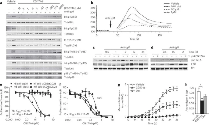
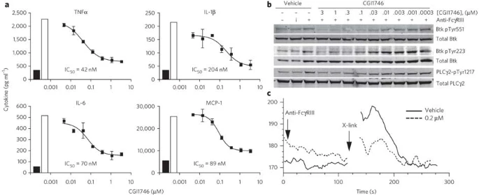
CGI1746 is selective for Btk, with approximately 1,000-fold selectivity over Tec and Src family kinases. In an ATP-free competition binding test, Btk's dissociation constant is 1.5 nM. CGI1746 suppresses Btk activity by a novel binding mechanism that stabilizes an inactive, nonphosphorylated enzyme structure. CGI1746 inhibits both the auto- and transphosphorylation processes required for enzyme activation. CGI1746 completely reduces anti-IgM-induced murine and human B cell proliferation at IC50s of 134 nM and 42 nM, respectively, but has no effect on anti-CD3- or anti-CD28-induced T cell proliferation. CGI1746 effectively inhibits the proliferation of CD27+IgG+ B cells isolated from the tonsils of four human donors, with an average IC50 of 112 nM. CGI1746 prevents macrophages from producing TNFα, IL-1β, and IL-6 triggered by FcγRIII. CGI1746 suppresses TNFα, IL-1β, and IL-6 production (with three- to eight-fold greater IC50) in human monocytes challenged with immobilized or soluble immune complexes [1]. CGI-1746 does not kill cells as effectively as irreversible BTK inhibitors at the same dose. CGI-1746 inhibits BTK phosphorylation at tyrosine 233 in the SH3 domain, but it does not kill LNCaP or DU145 prostate cancer cells at the same concentrations as Ibrutinib or AVL-292 [2]. However, it significantly reduces phosphorylation of both the BTK-A and BTK-C proteins, indicating that auto-phosphorylation of the BTK-C isoform is inhibited in a manner similar to that of BTK-A.
Inhibited B-cell-mediated inflammation: 20 nM CGI1746 reduced anti-IgM-induced mouse splenic B-cell proliferation by 85% (72 hours); decreased B-cell activation marker CD86 expression by 82% (flow cytometry) [1] - Suppressed myeloid cell function: 50 nM CGI1746 reduced LPS-induced TNF-α secretion by human monocytes by 75% (24 hours); inhibited osteoclast differentiation from bone marrow macrophages by 80% (14-day culture) [1] - Inhibited prostate cancer cell growth: Human prostate cancer PC-3 cells (IC50 = 15.2 nM), DU145 cells (IC50 = 18.7 nM); 100 nM CGI1746 reduced PC-3 cell clone formation by 80% (10-day culture); p-BTK (Tyr223) and p-AKT (Ser473) downregulated by >90% (Western blot) [2] |
| ln Vivo |
CGI1746 inhibits B cell-dependent arthritis. CGI1746 treatment (100 mg/kg, sc, twice-daily dosing) results in a 97% inhibition of overall clinical arthritis scores. CGI1746 treatment significantly reduces TNFα, IL-1β, and IL-6, as well as MCP1 and MIP-1α, on both the mRNA and protein levels in the passive anti-collagen II antibody-induced arthritis (CAIA) model. CGI1746 effectively reduces clinical scores and joint inflammation in mice and rats with established arthritis, similar to TNFα blockade [1].
In collagen-induced arthritis (CIA) mice (C57BL/6, [1]): Oral CGI1746 (30 mg/kg/day) for 21 days reduced arthritis score from 8.5 (vehicle) to 2.3; serum IL-6/TNF-α levels decreased by 70%/68%; splenic B-cell and myeloid cell infiltration in joints reduced by 75%/72% (immunohistochemistry) [1] - In PC-3 prostate cancer xenograft mice (nude mice, [2]): CGI1746 (25 mg/kg/day, oral) for 28 days achieved 72% tumor growth inhibition (TGI); tumor Ki-67 (proliferation marker) positive cells reduced by 65% vs. vehicle [2] |
| Enzyme Assay |
BTK kinase activity assay (literature 1): Recombinant human BTK kinase domain (50 ng/well) was incubated with CGI1746 (0.01-100 nM) in reaction buffer (25 mM HEPES pH 7.5, 10 mM MgCl₂, 1 mM DTT, 0.1 mM Na₃VO₄) at 37°C for 20 minutes. 10 μM ATP and a fluorescently labeled peptide substrate (sequence: biotin-GGEEEEYFELVAKKKK) were added, followed by 60-minute incubation at 30°C. Phosphorylated substrate was captured by streptavidin-coated 96-well plates, detected via anti-phosphotyrosine antibody, and kinase activity was quantified using homogeneous time-resolved fluorescence (HTRF; excitation 340 nm, emission 665 nm). IC50 was calculated via nonlinear regression analysis [1]
|
| Cell Assay |
Mouse B-cell proliferation assay (literature 1): Splenic B cells were isolated from C57BL/6 mice and seeded in 96-well plates (4×10³ cells/well). Cells were treated with CGI1746 (1-200 nM) for 1 hour, then stimulated with anti-mouse IgM (10 μg/mL) for 72 hours. Proliferation was measured via [³H]-thymidine incorporation assay; CD86 expression was analyzed by flow cytometry with a FITC-conjugated anti-CD86 antibody [1]
- Human monocyte cytokine assay (literature 1): Human peripheral blood monocytes were seeded in 24-well plates (1×10⁵ cells/well) and treated with CGI1746 (5-100 nM) for 2 hours, then stimulated with LPS (1 μg/mL) for 24 hours. Supernatants were collected; TNF-α levels were detected via ELISA [1] - Prostate cancer cell assay (literature 2): PC-3/DU145 cells were seeded in 96-well plates (5×10³ cells/well) and treated with CGI1746 (0.1 nM-1 μM) for 72 hours. Viability was measured via MTT assay; IC50 was calculated via four-parameter logistic fitting. For clone formation, cells were seeded in 6-well plates (2×10³ cells/well) with CGI1746 (100 nM) for 10 days, then stained with crystal violet and counted [2] |
| Animal Protocol |
100 mg/kg; s.c. administration
mouse models CIA mouse model (C57BL/6 mice, [1]): Arthritis was induced by intradermal injection of bovine type II collagen (200 μg/mouse) emulsified in complete Freund’s adjuvant. 14 days post-induction, mice received CGI1746 (30 mg/kg/day, oral gavage) for 21 days. Drug was dissolved in 0.5% methylcellulose + 0.2% Tween 80. Arthritis score (0-10, based on joint redness/swelling) was recorded every 3 days; serum cytokines and joint histopathology were analyzed at study end [1] - PC-3 prostate cancer xenograft model (nude mice, [2]): 6-week-old female nude mice were subcutaneously injected with 2×10⁶ PC-3 cells. When tumors reached 100 mm³, mice received CGI1746 (25 mg/kg/day, oral gavage) for 28 days. Drug was dissolved in 0.5% methylcellulose; tumor volume (length × width² / 2) was measured every 3 days. Tumor tissues were collected for Ki-67 immunohistochemistry [2] |
| ADME/Pharmacokinetics |
In mice (Reference 1): the oral bioavailability of CGI1746 was 55% (30 mg/kg); the plasma half-life (t₁/₂) was 4.0 h; and the peak plasma concentration (Cmax) 1.2 h after oral administration was 4.3 μM [1]. Plasma protein binding: the binding rate to human plasma proteins was 99.3% (determined by ultrafiltration) [1].
|
| Toxicity/Toxicokinetics |
In the 21-day CIA study ([1]): no significant weight loss (>8%); serum ALT (25 ± 3 U/L), AST (48 ± 5 U/L), and BUN (17 ± 2 mg/dL) were all within the normal range; 1/10 mice experienced mild diarrhea (which resolved on day 7) [1]
- In the 28-day prostate cancer study ([2]): no treatment-related deaths; no abnormalities were found in liver and kidney histopathological examination; peripheral blood leukocyte count was normal [2] |
| References | |
| Additional Infomation |
CGI1746 is an irreversible covalent Bruton's tyrosine kinase (BTK) inhibitor that binds to the Cys481 residue of BTK, blocking BTK activation and its downstream signaling in B cells and myeloid cells [1]. Its therapeutic potential covers autoimmune diseases (e.g., rheumatoid arthritis) by inhibiting B cell activation and myeloid cell pro-inflammatory function; and solid tumors (e.g., prostate cancer) by inhibiting BTK-dependent cancer cell proliferation [1][2]. Preclinical data have confirmed its efficacy in alleviating joint inflammation (arthritis) and prostate tumor growth, supporting its potential as a dual-target drug for BTK-dependent inflammatory and neoplastic diseases [1][2].
|
| Molecular Formula |
C34H37N5O4
|
|
|---|---|---|
| Molecular Weight |
579.69
|
|
| Exact Mass |
579.284
|
|
| CAS # |
910232-84-7
|
|
| Related CAS # |
|
|
| PubChem CID |
24857323
|
|
| Appearance |
White to light yellow solid powder
|
|
| Density |
1.2±0.1 g/cm3
|
|
| Index of Refraction |
1.627
|
|
| LogP |
3.42
|
|
| Hydrogen Bond Donor Count |
2
|
|
| Hydrogen Bond Acceptor Count |
5
|
|
| Rotatable Bond Count |
7
|
|
| Heavy Atom Count |
43
|
|
| Complexity |
1070
|
|
| Defined Atom Stereocenter Count |
0
|
|
| InChi Key |
JIFCFQDXHMUPGP-UHFFFAOYSA-N
|
|
| InChi Code |
InChI=1S/C34H37N5O4/c1-22-27(7-6-8-28(22)37-31(40)23-9-13-25(14-10-23)34(2,3)4)29-21-38(5)33(42)30(36-29)35-26-15-11-24(12-16-26)32(41)39-17-19-43-20-18-39/h6-16,21H,17-20H2,1-5H3,(H,35,36)(H,37,40)
|
|
| Chemical Name |
4-(tert-butyl)-N-(2-methyl-3-(4-methyl-6-((4-(morpholine-4-carbonyl)phenyl)amino)-5-oxo-4,5-dihydropyrazin-2-yl)phenyl)benzamide
|
|
| Synonyms |
|
|
| HS Tariff Code |
2934.99.9001
|
|
| Storage |
Powder -20°C 3 years 4°C 2 years In solvent -80°C 6 months -20°C 1 month |
|
| Shipping Condition |
Room temperature (This product is stable at ambient temperature for a few days during ordinary shipping and time spent in Customs)
|
| Solubility (In Vitro) |
|
|||
|---|---|---|---|---|
| Solubility (In Vivo) |
Solubility in Formulation 1: ≥ 2.5 mg/mL (4.31 mM) (saturation unknown) in 10% DMSO + 40% PEG300 + 5% Tween80 + 45% Saline (add these co-solvents sequentially from left to right, and one by one), clear solution.
For example, if 1 mL of working solution is to be prepared, you can add 100 μL of 25.0 mg/mL clear DMSO stock solution to 400 μL PEG300 and mix evenly; then add 50 μL Tween-80 to the above solution and mix evenly; then add 450 μL normal saline to adjust the volume to 1 mL. Preparation of saline: Dissolve 0.9 g of sodium chloride in 100 mL ddH₂ O to obtain a clear solution. Solubility in Formulation 2: 2.5 mg/mL (4.31 mM) in 10% DMSO + 90% (20% SBE-β-CD in Saline) (add these co-solvents sequentially from left to right, and one by one), suspension solution; with ultrasonication. For example, if 1 mL of working solution is to be prepared, you can add 100 μL of 25.0 mg/mL clear DMSO stock solution to 900 μL of 20% SBE-β-CD physiological saline solution and mix evenly. Preparation of 20% SBE-β-CD in Saline (4°C,1 week): Dissolve 2 g SBE-β-CD in 10 mL saline to obtain a clear solution. View More
Solubility in Formulation 3: ≥ 2.5 mg/mL (4.31 mM) (saturation unknown) in 10% DMSO + 90% Corn Oil (add these co-solvents sequentially from left to right, and one by one), suspension solution. |
| Preparing Stock Solutions | 1 mg | 5 mg | 10 mg | |
| 1 mM | 1.7251 mL | 8.6253 mL | 17.2506 mL | |
| 5 mM | 0.3450 mL | 1.7251 mL | 3.4501 mL | |
| 10 mM | 0.1725 mL | 0.8625 mL | 1.7251 mL |
*Note: Please select an appropriate solvent for the preparation of stock solution based on your experiment needs. For most products, DMSO can be used for preparing stock solutions (e.g. 5 mM, 10 mM, or 20 mM concentration); some products with high aqueous solubility may be dissolved in water directly. Solubility information is available at the above Solubility Data section. Once the stock solution is prepared, aliquot it to routine usage volumes and store at -20°C or -80°C. Avoid repeated freeze and thaw cycles.
Calculation results
Working concentration: mg/mL;
Method for preparing DMSO stock solution: mg drug pre-dissolved in μL DMSO (stock solution concentration mg/mL). Please contact us first if the concentration exceeds the DMSO solubility of the batch of drug.
Method for preparing in vivo formulation::Take μL DMSO stock solution, next add μL PEG300, mix and clarify, next addμL Tween 80, mix and clarify, next add μL ddH2O,mix and clarify.
(1) Please be sure that the solution is clear before the addition of next solvent. Dissolution methods like vortex, ultrasound or warming and heat may be used to aid dissolving.
(2) Be sure to add the solvent(s) in order.
|
|
|
Products are for research use only; We do not sell to patients
Copyright 2020 InvivoChem LLC | All Rights Reserved


 COA
COA