| Size | Price | Stock | Qty |
|---|---|---|---|
| 5mg |
|
||
| 10mg |
|
||
| 25mg |
|
||
| 50mg |
|
||
| 100mg |
|
||
| Other Sizes |


Purity: =100%
| Targets |
HDAC6 ( IC50 = 0.002 nM ); HDAC3 ( IC50 = 0.42 nM ); HDAC10 ( IC50 = 90.7 nM ); HDAC2 ( IC50 = 252 nM ); HDAC1 ( IC50 = 271 nM ); HDAC8 ( IC50 = 6851 nM )
CAY10603 is a selective inhibitor of histone deacetylase 6 (HDAC6), with weak inhibitory activity against other HDAC isoforms. The IC50 values (measured by fluorogenic enzyme assay) are: HDAC6 = 11 nM; HDAC1, HDAC2, HDAC3 = >10 μM (no significant inhibition at concentrations up to 10 μM) [3] |
|---|---|
| ln Vitro |
In vitro activity: CAY10603 (Compound 7) exhibits strong inhibitory effects on pancreatic cancer cell lines; its IC50 values for BxPC-3, HupT3, Mia Paca-2, Panc 04.03, SU.86.86, HMEC, and HPDE6c7 are 1, 0.3, 0.1, 0.1, 0.6, <1, 0.5 μM, respectively. Both the Panc04.03 and Mia Paca-2 cell lines are susceptible to the effects of CAY10603 (100 nM, 200-300 nM)[1]. Inhibiting HDAC6 deacetylase activity, CAY10603 suppresses lung adenocarcinoma cell proliferation. Lung adenocarcinoma cells undergo apoptosis in response to CAY10603. Moreover, CAY10603 and gefitinib work together to cause lung adenocarcinoma cell lines to undergo apoptosis, in part by destabilizing EGFR and inactivating the EGFR pathway[2].
1. Selective HDAC6 inhibition: CAY10603 potently inhibited recombinant human HDAC6 activity with an IC50 of 11 nM, while showing no significant inhibitory effect on class I HDACs (HDAC1, HDAC2, HDAC3) even at 10 μM, confirming its HDAC6 selectivity [3] 2. Alleviation of renal cell injury: - In human proximal tubular epithelial cells (HK-2) induced by high glucose (25 mM, 48 hours), treatment with CAY10603 (0.5–5 μM) reduced cell apoptosis: Annexin V/PI staining showed the apoptosis rate decreased from 32% (high glucose group) to 12% (5 μM CAY10603 group) [3] - In HK-2 cells injured by cisplatin (20 μM, 24 hours), CAY10603 (1–5 μM) dose-dependently increased the level of acetylated α-tubulin (Ac-α-tubulin, a specific substrate of HDAC6) by 2.1–3.5 folds (Western blot), indicating effective HDAC6 inhibition [3] 3. Suppression of the ATF6 branch of UPR: - Western blot analysis revealed that CAY10603 (5 μM, 24 hours) downregulated the expression of ATF6 (activating transcription factor 6) and its downstream target CHOP (C/EBP homologous protein) in injured HK-2 cells: ATF6 protein level decreased by 45%, and CHOP protein level decreased by 52% compared to the injury group [3] - Real-time PCR showed that CAY10603 (5 μM) reduced the mRNA levels of pro-inflammatory cytokines in injured HK-2 cells: IL-6 mRNA decreased by 60%, TNF-α mRNA decreased by 55% [3] |
| ln Vivo |
HDAC6 was significantly upregulated in renal tubular epithelial cells (RTECs) of both AKI and CKD patients as well as mice. In the murine models of AKI induced by LPS and adenine-induced nephropathy, CAY10603 exhibited notable protective effects, including improvement in biochemical indices and pathological changes. In vivo and in vitro studies revealed that CAY10603 effectively suppressed the activation of activating transcription factor 6 (ATF6) branch of UPR triggered by thapsigargin (Tg), a commonly employed endoplasmic reticulum (ER) stressor. Consistent with these findings, CAY10603 also displayed substantial inhibition of ATF6 activation in RTECs from both murine models of LPS-induced AKI and adenine-induced nephropathy.Conclusions: Collectively, these results suggest that CAY10603 holds promise as a potential therapeutic agent for both acute and chronic kidney injury.[3]
1. Amelioration of acute kidney injury (AKI): - Animal model: C57BL/6 mice were induced to develop AKI by a single intraperitoneal injection of cisplatin (20 mg/kg). CAY10603 was administered intraperitoneally at a dose of 10 mg/kg once daily for 5 consecutive days, starting 1 hour after cisplatin injection [3] - Efficacy results: Compared to the AKI model group, CAY10603 treatment significantly reduced serum creatinine (Scr) from 185 μmol/L to 92 μmol/L and blood urea nitrogen (BUN) from 35 mmol/L to 18 mmol/L. Renal histopathological examination (HE staining) showed that the tubular injury score decreased from 4.2 to 1.8 (scale: 0–5) [3] - Mechanism verification: Western blot of renal cortex tissues showed that CAY10603 decreased ATF6 and CHOP protein levels by 40% and 48%, respectively, and increased Ac-α-tubulin level by 2.3 folds [3] 2. Amelioration of chronic kidney injury (CKI): - Animal model: C57BL/6 mice were induced to develop diabetic nephropathy (a type of CKI) by intraperitoneal injection of streptozotocin (STZ, 50 mg/kg for 5 consecutive days). CAY10603 was administered intraperitoneally at 10 mg/kg, 5 times per week for 12 weeks, starting 1 week after STZ injection [3] - Efficacy results: CAY10603 treatment reduced renal fibrosis (Masson trichrome staining): the fibrotic area ratio decreased from 35% (model group) to 12%. It also decreased the protein levels of fibrogenic markers (Col IV, α-SMA) by 42% and 38%, respectively (Western blot) [3] |
| Enzyme Assay |
The acetylated peptide substrate and test compound, labeled with 1 mm carboxyfluorescein (FAM), are incubated with purified HDACs for 17 hours at 25°C in an HDAC assay buffer that contains 100 mm HEPES (pH 7.5), 25 mm KCl, 0.1% BSA, and 0.01% Triton X-100. The addition of buffer containing 0.078% SDS for a final SDS concentration of 0.05% ends the reaction. Using a Caliper LabChip 3000 system with blue laser excitation and green fluorescence detection (CCD2), the substrate and product are separated electrophoretically. The Caliper system's Well Analyzer software is used to calculate the fluorescence intensity in the substrate and product peaks. For every sample, the reactions are carried out in duplicate. The XLFit 4-Parameter Logistic Model (sigmoidal dose-response model) and the IDBS XLFit version 4.2.1 plug-in for Microsoft Excel are used to automatically calculate IC50 values: ((A+ ((B_A)/1+((C/x)D)))), where x is the compound concentration, A and B denote the estimated minimum and maximum percent inhibition, respectively, C is the sigmoidal curve's inflection point, and D is its hill slope.
1. Recombinant HDAC6 activity assay (fluorogenic method): - Reaction system preparation: Recombinant human HDAC6 (0.5 nM) was mixed with serial concentrations of CAY10603 (0.1 nM–20 μM) and a fluorogenic substrate (Boc-Lys(Ac)-AMC, 50 μM) in reaction buffer (50 mM Tris-HCl pH 8.0, 137 mM NaCl, 2.7 mM KCl, 1 mM MgCl₂, 1 mM DTT, 0.1 mg/mL BSA) [3] - Incubation and termination: The mixture was incubated at 37°C for 60 minutes. The reaction was stopped by adding 1 M trichloroacetic acid (final concentration 0.1 M), followed by neutralization with 1 M NaOH to adjust the pH to 7.0 [3] - Fluorescence detection and data analysis: The fluorescence intensity of the released 7-amino-4-methylcoumarin (AMC) was measured using a microplate reader with excitation at 360 nm and emission at 460 nm. The inhibition rate was calculated as [(control fluorescence – sample fluorescence)/control fluorescence] × 100%. The IC50 value was derived from the dose-response curve using four-parameter logistic regression [3] |
| Cell Assay |
The ATCC provides the pancreatic cancer cell lines BxPc-3, HupT3, Mia Paca-2, Panc 04.03, and SU 86.86. The cell lines are cultured in DMEM or RPMI medium supplemented with 10% fetal calf serum and l-glutamine. In six wells of a 96-well microtiter plate, duplicate pancreatic cancer cells are plated out at a density of 2.5–4P103 cells per well. Individual wells are treated with diluent (DMSO), different concentrations of SAHA, or the specified HDACIs at a concentration of 1 nm to 50 mm four hours after plating. The colorimetric MTT assay is used to measure cytotoxicity at time "0" and 72 hours after treatment. XLfit is used to compute the IC50 values.
1. Cell culture and injury induction: - Human proximal tubular epithelial cells (HK-2) were cultured in DMEM/F12 medium supplemented with 10% fetal bovine serum and 1% penicillin-streptomycin at 37°C with 5% CO₂. For injury models: - High glucose-induced injury: Cells were treated with 25 mM D-glucose for 48 hours; - Cisplatin-induced injury: Cells were treated with 20 μM cisplatin for 24 hours [3] - Drug treatment: CAY10603 was dissolved in DMSO (stock concentration 10 mM) and diluted with medium to final concentrations of 0.1–10 μM (DMSO final concentration <0.5%). Control cells were treated with 0.5% DMSO [3] 2. Apoptosis detection (Annexin V/PI staining): - Injured HK-2 cells were treated with CAY10603 for 24 hours, harvested, and washed with PBS. Cells were stained with Annexin V-FITC and PI for 15 minutes at room temperature in the dark, then analyzed by flow cytometry. The percentage of apoptotic cells (Annexin V-positive/PI-negative and Annexin V-positive/PI-positive) was quantified [3] 3. Western blot analysis: - Cells were lysed in RIPA buffer containing protease inhibitors 24 hours after treatment. Total protein (25 μg per lane) was separated by 10% SDS-PAGE and transferred to PVDF membranes. Membranes were blocked with 5% non-fat milk in TBST for 1 hour at room temperature, then incubated with primary antibodies against Ac-α-tubulin, ATF6, CHOP, and β-actin (loading control) overnight at 4°C [3] - After washing with TBST, membranes were incubated with HRP-conjugated secondary antibodies for 1 hour at room temperature. Signals were detected using enhanced chemiluminescence (ECL) reagent, and band intensity was quantified using densitometry software (normalized to β-actin) [3] 4. Real-time PCR for inflammatory cytokines: - Total RNA was extracted from treated HK-2 cells using TRIzol reagent, reverse-transcribed into cDNA. Real-time PCR was performed using primers for IL-6, TNF-α, and GAPDH (internal control). The relative mRNA levels were calculated using the 2⁻ΔΔCt method [3] |
| Animal Protocol |
Histone deacetylase 6 (HDAC6) inhibitor CAY10603 has been identified as a potential therapeutic agent for the treatment of diabetic kidney disease (DKD). The objective of this study was to investigate the therapeutic effects of CAY10603 in mice with acute kidney injury (AKI) and chronic kidney diseases (CKD).
Methods: Renal immunohistology was performed to assess the expression levels of HDAC6 in both human and mouse kidney samples. C57BL/6J mice were intraperitoneal injected with lipopolysaccharide (LPS) to induce AKI; CD-1 mice were fed with adenine diet to induce adenine-nephropathy as CKD model. Serum creatinine, blood urea nitrogen and uric acid were measured to reflect renal function; renal histology was applied to assess kidney damage. Western blot and immunohistology were used to analyze the unfolded protein response (UPR) level.[3]
1. Animal model establishment: - Acute kidney injury (AKI) model: 8-week-old male C57BL/6 mice were intraperitoneally injected with cisplatin (20 mg/kg) to induce AKI; - Chronic kidney injury (CKI) model: 8-week-old male C57BL/6 mice were intraperitoneally injected with streptozotocin (STZ, 50 mg/kg) once daily for 5 consecutive days to induce type 1 diabetes, which progresses to diabetic nephropathy (CKI) within 12 weeks [3] 2. Drug preparation and administration: - CAY10603 was dissolved in DMSO (10% v/v) and diluted with physiological saline to a final concentration of 1 mg/mL (DMSO final concentration <5%). The vehicle control was 5% DMSO in physiological saline [3] - AKI model mice: CAY10603 was administered intraperitoneally at 10 mg/kg once daily for 5 days, starting 1 hour after cisplatin injection; - CKI model mice: CAY10603 was administered intraperitoneally at 10 mg/kg 5 times per week for 12 weeks, starting 1 week after the last STZ injection [3] 3. Sample collection and analysis: - Mice were euthanized at the end of treatment. Blood was collected via orbital plexus to measure serum creatinine (Scr) and blood urea nitrogen (BUN) using commercial biochemical kits; - Kidneys were removed: one kidney was fixed in 4% paraformaldehyde for histopathological staining (HE staining for AKI, Masson trichrome staining for CKI), and the other kidney was dissected into renal cortex for Western blot analysis [3] |
| Toxicity/Toxicokinetics |
1. In vitro cytotoxicity to normal cells: CAY10603 (0.1–10 μM, 48 hours) had no significant effect on the viability (without induced damage) of normal HK-2 cells, with cell viability >90% (MTT assay) [3] 2. In vivo safety: - During the 5-day treatment with AKI and the 12-week treatment with CKI, the body weight of mice treated with CAY10603 did not change significantly (e.g., CKI model: final body weight 24.5 ± 1.3 g, compared to 23.8 ± 1.5 g in the vector control group); - Serum liver function indicators (ALT, AST) levels were within the normal range and showed no significant difference from the vector control group (ALT: 26 ± 4 U/L vs. 24 ± 3 U/L; AST: 38 ± 5 U/L vs. 36 ± 4 U/L) [3]
|
| References |
|
| Additional Infomation |
N-[4-[3-[[[7-(hydroxyamino)-7-oxohepyl]amino]-oxomethyl]-5-isoxazolyl]phenyl]carbamate tert-butyl is a carbamate.
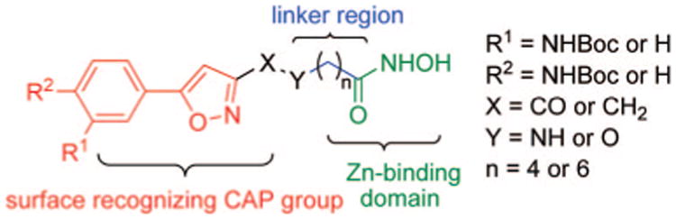
A series of hydroxyoxime acid HDAC inhibitors with phenylisoxazole as the CAP group were synthesized by nitrile oxide cycloaddition reaction. Among them, an HDAC6 selective inhibitor was identified with a potency of about 2 picomoles. Some compounds were tested for their ability to inhibit the growth of pancreatic cancer cells and found to be about 10 times more potent than SAHA. This study provides valuable new molecular probes for exploring the biology of HDAC. [1] Histone deacetylases (HDACs) are potential targets for cancer therapy, and first-generation HDAC inhibitors are currently undergoing clinical trials in cancer patients. HDAC6 is a key regulator of many cancer-related signaling pathways and has become a very attractive target for cancer therapy in recent years. This study found that HDAC6 is overexpressed in lung adenocarcinoma cell lines and its expression level is negatively correlated with the prognosis of lung adenocarcinoma patients. Overexpression of HDAC6 promotes the proliferation of lung adenocarcinoma cells in a deacetylase activity-dependent manner. Overexpression of HDAC6 confers resistance to gefitinib by stabilizing epidermal growth factor receptor (EGFR). CAY10603 is a potent and selective HDAC6 inhibitor that inhibits the proliferation of lung adenocarcinoma cells and induces apoptosis by inhibiting HDAC6 activity. CAY10603 downregulates the expression level of EGFR protein, thereby inhibiting the activation of the EGFR signaling pathway. In addition, CAY10603 synergizes with gefitinib to induce apoptosis in lung adenocarcinoma cell lines through EGFR instability. In summary, our results suggest that inhibiting HDAC6 may be a promising strategy for the treatment of lung adenocarcinoma. [2] Based on compound 12c (a recently discovered dual inhibitor of HDAC6/tubulin) and CAY10603 (a known HDAC6 inhibitor), we designed and synthesized a series of 2-phenylthiazole analogs as potential histone deacetylase 6 (HDAC6) inhibitors. Among the various HDAC6 inhibitors, XP5 was the most potent, with an IC50 of 31 nM and excellent HDAC6 selectivity (selectivity index SI = 338 for HDAC6 over HDAC3). XP5 exhibited high antiproliferative activity (IC50 = 0.16-2.31 μM) against multiple cancer cell lines, including HDACi-resistant YCC3/7 gastric cancer cells, superior to CAY10603. Furthermore, XP5 (50 mg/kg) demonstrated significant antitumor efficacy in a melanoma tumor model, achieving a tumor growth inhibition rate (TGI) of 63%, with no significant toxicity observed. In addition, the combination of XP5 and a small molecule PD-L1 inhibitor effectively enhanced the in vivo antitumor immune response, manifested as an increase in the number of tumor-infiltrating lymphocytes and a decrease in PD-L1 expression levels. In conclusion, these results indicate that XP5 is a promising HDAC6 inhibitor worthy of further investigation. J Med Chem. 2022 Feb 10;65(3):2434-2457. 1. Mechanism of action: CAY10603 alleviates acute and chronic kidney injury by selectively inhibiting HDAC6. HDAC6 inhibition increases the level of acetylated α-tubulin (Ac-α-tubulin), thereby reducing the nuclear translocation of ATF6 and inhibiting the activation of the ATF6 branch in the unfolded protein response (UPR). This leads to reduced endoplasmic reticulum stress, reduced apoptosis, and inhibition of inflammation and fibrosis in kidney tissue [3] 2. Background: HDAC6 is upregulated in kidney injury models (e.g., cisplatin-induced AKI, diabetic CKI), and activation of the ATF6-UPR pathway promotes renal cell death and fibrosis. CAY10603, as a selective HDAC6 inhibitor, offers a potential treatment strategy for kidney injury [3]. 3. Clinical status: Literature published in 2024 indicates that CAY10603 is currently in the preclinical research stage for kidney injury; no clinical trials, FDA approvals, or indications have been reported [3]. |
| Molecular Formula |
C22H30N4O6
|
|
|---|---|---|
| Molecular Weight |
446.5
|
|
| Exact Mass |
446.216
|
|
| Elemental Analysis |
C, 59.18; H, 6.77; N, 12.55; O, 21.50
|
|
| CAS # |
1045792-66-2
|
|
| Related CAS # |
|
|
| PubChem CID |
24951314
|
|
| Appearance |
White to light yellow solid powder
|
|
| Density |
1.2±0.1 g/cm3
|
|
| Index of Refraction |
1.563
|
|
| LogP |
1.94
|
|
| Hydrogen Bond Donor Count |
4
|
|
| Hydrogen Bond Acceptor Count |
7
|
|
| Rotatable Bond Count |
12
|
|
| Heavy Atom Count |
32
|
|
| Complexity |
616
|
|
| Defined Atom Stereocenter Count |
0
|
|
| SMILES |
O(C(N([H])C1C([H])=C([H])C(=C([H])C=1[H])C1=C([H])C(C(N([H])C([H])([H])C([H])([H])C([H])([H])C([H])([H])C([H])([H])C([H])([H])C(N([H])O[H])=O)=O)=NO1)=O)C(C([H])([H])[H])(C([H])([H])[H])C([H])([H])[H]
|
|
| InChi Key |
WWGBHDIHIVGYLZ-UHFFFAOYSA-N
|
|
| InChi Code |
InChI=1S/C22H30N4O6/c1-22(2,3)31-21(29)24-16-11-9-15(10-12-16)18-14-17(26-32-18)20(28)23-13-7-5-4-6-8-19(27)25-30/h9-12,14,30H,4-8,13H2,1-3H3,(H,23,28)(H,24,29)(H,25,27)
|
|
| Chemical Name |
tert-butyl N-[4-[3-[[7-(hydroxyamino)-7-oxoheptyl]carbamoyl]-1,2-oxazol-5-yl]phenyl]carbamate
|
|
| Synonyms |
CAY-10603; CAY10603; BML-281; tert-Butyl (4-(3-((7-(hydroxyamino)-7-oxoheptyl)carbamoyl)isoxazol-5-yl)phenyl)carbamate; CAY-10603; tert-butyl N-[4-[3-[[7-(hydroxyamino)-7-oxoheptyl]carbamoyl]-1,2-oxazol-5-yl]phenyl]carbamate; CHEMBL511749; compound 3 [PMID: 18642892]; CAY 10603
|
|
| HS Tariff Code |
2934.99.9001
|
|
| Storage |
Powder -20°C 3 years 4°C 2 years In solvent -80°C 6 months -20°C 1 month |
|
| Shipping Condition |
Room temperature (This product is stable at ambient temperature for a few days during ordinary shipping and time spent in Customs)
|
| Solubility (In Vitro) |
DMSO: ~89 mg/mL (~199.3 mM)
Water: 1 mg/mL (Insoluble) Ethanol: ~5 mg/mL (~11.2 mM) |
|---|---|
| Solubility (In Vivo) |
Solubility in Formulation 1: ≥ 2.5 mg/mL (5.60 mM) (saturation unknown) in 10% DMSO + 40% PEG300 + 5% Tween80 + 45% Saline (add these co-solvents sequentially from left to right, and one by one), clear solution.
For example, if 1 mL of working solution is to be prepared, you can add 100 μL of 25.0 mg/mL clear DMSO stock solution to 400 μL PEG300 and mix evenly; then add 50 μL Tween-80 to the above solution and mix evenly; then add 450 μL normal saline to adjust the volume to 1 mL. Preparation of saline: Dissolve 0.9 g of sodium chloride in 100 mL ddH₂ O to obtain a clear solution. Solubility in Formulation 2: ≥ 2.5 mg/mL (5.60 mM) (saturation unknown) in 10% DMSO + 90% (20% SBE-β-CD in Saline) (add these co-solvents sequentially from left to right, and one by one), clear solution. For example, if 1 mL of working solution is to be prepared, you can add 100 μL of 25.0 mg/mL clear DMSO stock solution to 900 μL of 20% SBE-β-CD physiological saline solution and mix evenly. Preparation of 20% SBE-β-CD in Saline (4°C,1 week): Dissolve 2 g SBE-β-CD in 10 mL saline to obtain a clear solution. View More
Solubility in Formulation 3: ≥ 2.5 mg/mL (5.60 mM) (saturation unknown) in 10% DMSO + 90% Corn Oil (add these co-solvents sequentially from left to right, and one by one), clear solution. Solubility in Formulation 4: 5% DMSO+50% PEG 300+ddH2O: 9mg/mL Solubility in Formulation 5: 5 mg/mL (11.20 mM) in 50% PEG300 50% Saline (add these co-solvents sequentially from left to right, and one by one), suspension solution; Need ultrasonic and warming and heat to 42°C. Preparation of saline: Dissolve 0.9 g of sodium chloride in 100 mL ddH₂ O to obtain a clear solution. |
| Preparing Stock Solutions | 1 mg | 5 mg | 10 mg | |
| 1 mM | 2.2396 mL | 11.1982 mL | 22.3964 mL | |
| 5 mM | 0.4479 mL | 2.2396 mL | 4.4793 mL | |
| 10 mM | 0.2240 mL | 1.1198 mL | 2.2396 mL |
*Note: Please select an appropriate solvent for the preparation of stock solution based on your experiment needs. For most products, DMSO can be used for preparing stock solutions (e.g. 5 mM, 10 mM, or 20 mM concentration); some products with high aqueous solubility may be dissolved in water directly. Solubility information is available at the above Solubility Data section. Once the stock solution is prepared, aliquot it to routine usage volumes and store at -20°C or -80°C. Avoid repeated freeze and thaw cycles.
Calculation results
Working concentration: mg/mL;
Method for preparing DMSO stock solution: mg drug pre-dissolved in μL DMSO (stock solution concentration mg/mL). Please contact us first if the concentration exceeds the DMSO solubility of the batch of drug.
Method for preparing in vivo formulation::Take μL DMSO stock solution, next add μL PEG300, mix and clarify, next addμL Tween 80, mix and clarify, next add μL ddH2O,mix and clarify.
(1) Please be sure that the solution is clear before the addition of next solvent. Dissolution methods like vortex, ultrasound or warming and heat may be used to aid dissolving.
(2) Be sure to add the solvent(s) in order.
|
|---|
|
|
Products are for research use only; We do not sell to patients
Copyright 2020 InvivoChem LLC | All Rights Reserved


 COA
COA