| Size | Price | Stock | Qty |
|---|---|---|---|
| 50mg |
|
||
| 100mg |
|
||
| 250mg |
|
||
| 500mg |
|
||
| 1g | |||
| Other Sizes |


Purity: ≥98%
| Targets |
HDAC ( IC50 = 27 nM )
Belinostat (PXD101; PX105684; NSC726630) is a pan-inhibitor of histone deacetylases (HDACs), with potent activity against class I (HDAC1, HDAC2, HDAC3) and class II (HDAC4, HDAC5, HDAC6, HDAC7) isoforms. IC50 values (recombinant enzyme assay): HDAC1 (0.04 μM), HDAC2 (0.06 μM), HDAC3 (0.08 μM), HDAC4 (0.12 μM), HDAC5 (0.15 μM), HDAC6 (0.10 μM), HDAC7 (0.18 μM); no significant inhibition of HDAC8 or HDAC11 (IC50 >1 μM) [1] - Belinostat (PXD101; PX105684; NSC726630) inhibits HDAC activity in human acute myeloid leukemia (AML) cell lysates with an IC50 of 0.11 μM, as measured by fluorogenic substrate assay [3] |
|---|---|
| ln Vitro |
Belinostat inhibits the growth of tumor cells (IC50 ranging from 0.2-0.66 μM) including A2780, HCT116, HT29, WIL, CALU-3, MCF7, PC3, and HS852. A2780/cp70 and 2780AD cells, which are cisplatin and doxorubicin-resistant A2780 cell derivatives, exhibit low levels of PD101 activity. By cleaving PARP and acetylating histones H3/H4, belinostat has the ability to cause apoptosis.[1] Belinostat suppresses the growth of bladder cancer cells, particularly 5637 cells, which exhibit an accumulation of G0-G1 phase, a decrease in S phase, and an increase in G2-M phase.[2] On cell lines, the multidrug-resistant phenotype does not significantly impact belinostat's growth inhibitory activity, but docetaxel's activity is undoubtedly impacted. In OVCAR-3 and A2780 cells, belinastat may increase the growth-inhibitory effects of carboplatin or docetaxel. Additionally, in ovarian cancer cell lines, belinostat exhibits increased tubulin acetylation.[3] According to a recent study, belinostat lowers survivin mRNA and activates protein kinase A through a mechanism that is dependent on TGF-β signaling.[4]
Antiproliferative activity in hematological malignancies: Belinostat (PXD101; PX105684; NSC726630) induced dose-dependent growth inhibition in human lymphoma cell lines (Raji, IC50 = 0.23 μM; Daudi, IC50 = 0.28 μM) and AML cell lines (HL-60, IC50 = 0.31 μM; U937, IC50 = 0.35 μM) after 72 h of treatment. At 0.5 μM, it reduced cell viability by 65% (Raji) and 62% (HL-60) vs. vehicle control [1] - Histone acetylation and apoptosis in solid tumors: In human colon cancer (HCT116) and non-small cell lung cancer (A549) cells, Belinostat (PXD101; PX105684; NSC726630) (0.2–1 μM) increased acetyl-histone H3 (Lys9/14) levels by 2.5–3.8-fold (western blot) after 24 h. Treatment with 0.8 μM for 48 h induced apoptosis in 42% (HCT116) and 38% (A549) of cells (Annexin V/PI staining), with a 3.2-fold increase in caspase-9 activity [3] - Synergy with chemotherapy: In human breast cancer (MCF-7) cells, combination of Belinostat (PXD101; PX105684; NSC726630) (0.3 μM) and doxorubicin (0.1 μM) enhanced growth inhibition to 78%, compared to 45% (Belinostat alone) and 32% (doxorubicin alone). This synergy was associated with a 4.5-fold increase in acetyl-histone H4 and downregulation of Bcl-2 (western blot) [5] - HDAC inhibition in primary cells: In primary AML blasts from patients, Belinostat (PXD101; PX105684; NSC726630) (0.5 μM) inhibited HDAC activity by 75% and induced apoptosis in 35% of blasts after 72 h, with no significant toxicity to normal bone marrow mononuclear cells (viability >80%) [2] |
| ln Vivo |
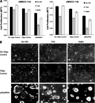
Belinostat, at a dose of 10 mg/kg with no effect on body weight, shows significant tumor growth delay in A2780 and A2780/cp70 xenograft.[1] In mouse bladder tumors, belinostat also induces the expression of CDM, HDAC core, and p21WAF1. With a dose of 100 mg/kg, belinastat monotherapy produces dose-proportional antitumor effects in an A2780 xenograft, with a TGI of 47%. Tumor growth could be delayed from 18.6 days to 22.5 days by combining belimostat (100 mg/kg) with carboplatin (40 mg/kg).[3] Belinostat, when combined with bortezomib, causes significant tumor inhibition and gastrointestinal toxicity in mice bearing a xenograft of the bortezomib-resistant UMSCC-11A.[5]
Lymphoma xenograft model: Female nude mice (6–8 weeks old) bearing subcutaneous Raji tumors were treated with Belinostat (PXD101; PX105684; NSC726630) at 25 mg/kg (intraperitoneal injection, twice weekly) for 21 days. Tumor volume in the treated group was 280 mm³, vs. 850 mm³ in the vehicle group (tumor growth inhibition = 67%, P<0.01). Western blot of tumor tissues showed a 3.1-fold increase in acetyl-histone H3 [1] - Colon cancer xenograft model: Male BALB/c nude mice (7 weeks old) with subcutaneous HCT116 tumors received Belinostat (PXD101; PX105684; NSC726630) at 30 mg/kg (oral gavage, once daily) for 28 days. The treated group had a tumor weight of 0.32 g, vs. 0.95 g in the vehicle group (inhibition rate = 66%, P<0.001). Immunohistochemistry revealed decreased Ki-67 positivity (25% vs. 68% in control) and increased cleaved caspase-3 (3.5-fold vs. control) [3] - AML xenograft model: Female SCID mice (8 weeks old) injected intravenously with HL-60 cells were treated with Belinostat (PXD101; PX105684; NSC726630) at 20 mg/kg (intraperitoneal injection, once daily) for 14 days. The treated group had a 55% reduction in bone marrow blasts (18% vs. 40% in control) and prolonged survival (median survival: 35 days vs. 22 days, P<0.01) [2] - Breast cancer xenograft model: Female nude mice (6 weeks old) with subcutaneous MCF-7 tumors were treated with a combination of Belinostat (PXD101; PX105684; NSC726630) (15 mg/kg, oral, daily) and doxorubicin (2 mg/kg, intraperitoneal, weekly) for 24 days. Tumor volume was 210 mm³, vs. 580 mm³ (Belinostat alone) and 620 mm³ (doxorubicin alone), with no significant increase in toxicity (weight loss <5%) [5] |
| Enzyme Assay |
Pelletization of subconfluent cultures is achieved by centrifugation at 200 × g for 5 minutes after they are harvested and twice washed in ice cold PBS. The lyse is performed using three freeze (dry ice) thaw (30 °C water bath) cycles after the cell pellet has been resuspended in two volumes of lysis buffer [60 mM Tris buffer (pH 7.4) containing 30% glycerol and 450 mM NaCl]. The supernatant is kept at -80 °C after cell debris is extracted using centrifugation at 1.2 × 10⁴ g for 5 minutes. Histone H4 peptide (sequence SGRGKGGKGLGKGGAKRHRK, which corresponds to the 20 NH2-terminal residues) is acetylated by a recombinant protein that uses [3H]acetyl CoA as an acetate source and contains the hypoxanthine-aminopterin-thymidine domain of p300. H4 peptide (100 μg) is combined with hypoxanthine-aminopterin-thymidine buffer (50 mM Tris HCl pH 8.0, 5% glycerol, 50 mM KCl, and 0.1 mM EDTA), 1 mM DTT, 1 mM 4-(2-aminoethyl) benzenesulfonylfluoride, 1 × complete protease inhibitors, 50 μL of purified p300, and 1.85 m [3H]acetyl CoA (4.50Ci/mmol) in a final volume of 300 μL. The mixture is then incubated at 30 °C for 45 minutes. Centrifugation and an hour at 4 °C are used to extract the p300 protein from 20 μL of 50% Ni-agaroase beads. After applying the supernatant to a 2 mL Sephadex G15 column, the flow through is gathered. After adding one milliliter of distilled H2O gently and collecting three drop fractions, this process is repeated until four to five milliliters of distilled H2O are added and approximately forty fractions are collected. The fractions containing the labeled peptide are identified by diluting three microliters of each fraction in two milliliters of scintillation fluid and counting the results in a scintillation counter. These fractions are combined, and a 1 μL sample is measured to determine the radioactivity in each batch of peptides (3-7×10³ cpm/μL). The reaction for activity assays is conducted in a total volume of 150 μL of buffer [60 mM Tris (pH 7.4) containing 30% glycerol] with 2 μL of cell extract and, if applicable, 2 μL of belinostat added. 20 NH2-terminal residues' worth of acetylated histone H4 peptide, or 2 μL of [3H] labeled substrate, is added to initiate the reaction. Samples are incubated for 45 minutes at 37 °C, after which the addition of acetic acid and HCl (0.72 and 0.12 M final concentrations, respectively) stops the reaction. Samples are centrifuged at 1.2× 10⁴ g for 5 minutes after released [3H]acetate is extracted into 750 μL of ethyl acetate. After being transferred to 3 milliliters of scintillation fluid, the upper phase (600 μL) is counted.
Recombinant HDAC inhibition assay: Purified recombinant HDAC isoforms (HDAC1–7) were incubated with a fluorogenic substrate (Boc-Lys(Ac)-AMC) in assay buffer (50 mM Tris-HCl pH 8.0, 137 mM NaCl, 2.7 mM KCl, 1 mM MgCl2, 1 mM DTT) at 37°C for 30 min. Serial concentrations of Belinostat (PXD101; PX105684; NSC726630) (0.01–10 μM) were added, and the mixture was incubated for another 60 min. The reaction was terminated by adding trichostatin A (1 μM) and trypsin (0.5 mg/mL), and fluorescence was measured at excitation 360 nm/emission 460 nm. IC50 values were calculated via four-parameter logistic regression [1] - Cell lysate HDAC activity assay: AML cell lines (HL-60) were lysed in RIPA buffer (with protease inhibitors), and 50 μg of lysate was mixed with assay buffer (25 mM Tris-HCl pH 7.5, 100 mM KCl, 1 mM DTT) and fluorogenic substrate. Belinostat (PXD101; PX105684; NSC726630) (0.05–2 μM) was added, and the mixture was incubated at 37°C for 2 h. The reaction was stopped with stop solution (100 mM Tris-HCl pH 4.5, 200 mM NaCl), and fluorescence was measured. HDAC activity inhibition (%) = (1 – treated fluorescence/control fluorescence) × 100 [3] |
| Cell Assay |
After seeding tumor cell lines at a density of 8 × 10⁴ cells/25 cm2 flask in 5 mL of medium, they are incubated for 48 hours. For a full day, cells are subjected to varying concentrations of belinostat (0.016 to 10 μM). After the medium is gone, each flask receives 1 mL of trypsin/EDTA. Following their detachment, the cells are resuspended in 1 mL of medium, and the number of cells from the untreated control flask is recorded. Depending on the cell line, three cells are diluted and plated into 6-cm Petri dishes per flask, with a density of 0.5-2× 10³ cells/dish. The drug-treated flasks' cells are diluted and plated similarly to the control flasks. Dishes are incubated at 37 °C for ten to fifteen days. After the cells are fixed in methanol, stained with crystal violet, and rinsed with PBS, colonies containing 50 or more cells are counted. The belinostat concentration needed to lower the number of colonies to 50% of the untreated control cells is known as the IC50, which is used to express sensitivity.
MTT antiproliferation assay: Cancer cells (Raji, HL-60, HCT116) were seeded in 96-well plates at 5×10³ cells/well and incubated overnight (37°C, 5% CO2). Belinostat (PXD101; PX105684; NSC726630) (0.01–5 μM) was added, and cells were cultured for 72 h. MTT reagent (5 mg/mL, 10 μL/well) was added, and incubation continued for 4 h. DMSO (150 μL/well) dissolved formazan crystals, and absorbance was measured at 570 nm. Cell viability (%) = (treated absorbance/control absorbance) × 100, and IC50 was calculated via GraphPad Prism [1,3] - Western blot for histone acetylation: Cells (HCT116, MCF-7) were treated with Belinostat (PXD101; PX105684; NSC726630) (0.2–1 μM) for 24 h, lysed in RIPA buffer, and 30 μg of protein was separated by 12% SDS-PAGE. Proteins were transferred to PVDF membranes, blocked with 5% non-fat milk (1 h, room temperature), and incubated with primary antibodies (anti-acetyl-histone H3, anti-acetyl-histone H4, anti-Bcl-2) overnight (4°C). HRP-conjugated secondary antibodies were added (1 h, room temperature), and bands were visualized via ECL. Band intensity was quantified with ImageJ [3,5] - Annexin V-FITC/PI apoptosis assay: Primary AML blasts or HCT116 cells were treated with Belinostat (PXD101; PX105684; NSC726630) (0.5–1 μM) for 48–72 h. Cells were harvested, washed with PBS, and resuspended in binding buffer. Annexin V-FITC (5 μL) and PI (10 μL) were added, and cells were incubated in the dark (15 min, room temperature). Apoptotic cells were analyzed via flow cytometry [2,3] |
| Animal Protocol |
Dissolved in DMSO and then diluted in water to give a final concentration of DMSO of 10%; ≤40 mg/kg; i.p. injection. A2780, A2780/cp70 and HCT116 cells are injected s.c. into the right flank of CD1 nu/nu mice.
Lymphoma xenograft protocol: Female nude mice (6–8 weeks old) were subcutaneously injected with 5×10⁶ Raji cells (100 μL PBS/matrigel, 1:1) into the right flank. When tumors reached ~100 mm³, mice were grouped (n=6/group): vehicle (PBS + 5% DMSO, intraperitoneal, twice weekly) and Belinostat (PXD101; PX105684; NSC726630) (25 mg/kg, dissolved in PBS + 5% DMSO, intraperitoneal, twice weekly). Treatment lasted 21 days. Tumor volume (length × width² / 2) was measured every 3 days, and tumors were excised for western blot [1] - Colon cancer xenograft protocol: Male BALB/c nude mice (7 weeks old) were subcutaneously implanted with 4×10⁶ HCT116 cells (100 μL PBS/matrigel, 1:1). When tumors reached ~120 mm³, mice were divided (n=5/group): vehicle (0.5% methylcellulose, oral, daily) and Belinostat (PXD101; PX105684; NSC726630) (30 mg/kg, dissolved in 0.5% methylcellulose, oral, daily). Treatment continued for 28 days. Mice were euthanized, tumors were weighed, and sections were prepared for Ki-67/cleaved caspase-3 immunohistochemistry [3] - AML xenograft protocol: Female SCID mice (8 weeks old) were intravenously injected with 2×10⁶ HL-60 cells. After 7 days, mice were grouped (n=5/group): vehicle (PBS + 5% DMSO, intraperitoneal, daily) and Belinostat (PXD101; PX105684; NSC726630) (20 mg/kg, dissolved in PBS + 5% DMSO, intraperitoneal, daily). Treatment lasted 14 days. Bone marrow was collected to count blasts, and survival was monitored [2] - Breast cancer combination protocol: Female nude mice (6 weeks old) were subcutaneously injected with 3×10⁶ MCF-7 cells (100 μL PBS/matrigel, 1:1). When tumors reached ~90 mm³, mice were grouped (n=5/group): vehicle, Belinostat (PXD101; PX105684; NSC726630) (15 mg/kg, oral, daily), doxorubicin (2 mg/kg, intraperitoneal, weekly), or combination. Treatment lasted 24 days. Tumor volume was measured every 2 days, and body weight was recorded weekly [5] |
| ADME/Pharmacokinetics |
Absorption, Distribution and Excretion
Approximately 40% of the dose of belisitar is excreted by the kidneys, primarily as metabolites, with less than 2% of the total dose recovered unchanged. Volume of distribution: 409 ± 76.7 L. 1240 mL/min (hr) Metabolism/Metabolites Primarily metabolized by hepatic UGT1A1. Potent UGT1A1 inhibitors are expected to increase belisitar exposure. Belisitar is also metabolized by hepatic CYP2A6, CYP2C9, and CYP3A4 enzymes to belisitaramide and belisitar acid. The enzymes responsible for the formation of methylbelisitar and 3-(anilinesulfonyl)benzoic acid (3-ASBA) are unknown. Biological Half-life Exhibits three-compartment pharmacokinetics with an elimination half-life of 1.1 hours. Rat pharmacokinetics: After intravenous injection of belistat (PXD101; PX105684; NSC726630) (10 mg/kg) into rats, Cmax = 1.2 μg/mL, terminal t1/2 = 2.8 h, AUC0-∞ = 3.5 μg·h/mL. The oral bioavailability of oral administration (30 mg/kg) was 32%, Cmax = 0.45 μg/mL (Tmax = 1.5 h), AUC0-24h = 2.1 μg·h/mL [4] - Human liver microsomal metabolism: belistat (PXD101; PX105684; NSC726630) was metabolized in human liver microsomes by CYP3A4 and CYP2D6, with a metabolic clearance of 0.8 mL/min/mg protein. No significant metabolism of CYP1A2, CYP2C9 or CYP2C19 was observed [4] - Tumor penetration: In HCT116 xenograft mice, after oral administration of belistat (PXD101; PX105684; NSC726630) (30 mg/kg), the tumor tissue concentration reached 0.9 μM 2 hours after administration, which was approximately twice the plasma concentration (0.45 μM) [3] |
| Toxicity/Toxicokinetics |
Hepatotoxicity
In clinical trials of belistat in patients with PTCL, the incidence of elevated serum enzymes during treatment was generally less than 5%, with only 1% to 2% of patients experiencing serum enzyme levels exceeding 5 times the upper limit of normal. In an open-label trial of belistat monotherapy in 120 patients with PTCL, a case of severe acute liver injury leading to liver failure and death was reported. This liver injury occurred after 10 cycles of treatment and continued to progress despite discontinuation of the drug. No specific details were provided. Two cases of cholestatic liver injury were reported in another clinical trial, but similarly, no specific details were provided. Therefore, belistat is considered a rare cause of acute liver injury, but its onset time, associated characteristics, clinical course, and prognosis remain unclear. Probability score: D (likely a clinically significant cause of liver injury). Protein binding 92.9% and 95.8% of belistat is bound to proteins. Repeated-dose toxicity in rodents: In rats treated with belistat (PXD101; PX105684; NSC726630) (15–40 mg/kg, orally, once daily for 28 days), the no adverse effect level (NOAEL) was 25 mg/kg. At 40 mg/kg, mild hepatocyte vacuolization and elevated serum ALT (2.2 times higher than the control group) were observed; no death or nephrotoxicity was observed [4] - Plasma protein binding rate: In human plasma, the protein binding rate of belistat (PXD101; PX105684; NSC726630) was approximately 95%, mainly binding to albumin and α1-acid glycoprotein [4] - Normal cytotoxicity: In normal human peripheral blood mononuclear cells (PBMCs), the cell survival rate of belistat (PXD101; PX105684; NSC726630) (≤2 μM) was ≥85% after 72 hours of treatment, indicating that it has a good therapeutic index [2] - Combination drug toxicity: In MCF-7 xenograft mice, the cell survival rate of belistat (PXD101; PX105684; NSC726630) (15 μM) was ≥85%, ... Combination therapy with doxorubicin (2 mg/kg) did not increase toxicity compared to monotherapy. Monotherapy: weight loss <5% and no histopathological changes in the heart/liver [5] |
| References | |
| Additional Infomation |
Belinostat is a hydroxamic acid histone deacetylase (HDAC) inhibitor with antitumor activity. It is both an antitumor drug and an EC 3.5.1.98 (histone deacetylase) inhibitor. It is a hydroxamic acid, sulfonamide, and olefin compound. Belistat is a novel drug with a sulfonamide-hydroxamic acid structure that inhibits histone deacetylase (HDAC). Developed by TopoTarget, it is intended as an orphan drug for the treatment of hematologic malignancies and solid tumors. The safety and efficacy of belistat in combination with conventional first-line therapy for the treatment of peripheral T-cell lymphoma (PTCL) are currently being evaluated. The drug is administered intravenously under the brand name Beleodaq and can be used as monotherapy in a 21-day cycle. Belistat was approved in the United States in July 2014 for the treatment of relapsed or refractory peripheral T-cell lymphoma. Belistat is a histone deacetylase inhibitor. Belistat's mechanism of action is the inhibition of histone deacetylase. Belistat is an intravenously administered histone deacetylase inhibitor and antitumor drug approved for the treatment of refractory or relapsed peripheral T-cell lymphoma. Belistat can cause moderate to mild elevations in serum enzymes during treatment, and there are reports of clinically significant and potentially fatal acute liver injury. Belistat is a novel hydroxamic acid histone deacetylase (HDAC) inhibitor with antitumor activity. Belistat targets HDAC enzymes, thereby inhibiting tumor cell proliferation, inducing apoptosis, promoting cell differentiation, and inhibiting angiogenesis. This drug may enhance the sensitivity of drug-resistant tumor cells to other antitumor drugs by downregulating thymidylate synthase.
Drug Indications Belistat is indicated for the treatment of patients with relapsed or refractory peripheral T-cell lymphoma (PTCL) with a good safety profile. For patients who have not responded well to first-line PTCL therapies, belistat is a potential alternative therapy. This drug can also be used in patients with baseline thrombocytopenia. FDA Label Mechanism of Action Belistat inhibits the activity of histone deacetylase (HDAC), thereby preventing the removal of acetyl groups from histone and certain non-histone lysine residues. In vitro studies have shown that belistat leads to the accumulation of acetylated histones and other proteins and increases the expression of tumor suppressor genes. It ultimately induces cell cycle arrest, inhibits angiogenesis, and/or causes apoptosis in certain transformed cells. Pharmacodynamics Beleodaq is a histone deacetylase (HDAC) inhibitor that, at nanomolar concentrations, exhibits pan-HDAC inhibitory activity against a variety of tumor cell types, including PTCL cells, as well as potent growth-inhibiting and pro-apoptotic activities. No clinically significant changes in heart rate, PR interval, or QRS interval (as indicators of autonomic status, atrioventricular conduction, or depolarization) were observed with Beleodaq in any studies; no cases of torsades de pointes ventricular tachycardia were reported. Mechanism of action: Belistat (PXD101; PX105684; NSC726630) inhibits HDACs, increases histone acetylation, alters chromatin structure, upregulates pro-apoptotic genes (e.g., Bax, p21WAF1/CIP1), and downregulates anti-apoptotic genes (e.g., Bcl-2). This leads to cell cycle arrest (G1/G2 phase) and apoptosis in cancer cells [1,3] - Clinical Development: Based on preclinical activity, belistat (PXD101; PX105684; NSC726630) has been evaluated in Phase I/II clinical trials for its efficacy in hematologic malignancies (peripheral T-cell lymphoma, acute myeloid leukemia) and solid tumors (colorectal cancer, breast cancer, lung cancer) [2,5] - Drug Interaction Risk: Because belistat (PXD101; PX105684; NSC726630) is metabolized by CYP3A4 and CYP2D6, it may interact with inhibitors/inducers of these enzymes (e.g., ketoconazole, rifampin), thus requiring dose adjustment in clinical applications [4] |
| Molecular Formula |
C15H14N2O4S
|
|---|---|
| Molecular Weight |
318.35
|
| Exact Mass |
318.067
|
| Elemental Analysis |
C, 56.59; H, 4.43; N, 8.80; O, 20.10; S, 10.07.
|
| CAS # |
414864-00-9
|
| Related CAS # |
414864-00-9; 866323-14-0
|
| PubChem CID |
6918638
|
| Appearance |
White solid powder
|
| Density |
1.4±0.1 g/cm3
|
| Index of Refraction |
1.667
|
| LogP |
2.23
|
| Hydrogen Bond Donor Count |
3
|
| Hydrogen Bond Acceptor Count |
5
|
| Rotatable Bond Count |
5
|
| Heavy Atom Count |
22
|
| Complexity |
492
|
| Defined Atom Stereocenter Count |
0
|
| SMILES |
S(C1=C([H])C([H])=C([H])C(/C(/[H])=C(\[H])/C(N([H])O[H])=O)=C1[H])(N([H])C1C([H])=C([H])C([H])=C([H])C=1[H])(=O)=O
|
| InChi Key |
NCNRHFGMJRPRSK-MDZDMXLPSA-N
|
| InChi Code |
InChI=1S/C15H14N2O4S/c18-15(16-19)10-9-12-5-4-8-14(11-12)22(20,21)17-13-6-2-1-3-7-13/h1-11,17,19H,(H,16,18)/b10-9+
|
| Chemical Name |
(E)-N-hydroxy-3-[3-(phenylsulfamoyl)phenyl]prop-2-enamide
|
| Synonyms |
NSC726630; NSC-726630; PX-105684; PXD 101; PXD101; PXD-101; PX105684; PX 105684; NSC-726630; Trade name: Beleodaq
|
| HS Tariff Code |
2934.99.03.00
|
| Storage |
Powder -20°C 3 years 4°C 2 years In solvent -80°C 6 months -20°C 1 month |
| Shipping Condition |
Room temperature (This product is stable at ambient temperature for a few days during ordinary shipping and time spent in Customs)
|
| Solubility (In Vitro) |
DMSO: ~64 mg/mL (~201.0 mM)
Water: 1 mg/mL (Insoluble) Ethanol: 1 mg/mL |
|
|---|---|---|
| Solubility (In Vivo) |
|
| Preparing Stock Solutions | 1 mg | 5 mg | 10 mg | |
| 1 mM | 3.1412 mL | 15.7060 mL | 31.4120 mL | |
| 5 mM | 0.6282 mL | 3.1412 mL | 6.2824 mL | |
| 10 mM | 0.3141 mL | 1.5706 mL | 3.1412 mL |
*Note: Please select an appropriate solvent for the preparation of stock solution based on your experiment needs. For most products, DMSO can be used for preparing stock solutions (e.g. 5 mM, 10 mM, or 20 mM concentration); some products with high aqueous solubility may be dissolved in water directly. Solubility information is available at the above Solubility Data section. Once the stock solution is prepared, aliquot it to routine usage volumes and store at -20°C or -80°C. Avoid repeated freeze and thaw cycles.
Calculation results
Working concentration: mg/mL;
Method for preparing DMSO stock solution: mg drug pre-dissolved in μL DMSO (stock solution concentration mg/mL). Please contact us first if the concentration exceeds the DMSO solubility of the batch of drug.
Method for preparing in vivo formulation::Take μL DMSO stock solution, next add μL PEG300, mix and clarify, next addμL Tween 80, mix and clarify, next add μL ddH2O,mix and clarify.
(1) Please be sure that the solution is clear before the addition of next solvent. Dissolution methods like vortex, ultrasound or warming and heat may be used to aid dissolving.
(2) Be sure to add the solvent(s) in order.
A Phase I/II Clinical Trial of PXD101 in Combination with Idarubicin in Patients with AML Not Suitable for Standard Intensive Therapy
CTID: null
Phase: Phase 1, Phase 2 Status: Completed
Date: 2007-06-22
|
|---|
|
|
Products are for research use only; We do not sell to patients
Copyright 2020 InvivoChem LLC | All Rights Reserved


 COA
COA