| Size | Price | Stock | Qty |
|---|---|---|---|
| 2mg |
|
||
| 5mg |
|
||
| 10mg |
|
||
| 25mg |
|
||
| 50mg |
|
||
| 100mg | |||
| 500mg | |||
| Other Sizes |


Purity: ≥98%
| Targets |
BACE1 (β-site amyloid precursor protein cleaving enzyme 1) with Ki = 26.1 nmol/L [1]
BACE2 with Ki = 372 nmol/L (14‑fold selectivity over BACE1) [1] Cathepsin D with Ki >25 μmol/L (>1000‑fold selectivity over BACE1) [1] AZD3839 targets BACE1 (β-secretase 1), an enzyme involved in the production of amyloid-beta peptides from amyloid precursor protein (APP). It is a potent and selective inhibitor with a Ki of 26.1 nM for human BACE1. It displays approximately 14-fold selectivity over BACE2 (Ki = 372.4 nM) and >1000-fold selectivity over cathepsin D. By inhibiting BACE1, AZD3839 reduces the generation of amyloid-beta peptides, which are implicated in the pathogenesis of Alzheimer's disease. Its brain permeability allows it to reach its target in the central nervous system. |
|---|---|
| ln Vitro |
In SH-SY5Y cells, AZD3839 efficiently decreases the Aβ40 levels with IC50 of 4.8 nM, and decreases the formation of sAPPβ with IC50 of 16.7 nM. AZD3839 also decreases the Aβ40 levels secreted from C57BL/6 mouse primary cortical neurons, N2A cells, and Dunkin-Hartley guinea pig primary cortical neurons with IC50 values of 50.9, 32.2, and 24.8 nM, respectively. AZD3839 causes in vitro BACE1 inhibition in the cell assay with IC50 value of 16.7 nM.
Kinase Assay: AZD3839 (free base) is a potent and selective BACE1 inhibitor with IC50 of 23.6 uM, about 14-fold selectivity over BACE2, also a β-secretase enzyme inhibitor. target: BACE1, β-secretase enzyme. Cell Assay: AZD3839 concentration-dependently inhibited BACE1 activity in a biochemical fluorescence resonance energy transfer assay, Aβ and sAPPβ release from modified and wild-type human SH-SY5Y cells and mouse N2A cells as well as from guinea pig and mouse primary cortical neurons. Selectivity against BACE2 and cathepsin D was 14 and >1000-fold, respectively. In biochemical FRET assay, AZD3839 achieved 100% inhibition of recombinant human BACE1 cleavage of a 10‑amino‑acid APP swe‑mutation sequence with Ki = 26.1 nmol/L. It showed 14‑fold selectivity against BACE2 (Ki = 372 nmol/L) and >1000‑fold selectivity against cathepsin D (Ki >25 μmol/L). [1] In SH‑SY5Y cells overexpressing APP695wt, AZD3839 inhibited Aβ40 and sAPPβ release in a concentration‑dependent manner. [1] In mouse N2A cells and wild‑type human SH‑SY5Y cells, as well as mouse and guinea pig primary cortical neurons, AZD3839 concentration‑dependently reduced Aβ40 and sAPPβ release. [1] AZD3839 completely abolished Aβ40 and sAPPβ production in vitro. [1] In vitro, AZD3839 is a potent BACE1 inhibitor with a Ki of 26 nM and a cell-based IC50 of 16.7 nM. It demonstrates 14-fold selectivity over BACE2 and >1000-fold selectivity over cathepsin D. Its activity has been characterized in enzyme assays using purified BACE1 and in cell-based assays measuring Aβ production. These studies confirm its potency and selectivity as a BACE1 inhibitor. It is a valuable tool for studying the role of BACE1 in Alzheimer's disease pathogenesis. |
| ln Vivo |
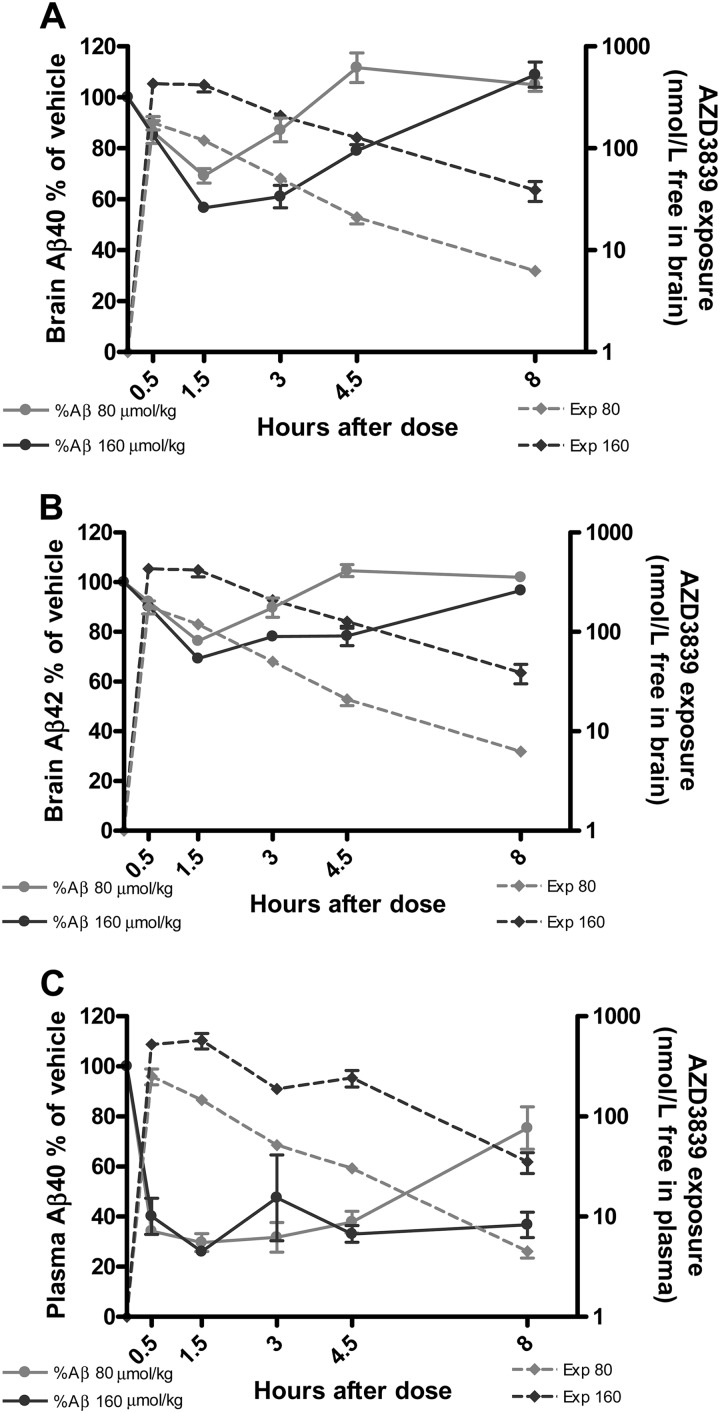
In C57BL/6 mice, AZD3839 (69 mg/kg, p.o.) causes a dose- and time-dependent reduction of plasma and brain Aβ. In guinea pig and non-human primates, AZD3839 also inhibits Aβ generation.
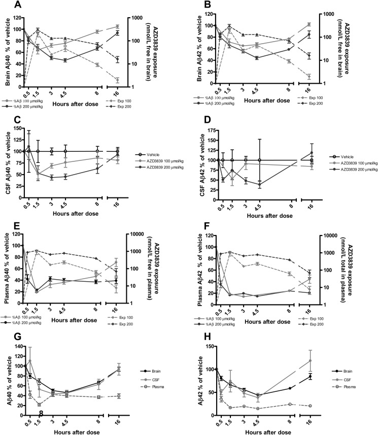
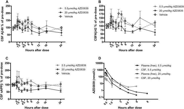
In C57BL/6 mice, oral administration of AZD3839 produced time‑ and dose‑dependent reductions of brain Aβ40 and Aβ42, as well as plasma Aβ40. A dose of 20 μmol/kg reduced brain Aβ40 by ~60‑70% at 4.5 h. The effect returned to baseline after 6‑8 h. [1] In guinea pigs, oral AZD3839 reduced Aβ40 and Aβ42 in brain, CSF, and plasma in a time‑ and dose‑dependent manner. The maximal inhibition in brain was ~60‑70% (except guinea pig brain where near 98% was achieved according to PKPD modeling). The effect in plasma was more pronounced than in brain and CSF. [1] In cynomolgus monkeys, a single 15‑min intravenous infusion of AZD3839 (5.5 and 20 μmol/kg) reduced CSF Aβ40, Aβ42, and sAPPβ in a time‑ and dose‑dependent manner. The higher dose produced a greater and more sustained reduction. [1] PKPD modeling showed that free plasma IC50 for Aβ40 reduction in mouse plasma was 5.9 nM, in mouse brain 380 nM; in guinea pig plasma 12 nM, in guinea pig brain 578 nM. [1] In vivo, AZD3839 exhibits dose- and time-dependent lowering of plasma, brain, and cerebrospinal fluid Aβ levels in mouse, guinea pig, and non-human primate models. As an orally active and brain-permeable compound, it effectively reduces Aβ production in the central nervous system. These in vivo findings support its potential for the treatment of Alzheimer's disease. It has been evaluated in Phase 1 clinical trials, but further development details are not extensively documented. |
| Enzyme Assay |
Human BACE1 and BACE2 time‑resolved FRET assay: The soluble part of recombinant hBACE1 (aa 1‑460) or hBACE2 (aa 1‑473) was mixed with compound in reaction buffer (sodium acetate, CHAPS, Triton X‑100, EDTA, pH 4.5) and preincubated for 10 min. Substrate (Europium)CEVNLDAAEFK(Qsy7) was added, and the reaction was allowed to proceed for 6.5 h in darkness at 22°C. The reaction was stopped by adding sodium acetate pH 9, and fluorescence was measured at excitation 340 nm and emission 615 nm. [1]
Human cathepsin D FRET assay: Cathepsin D enzyme and substrate (Ac‑Glu‑Asp(EDANS)‑Lys‑Pro‑Ile‑Leu‑Phe‑Phe‑Arg‑Leu‑Gly‑Lys(DABCYL)‑Glu‑NH2) were separately diluted in glycine‑HCl buffer. Cathepsin D was mixed with compound dissolved in DMSO and preincubated for 10 min. Substrate was added, and the mixture incubated for 15 min in darkness at 22°C. Fluorescence was measured at excitation 355 nm and emission 460 nm. [1] Non-cellular enzyme assays for AZD3839 typically involve measuring its inhibition of BACE1 activity using purified enzyme and a fluorogenic substrate. The compound's Ki of 26 nM is determined through these assays. Selectivity over BACE2 and cathepsin D is assessed using similar enzyme assays with the respective enzymes. These assays are essential for characterizing the compound's potency and selectivity as a BACE1 inhibitor. Dose-response curves are generated to calculate IC50 or Ki values. |
| Cell Assay |
SH‑SY5Y sAPPβ release assay: SH‑SY5Y cells were cultured in DMEM/F‑12 with Glutamax, 10% FCS, and 1% nonessential amino acids. Compound was incubated with cells for 16 h at 37°C in 5% CO2. Detection of sAPPβ release was performed using MSD plates according to manufacturer’s instructions, and plates were read in a SECTOR Imager. Cytotoxicity was assessed using a ViaLight Plus kit. [1]
SH‑SY5Y Aβ40 release assay: SH‑SY5Y cells overexpressing APP695wt were cultured in the same medium. Compound was incubated with cells for 16 h at 37°C, 5% CO2. Human Aβ40 secreted into medium was measured using ELISA strips (Invitrogen KHB3482) and read with a Spektramax microplate reader. [1] N2A Aβ40 release assay: N2A cells were cultured in minimum Eagle’s medium with 10% FCS, 10 mM HEPES, nonessential amino acids, and penicillin‑streptomycin. Compound was incubated with cells for 16 h at 37°C, 5% CO2. Mouse Aβ40 was measured using ELISA strips (Invitrogen KMB3481). [1] Mouse primary neuron Aβ40 release assay: Primary cortical cells from fetal C57BL/6 mice (E16) were isolated, dissociated, and plated on poly‑D‑lysine‑coated 96‑well plates at 200,000 cells/well. After 5 days, medium was replaced with medium containing AZD3839 (1% DMSO final) and incubated overnight. Released Aβ40 in extracellular medium was measured by ELISA. [1] In vitro cell-based assays for AZD3839 are used to measure its ability to inhibit Aβ production in cells. These assays typically use cell lines expressing APP, such as HEK293 cells, and measure Aβ levels in the culture medium using ELISA or other detection methods. The compound has a cell-based IC50 of 16.7 nM. These experiments are crucial for confirming its activity in a cellular context and for evaluating its potential to reduce Aβ production in vivo. |
| Animal Protocol |
Dissolved in 5% dimethylacetamide and 20% hydroxypropyl-β-cyclodextrin in 0.3 M gluconic acid, pH 3, or 0.3 M gluconic acid, pH 3.; 69 mg/kg; p.o. administration C57BL/6 mice.
Mouse in vivo experiments: C57BL/6 mice received oral administration of AZD3839 at doses of 5.5 and 20 μmol/kg (formulation not specified). Brain, plasma, and CSF were collected at various time points up to 24 h for Aβ measurement. For intravenous infusion, mice received 5.5 or 20 μmol/kg over 15 min. [1] Guinea pig in vivo experiments: Guinea pigs received oral AZD3839 (doses not explicitly given but referred to as dose‑dependent, with PKPD analysis using data from multiple dose groups). Brain, CSF, and plasma were collected for Aβ analysis. [1] Non‑human primate (cynomolgus monkey) experiment: Animals received a single 15‑min intravenous infusion of AZD3839 at 5.5 or 20 μmol/kg (vehicle not specified). CSF and plasma samples were collected pre‑dose and at multiple time points post‑infusion (up to 12 h). Aβ40, Aβ42, and sAPPβ were measured in CSF; drug concentrations were measured in plasma and CSF. [1] In vivo animal studies for AZD3839 have been conducted in mouse, guinea pig, and non-human primate models. The compound is typically administered orally, and its effects on Aβ levels in plasma, brain, and cerebrospinal fluid are measured over time. These studies demonstrate dose- and time-dependent lowering of Aβ levels. Such experiments are essential for validating its in vivo efficacy as a BACE1 inhibitor and for assessing its brain penetration. |
| ADME/Pharmacokinetics |
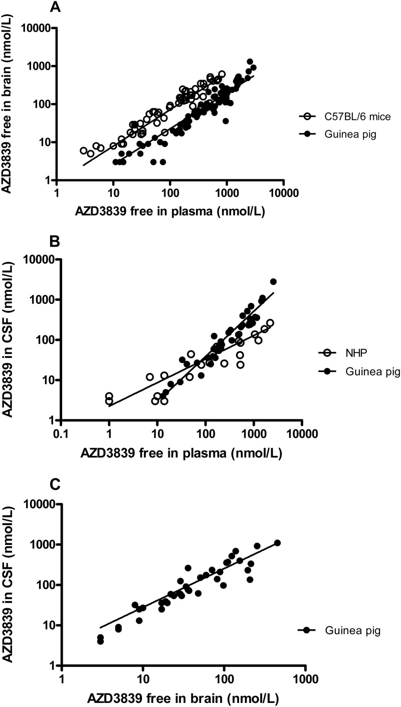
In C57BL/6 mice, the free brain/plasma concentration ratio of AZD3839 was 0.7, indicating unrestricted CNS access. The free plasma‑brain correlation had a slope of 0.96 (R²=0.78). [1]
In guinea pigs, free brain/plasma ratio was 0.3 (slope 0.94, R²=0.68); free CSF/plasma ratio was 0.7 (slope 1.1, R²=0.76); free CSF/brain ratio was 2.5 (slope 0.96, R²=0.76). [1] In non‑human primates, the correlation between free plasma and CSF concentrations had a slope of 0.59 (R²=0.75), indicating slower clearance in CSF compartment. [1] Following intravenous infusion in mice, a two‑compartment model estimated plasma half‑life of AZD3839 as 18 minutes. [1] Caco‑2 permeability (Papp) and efflux ratio: For the isoindole series leading to AZD3839, the efflux ratio was reduced to 3.5 (compared to >35 for earlier compounds). [1] AZD3839 has a molecular weight of 431.41 g/mol and a molecular formula of C24H16F3N5. It is soluble in DMSO at 125 mg/mL (289.75 mM). The compound should be stored in a dry, dark place at -20°C for up to 1 year. As an orally active and brain-permeable compound, it is expected to be well-absorbed and to distribute to the central nervous system. Its pharmacokinetic properties have been studied in preclinical models and clinical trials. |
| Toxicity/Toxicokinetics |
In 1‑month rodent and dog toxicology studies, no adverse target‑related effects were observed for AZD3839. No tolerance or sensitization to Aβ40 lowering in brain was seen after repeated dosing in mice. [1]
Comprehensive toxicological data for AZD3839 are not extensively detailed in standard summaries, as it is a research compound that has undergone Phase 1 clinical trials. As with all investigational drugs, its safety profile would have been evaluated in preclinical studies. However, specific toxicological findings are not publicly available. It is not an approved drug and is available only as a research compound for Alzheimer's disease research. |
| References |
J Biol Chem.2012 Nov 30;287(49):41245-57;J Med Chem.2012 Nov 8;55(21):9346-61.
|
| Additional Infomation |
AZD3839 has been used in basic science trials to study its safety, tolerability, Alzheimer's disease, and blood concentration.
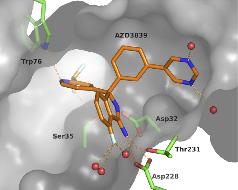
AZD3839 (CAS# 1227163-84-9) is a BACE1 inhibitor developed for Alzheimer disease. It targets the first step in Aβ formation by inhibiting BACE1, thereby reducing production of Aβ peptides, particularly Aβ42, which is implicated in amyloid plaque formation. The compound was discovered via fragment‑based lead generation and structure‑based design, with optimization focusing on improving permeability and reducing P‑glycoprotein‑mediated efflux to achieve brain exposure. It has been progressed into Phase 1 clinical trials based on its preclinical pharmacological profile and drug‑like properties. [1] AZD3839 is a potent and selective BACE1 inhibitor with a Ki of 26 nM and a cell-based IC50 of 16.7 nM. It shows 14-fold selectivity over BACE2 and >1000-fold selectivity over cathepsin D. As an orally active, brain-permeable compound, it reduces Aβ levels in plasma, brain, and cerebrospinal fluid in animal models. It has been investigated for the treatment of Alzheimer's disease and reached Phase 1 clinical trials. AZD3839 is not an approved drug and is available as a research compound. |
| Molecular Formula |
C24H16F3N5
|
|
|---|---|---|
| Molecular Weight |
431.41
|
|
| Exact Mass |
431.135
|
|
| CAS # |
1227163-84-9
|
|
| Related CAS # |
|
|
| PubChem CID |
46202416
|
|
| Appearance |
White to off-white solid powder
|
|
| Density |
1.4±0.1 g/cm3
|
|
| Boiling Point |
596.3±60.0 °C at 760 mmHg
|
|
| Flash Point |
314.4±32.9 °C
|
|
| Vapour Pressure |
0.0±1.7 mmHg at 25°C
|
|
| Index of Refraction |
1.674
|
|
| LogP |
1.35
|
|
| Hydrogen Bond Donor Count |
1
|
|
| Hydrogen Bond Acceptor Count |
7
|
|
| Rotatable Bond Count |
4
|
|
| Heavy Atom Count |
32
|
|
| Complexity |
685
|
|
| Defined Atom Stereocenter Count |
1
|
|
| SMILES |
C1=CC(=CC(=C1)[C@@]2(C3=C(C(=CC=C3)F)C(=N2)N)C4=CC(=NC=C4)C(F)F)C5=CN=CN=C5
|
|
| InChi Key |
MRXBCEQZNKUUIP-DEOSSOPVSA-N
|
|
| InChi Code |
InChI=1S/C24H16F3N5/c25-19-6-2-5-18-21(19)23(28)32-24(18,17-7-8-31-20(10-17)22(26)27)16-4-1-3-14(9-16)15-11-29-13-30-12-15/h1-13,22H,(H2,28,32)/t24-/m0/s1
|
|
| Chemical Name |
(3S)-3-[2-(difluoromethyl)pyridin-4-yl]-7-fluoro-3-(3-pyrimidin-5-ylphenyl)isoindol-1-amine
|
|
| Synonyms |
|
|
| HS Tariff Code |
2934.99.9001
|
|
| Storage |
Powder -20°C 3 years 4°C 2 years In solvent -80°C 6 months -20°C 1 month |
|
| Shipping Condition |
Room temperature (This product is stable at ambient temperature for a few days during ordinary shipping and time spent in Customs)
|
| Solubility (In Vitro) |
DMSO: 86 mg/mL (199.3 mM)
Water: 1 mg/mL (Insoluble) Ethanol: 86 mg/mL (199.3 mM) |
|---|---|
| Solubility (In Vivo) |
Solubility in Formulation 1: ≥ 2.08 mg/mL (4.82 mM) (saturation unknown) in 10% DMSO + 40% PEG300 + 5% Tween80 + 45% Saline (add these co-solvents sequentially from left to right, and one by one), clear solution.
For example, if 1 mL of working solution is to be prepared, you can add 100 μL of 20.8 mg/mL clear DMSO stock solution to 400 μL PEG300 and mix evenly; then add 50 μL Tween-80 to the above solution and mix evenly; then add 450 μL normal saline to adjust the volume to 1 mL. Preparation of saline: Dissolve 0.9 g of sodium chloride in 100 mL ddH₂ O to obtain a clear solution. Solubility in Formulation 2: ≥ 2.08 mg/mL (4.82 mM) (saturation unknown) in 10% DMSO + 90% (20% SBE-β-CD in Saline) (add these co-solvents sequentially from left to right, and one by one), clear solution. For example, if 1 mL of working solution is to be prepared, you can add 100 μL of 20.8 mg/mL clear DMSO stock solution to 900 μL of 20% SBE-β-CD physiological saline solution and mix evenly. Preparation of 20% SBE-β-CD in Saline (4°C,1 week): Dissolve 2 g SBE-β-CD in 10 mL saline to obtain a clear solution. View More
Solubility in Formulation 3: ≥ 2.08 mg/mL (4.82 mM) (saturation unknown) in 10% DMSO + 90% Corn Oil (add these co-solvents sequentially from left to right, and one by one), clear solution. Solubility in Formulation 4: 5% dimethylacetamide and 20% hydroxypropyl-β-cyclodextrin in 0.3 M gluconic acid, pH 3, or 0.3 M gluconic acid, pH 3:2 mg/mL |
| Preparing Stock Solutions | 1 mg | 5 mg | 10 mg | |
| 1 mM | 2.3180 mL | 11.5899 mL | 23.1798 mL | |
| 5 mM | 0.4636 mL | 2.3180 mL | 4.6360 mL | |
| 10 mM | 0.2318 mL | 1.1590 mL | 2.3180 mL |
*Note: Please select an appropriate solvent for the preparation of stock solution based on your experiment needs. For most products, DMSO can be used for preparing stock solutions (e.g. 5 mM, 10 mM, or 20 mM concentration); some products with high aqueous solubility may be dissolved in water directly. Solubility information is available at the above Solubility Data section. Once the stock solution is prepared, aliquot it to routine usage volumes and store at -20°C or -80°C. Avoid repeated freeze and thaw cycles.
Calculation results
Working concentration: mg/mL;
Method for preparing DMSO stock solution: mg drug pre-dissolved in μL DMSO (stock solution concentration mg/mL). Please contact us first if the concentration exceeds the DMSO solubility of the batch of drug.
Method for preparing in vivo formulation::Take μL DMSO stock solution, next add μL PEG300, mix and clarify, next addμL Tween 80, mix and clarify, next add μL ddH2O,mix and clarify.
(1) Please be sure that the solution is clear before the addition of next solvent. Dissolution methods like vortex, ultrasound or warming and heat may be used to aid dissolving.
(2) Be sure to add the solvent(s) in order.
|
|---|
|
|
|
|---|
|
Products are for research use only; We do not sell to patients
Copyright 2020 InvivoChem LLC | All Rights Reserved


 COA
COA