| Size | Price | Stock | Qty |
|---|---|---|---|
| 10mg |
|
||
| 25mg |
|
||
| 50mg |
|
||
| 100mg |
|
||
| 250mg |
|
||
| Other Sizes |


Purity: =99.58%
| Targets |
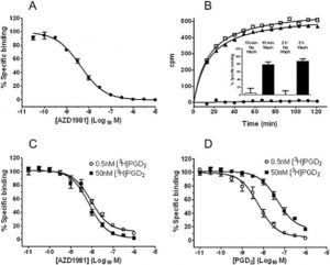
DP2 (chemoattractant receptor-homologous molecule expressed on Th2 cells, also called CRTH2 or GPR44) with pIC50 = 8.4 ± 0.1 (geometric mean IC50 = 4 nM) in human recombinant binding assay[1]
DP1: no significant affinity, mean 27% displacement of [3H]PGD2 at 10 μM (>1000-fold selective over DP2)[1] Rat aldose reductase: pIC50 = 7.4 (98% inhibition at 10 μM)[1] Rat steroid 5α-reductase: pIC50 = 5.2 (88% inhibition at 10 μM)[1] Human aldose reductase: pIC50 = 5.2 ± 0.1[1] Human aldehyde reductase: pIC50 = 5.8 ± 0.1[1] AZD1981 targets the CRTh2 (DP2) receptor, which is a G-protein coupled receptor for prostaglandin D2. It acts as a potent and selective antagonist at this receptor. It has an IC50 of 4 nM. AZD1981 blocks DP2-mediated shape change in human eosinophils and basophils, as well as DP2-mediated chemotaxis of human Th2 cells and eosinophils. |
|---|---|
| ln Vitro |
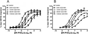
AZD1981, as a potent antagonist in a disease relevant cell system, inhibits DK-PGD2-induced CD11b expression in human eosinophils with IC50 of 10 nM. AZD1981 blocks DP2-mediated shape change in human eosinophils and basophils in blood, as well as DP2-mediated chemotaxis of human Th2 cells and eosinophils. Moreover, AZD1981 also blocks the binding of [3H]PGD2 to mouse, rat, guinea pig, rabbit and dog recombinant DP2.
Kinase Assay: AZD1981 is a novel potent and selective CRTh2 (DP2) receptor antagonist with IC50 of 4 nM, it displayed >1000-fold selectivity over more than 340 other enzymes and receptors, including DP1. Cell Assay: AZD1981 could block chemotaxis of DP2+ T-cell lines with a pIC50 value of 7.5±0.1. AZD1981 has been demonstrated to inhibit PGD2 binding to mouse, rat, rabbit, dog, guinea pig and human DP2. AZD1981 could inhibit shape change induced by DP2 in dog and guinea pig granulocytes as well as in human eosinophils and basophils in blood. AZD1981 displaced [3H]PGD2 from human recombinant DP2 with high potency (pIC50 = 8.4, mean IC50 = 4 nM); binding was reversible and non-competitive (pIC50 unchanged from 0.5 nM to 50 nM radioligand).[1] AZD1981 inhibited DP2-mediated CD11b up-regulation in human eosinophils with non-competitive antagonism, yielding pKB = 8.55 ± 0.03 (n=3) from operational model fitting.[1] AZD1981 inhibited DP2-mediated shape change in human eosinophils in blood (1 μM caused 20-fold rightward shift, pA2 = 7.3 ± 0.02, n=4) and in human basophils (1 μM caused 70-fold rightward shift, pA2 = 7.5 ± 0.47, n=5).[1] AZD1981 inhibited DP2-mediated chemotaxis of human eosinophils (pIC50 = 7.6 ± 0.1, n=4) and human Th2 cells (pIC50 = 7.5 ± 0.1, n=5) using single submaximal agonist concentrations.[1] AZD1981 showed cross-species binding to mouse, rat, guinea pig, rabbit and dog DP2 with pIC50 values: human 8.4, rat 8.5, mouse 8.1, dog 8.1, guinea pig 7.8, rabbit 8.7.[1] AZD1981 blocked DP2-mediated shape change in guinea pig eosinophils (pA2 = 6.9 ± 0.12, n=5) and dog eosinophils (pIC50 = 7.5 ± 0.4, n=6) in whole blood.[1] In vitro, AZD1981 produced a concentration-dependent displacement of [3H]PGD2-specific binding with a mean pIC50 of 8.4, corresponding to an IC50 of 4 nM. It inhibits DK-PGD2-induced CD11b expression in human eosinophils with an IC50 of 10 nM. It shows >1000-fold selectivity over DP1 and other targets. |
| ln Vivo |
Using the previously described guinea pig hind limb model , 10 nM AZD1981 significantly inhibited DK-PGD2-induced eosinophil mobilization by approximately 50%, and the response was completely inhibited with 100 nM AZD1981. AZD1981 exhibited good cross-species binding activity against mouse, rat, guinea pig, rabbit and dog DP2 . Evaluation in mouse, rat or rabbit cell systems was not possible as they did not respond to DP2 agonists. Agonist responses were seen in guinea pig and dog, and AZD1981 blocked DP2 -mediated eosinophil shape change. Such responses were more robust in the guinea pig, where AZD1981 also blocked DP2 -dependent eosinophil emigration from bone marrow. AZD1981 has high oral bioavailability in male sprague dawley rats. In guinea pig hind limb model, AZD1981 (100 nM) completely inhibits DK-PGD2-induced eosinophil mobilization.
In isolated perfused guinea pig hind limb, AZD1981 at 10 nM significantly inhibited DK-PGD2 (30 nM)-induced eosinophil mobilization from bone marrow by approximately 50%, and 100 nM completely inhibited the response (p<0.05 vs vehicle by two-way ANOVA for repeated measurements, n=5-7).[1] In vivo, AZD1981 has been used in research on the treatment of asthma, postmenopause, pharmacokinetics, and drug interactions. It exhibits immunomodulatory effects. It has been investigated in clinical trials for chronic spontaneous urticaria. As a potent and selective CRTh2 antagonist, it is a valuable tool for studying the role of the PGD2-CRTh2 pathway in allergic and inflammatory diseases. |
| Enzyme Assay |
Radioligand binding assay using scintillation proximity assay (SPA): Membranes from HEK293 cells expressing recombinant human DP2 were prebound to wheat germ agglutinin-coated PVT-SPA beads for 18 h at 4°C. Assays started by adding membrane-coated beads (10 mg/mL) to assay buffer (50 mM HEPES pH7.4, 5 mM MgCl2) containing 2-5 nM [3H]PGD2 in absence or presence of increasing compound concentrations (50 μL final volume). Non-specific binding determined with 10 μM DK-PGD2. Plates incubated 2 h at room temperature, bead-associated radioactivity measured. IC50 was calculated.[1]
Reversibility binding assay: HEK-membrane-coated beads incubated with AZD1981 for 2 h at room temperature, then centrifuged and washed four times with assay buffer. Aliquots transferred to 96-well plates, [3H]PGD2 binding evaluated as above. Recovery of binding observed within 13 min after compound removal.[1] Competition binding across species: Same methodology used for recombinant human, murine, rat, guinea pig, dog and rabbit DP2.[1] Enzyme inhibition assays for aldose and aldehyde reductase: Human recombinant enzymes were used. Assays performed in UV-clear 96-well plates (200 μL final volume). Each well contained AZD1981, recombinant enzyme (10 μg/mL aldose reductase in 5 mM sodium phosphate pH7.5 with 5 mM 2-mercaptoethanol, or 2.5 μg/mL aldehyde reductase in 5 mM sodium phosphate pH7.5), substrate (0.2 mM DL-glyceraldehyde for aldose reductase or 2 mM D-glucuronic acid for aldehyde reductase) and 0.2 mM NADPH in 0.1 M sodium phosphate pH7.0. Reaction rate measured by monitoring decrease in absorbance at 340 nm.[1] Non-cellular receptor binding assays for AZD1981 involve measuring its affinity for the CRTh2 receptor. These assays use membrane preparations from cells expressing the receptor to study the displacement of a labeled ligand, such as [3H]PGD2. The IC50 of 4 nM and pIC50 of 8.4 are key parameters determined in these experiments. Such assays are essential for characterizing its potency and selectivity. |
| Cell Assay |
CD11b up-regulation assay in human eosinophils from mixed leukocyte preparation: Granulocytes from healthy volunteers were washed and resuspended at 3.5×10^6 cells/mL in HBSS/HEPES. AZD1981 or vehicle pre-incubated with cells for 15 min, then antibody mix (FITC-anti-human CD11b and PE-anti-human CD16) and agonist (DK-PGD2) added. Incubated 15 min at 37°C, fixed with ice-cold autologous plasma and formaldehyde, then red blood cell lysis. CD11b expression measured by flow cytometry (median peak fluorescence). Eosinophils gated by forward/side scatter and low CD16 expression.[1]
Human eosinophil shape change assay in whole blood: Blood from healthy volunteers pre-treated with AZD1981 or vehicle for 60 min at room temperature. Then 15R-methyl PGD2 or vehicle added, incubated 15 min at 37°C, fixed with Optilys B, lysed with water, centrifuged, resuspended in PBS with Cyto-Chex. Shape change analysed by flow cytometry; eosinophils gated by forward/side scatter and high autofluorescence.[1] Human basophil shape change assay: Whole blood labelled with FITC-HLA-DR and PE-CD123 monoclonal antibodies, pre-incubated with vehicle or AZD1981 for 10 min at 37°C, then stimulated with PGD2 for 4 min at 37°C. Samples fixed, red blood cells lysed with NH4Cl, washed, resuspended. Basophils gated as CD123-positive and HLA-DR-negative, responses quantified as percentage of cells moving into higher forward scatter gate.[1] Chemotaxis assay for human eosinophils: Purified human eosinophils used. AZD1981 tested against single concentration of PGD2 (1 μM). Cells applied to upper surface of 96-well ChemoTx plates (5 μm pore size). Lower wells contained agonist. Incubated 1 h at 37°C/5% CO2, migrated cells quantified by cell-associated LDH using a commercial kit.[1] Chemotaxis assay for human Th2 cells: DP2+ T-cell lines expanded from peripheral blood of healthy volunteers using anti-CD3/anti-CD28 beads and IL-2. Cells applied to upper surface of ChemoTx plates, lower wells contained DK-PGD2 (330 nM). AZD1981 present in both upper and lower solutions. Incubated 1 h at 37°C/5% CO2, migrated cells quantified by LDH.[1] In vitro cell-based assays for AZD1981 are used to study its functional effects on immune cells. For example, its ability to inhibit DK-PGD2-induced CD11b expression in human eosinophils has been measured. It also blocks DP2-mediated shape change in human eosinophils and basophils, and DP2-mediated chemotaxis of human Th2 cells and eosinophils. These studies help to confirm its mechanism of action. |
| Animal Protocol |
10 nM Guinea pig hind limb model
Isolated perfused guinea pig hind limb model: Eosinophil mobilization measured as described previously (Royer et al., 2008). Briefly, guinea pig hind limb was perfused with buffer containing AZD1981 or vehicle throughout the experiment. DK-PGD2 (30 mM) was added during the 20 to 40 min period. Eosinophil release was measured over time (0-80 min). AZD1981 at 10 nM and 100 nM significantly inhibited DK-PGD2-induced eosinophil mobilization in a concentration-dependent manner.[1] In vivo animal studies for AZD1981 would typically involve models of allergic airway inflammation or other diseases where the PGD2-CRTh2 pathway is implicated. The compound would be administered orally or by other routes to evaluate its efficacy in reducing inflammation. These studies are crucial for validating its potential as a therapeutic agent. |
| ADME/Pharmacokinetics |
Log D7.4 (distribution coefficient between 1-octanol and aqueous buffer at pH7.4) = -0.22[1]
Plasma protein binding (% bound): human 97.2%, rat 98.3%, mouse 97.5%, dog 97.4%, rabbit 98.4%, guinea pig 96.5%[1] Solubility in 10 mM sodium phosphate pH7.4 at 20°C: 1.87 mM[1] pKa (acid dissociation constant): 2.64[1] AZD1981 is a small molecule with a molecular weight of 452.45 g/mol. It is an orally available compound. As a research compound, its pharmacokinetic profile has been studied in preclinical models and clinical trials. It is a research compound that has been investigated in clinical trials. |
| Toxicity/Toxicokinetics |
Comprehensive toxicological data for AZD1981 are not extensively detailed in standard summaries, as it is a research compound. As with all investigational drugs, its safety profile would have been evaluated in preclinical and clinical studies. AZD1981 is not an approved drug and is available only as a research compound.
|
| References |
Br J Pharmacol.2013 Apr;168(7):1626-38;Bioorg Med Chem Lett.2011 Nov 1;21(21):6288-92.
|
| Additional Infomation |
AZD1981 has been used in research on the treatment and basic scientific trials of asthma, postmenopause, pharmacokinetics, asthma patients, and drug interactions.
AZD1981 is a reversible, non-competitive DP2 antagonist derived from indomethacin scaffold (indomethacin showed partial agonist activity at DP2). It binds to a site distinct from PGD2, precluding agonist binding and activation.[1] The compound is an indole acetic acid with high aqueous solubility, moderately low logD, and high plasma protein binding.[1] AZD1981 shows no inhibition of COX-1 or COX-2, and no significant activity at other receptors/enzymes (including CCR3, CCR4, TP receptor) in a panel of >338 targets.[1] Insurmountable antagonism observed in eosinophil CD11b assay, while surmountable antagonism in shape change assays due to higher receptor reserve or higher efficacy agonist (15R-methyl PGD2).[1] AZD1981 is under clinical evaluation as a potential therapeutic agent in respiratory diseases including asthma.[1] AZD1981 is a potent and selective CRTh2 (DP2) receptor antagonist with an IC50 of 4 nM. It is highly selective, showing >1000-fold selectivity over DP1 and other targets. It has been researched for the treatment of asthma and chronic spontaneous urticaria. AZD1981 is not an approved drug and is available only as a research compound. |
| Molecular Formula |
C19H17CLN2O3S
|
|
|---|---|---|
| Molecular Weight |
388.87
|
|
| Exact Mass |
388.065
|
|
| CAS # |
802904-66-1
|
|
| Related CAS # |
|
|
| PubChem CID |
11292191
|
|
| Appearance |
White to gray solid powder
|
|
| LogP |
5.446
|
|
| Hydrogen Bond Donor Count |
2
|
|
| Hydrogen Bond Acceptor Count |
4
|
|
| Rotatable Bond Count |
5
|
|
| Heavy Atom Count |
26
|
|
| Complexity |
527
|
|
| Defined Atom Stereocenter Count |
0
|
|
| SMILES |
O=C(O)CN1C(C)=C(SC2=CC=C(Cl)C=C2)C3=C1C=CC=C3NC(C)=O
|
|
| InChi Key |
JWYIGNODXSRKGP-UHFFFAOYSA-N
|
|
| InChi Code |
InChI=1S/C19H17ClN2O3S/c1-11-19(26-14-8-6-13(20)7-9-14)18-15(21-12(2)23)4-3-5-16(18)22(11)10-17(24)25/h3-9H,10H2,1-2H3,(H,21,23)(H,24,25)
|
|
| Chemical Name |
2-[4-acetamido-3-(4-chlorophenyl)sulfanyl-2-methylindol-1-yl]acetic acid
|
|
| Synonyms |
|
|
| HS Tariff Code |
2934.99.9001
|
|
| Storage |
Powder -20°C 3 years 4°C 2 years In solvent -80°C 6 months -20°C 1 month |
|
| Shipping Condition |
Room temperature (This product is stable at ambient temperature for a few days during ordinary shipping and time spent in Customs)
|
| Solubility (In Vitro) |
DMSO: 11 mg/mL (28.3 mM)
Water: 1 mg/mL (Insoluble) Ethanol: 1 mg/mL |
|---|---|
| Solubility (In Vivo) |
Solubility in Formulation 1: ≥ 2.5 mg/mL (6.43 mM) (saturation unknown) in 10% DMSO + 90% Corn Oil (add these co-solvents sequentially from left to right, and one by one), clear solution.
For example, if 1 mL of working solution is to be prepared, you can add 100 μL of 25.0 mg/mL clear DMSO stock solution to 900 μL of corn oil and mix evenly. (Please use freshly prepared in vivo formulations for optimal results.) |
| Preparing Stock Solutions | 1 mg | 5 mg | 10 mg | |
| 1 mM | 2.5716 mL | 12.8578 mL | 25.7155 mL | |
| 5 mM | 0.5143 mL | 2.5716 mL | 5.1431 mL | |
| 10 mM | 0.2572 mL | 1.2858 mL | 2.5716 mL |
*Note: Please select an appropriate solvent for the preparation of stock solution based on your experiment needs. For most products, DMSO can be used for preparing stock solutions (e.g. 5 mM, 10 mM, or 20 mM concentration); some products with high aqueous solubility may be dissolved in water directly. Solubility information is available at the above Solubility Data section. Once the stock solution is prepared, aliquot it to routine usage volumes and store at -20°C or -80°C. Avoid repeated freeze and thaw cycles.
Calculation results
Working concentration: mg/mL;
Method for preparing DMSO stock solution: mg drug pre-dissolved in μL DMSO (stock solution concentration mg/mL). Please contact us first if the concentration exceeds the DMSO solubility of the batch of drug.
Method for preparing in vivo formulation::Take μL DMSO stock solution, next add μL PEG300, mix and clarify, next addμL Tween 80, mix and clarify, next add μL ddH2O,mix and clarify.
(1) Please be sure that the solution is clear before the addition of next solvent. Dissolution methods like vortex, ultrasound or warming and heat may be used to aid dissolving.
(2) Be sure to add the solvent(s) in order.
|
|---|
|
Products are for research use only; We do not sell to patients
Copyright 2020 InvivoChem LLC | All Rights Reserved


 COA
COA