| Size | Price | Stock | Qty |
|---|---|---|---|
| 250mg |
|
||
| 500mg |
|
||
| Other Sizes |


Purity: ≥98%
| Targets |
Muscarinic acetylcholine receptor/mAChR
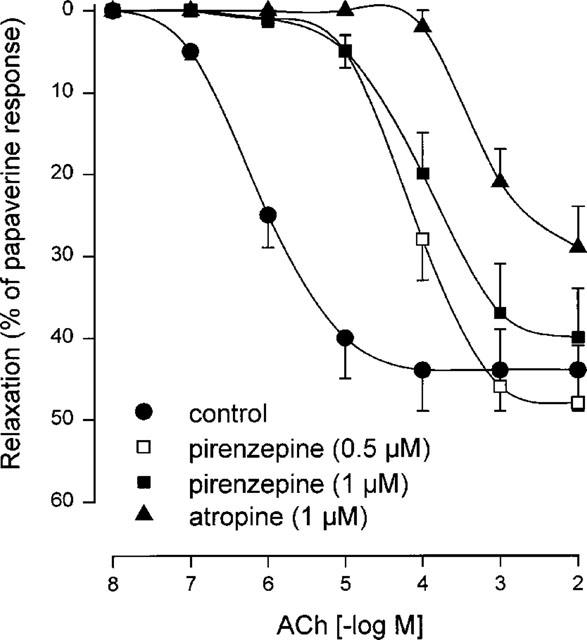
Human mAChR M4: Atropine acts as an antagonist with an IC50 of 390 pM and a calculated Ki(CP) of 140 pM in a CRE-luciferase assay using transfected HEK293T cells. [3] Chicken mAChR cM4: Atropine acts as an antagonist with an IC50 of 710 pM and a calculated Ki(CP) of 120 pM in a CRE-luciferase assay using transfected HEK293T cells. [3] Human alpha2A-adrenoceptor (hADRA2A): Atropine acts as an antagonist with an IC50 of 45 μM and a calculated Ki(CP) of 14 μM in a CRE-luciferase assay using transfected HEK293T cells. [3] Atropine free base targets muscarinic acetylcholine receptors (mAChRs), acting as a competitive, reversible antagonist. It has IC₅₀ values of 0.39 nM and 0.71 nM for Human mAChR M4 and Chicken mAChR M4, respectively. It inhibits acetylcholine-induced relaxations in human pulmonary veins. By blocking muscarinic receptors, atropine inhibits parasympathetic nervous system activity. |
|---|---|
| ln Vitro |
In a CRE-luciferase assay using CRISPR-M3 HEK293T cells transfected with the human M4 mAChR, atropine inhibited carbachol (10 μM)-induced luminescence with an IC50 of 390 pM. The inhibitory constant (Ki) calculated from this data was 140 pM. [3]
In a CRE-luciferase assay using CRISPR-M3 HEK293T cells transfected with the chicken M4 mAChR (cM4), atropine inhibited carbachol (10 μM)-induced luminescence with an IC50 of 710 pM. The inhibitory constant (Ki) calculated from this data was 120 pM. [3] In a CRE-luciferase assay using CRISPR-M3 HEK293T cells transfected with the human alpha2A-adrenoceptor (hADRA2A), atropine inhibited clonidine (1 μM)-induced luminescence with an IC50 of 45 μM. The inhibitory constant (Ki) calculated from this data was 14 μM. [3] The study notes that at high concentrations (1-100 μM), atropine has been reported by others to have antagonist activity at alpha-adrenoceptors. [3] Acetylcholine-induced pulmonary vein dilatation in humans is inhibited by atropine (tropine; 1 μM; pulmonary veins and arteries) [4]. In vitro, Atropine (1 μM) inhibits ACh-induced relaxations in human pulmonary veins and arteries. It is a competitive nonselective antagonist at central and peripheral muscarinic acetylcholine receptors. Atropine effectively inhibits acetylcholine-induced relaxations in human pulmonary veins. Its activity at muscarinic receptors has been extensively characterized in various cell-based and tissue-based assays. |
| ln Vivo |
Usually occurring during periods of torpor, atropine (tropine; 10 mg/kg; intraperitoneally; once over 40 minutes; Peromyscus sp.) inhibits cardiac arrhythmias [2].
The article discusses that topical atropine is effective against myopia in children, but the 1% dosage can induce side effects. A 0.01% concentration has been shown in recent studies to retain effectiveness for inhibiting myopia with reduced side effects. [3] In the chick model of form-deprivation myopia (FDM), atropine is known to inhibit myopia, requiring estimated vitreal concentrations in the range of 0.1-10 mM to be effective. [3] The paper references findings that ablating cholinergic amacrine cells in chicks does not impair myopia inhibition by atropine, suggesting its site of action might be non-retinal. [3] The paper also notes that treatment with myopia-inhibiting concentrations of atropine in chick retina-RPE-choroid-sclera preparations causes a massive, nonspecific release of retinal neurotransmitters. [3] In in vitro preparations of mouse scleral fibroblasts, the inhibitory action of atropine on carbachol-induced proliferation was observed only at high concentrations (0.5-100 μM), which are 500-1000 fold higher than its Ki for mAChRs. [3] In vivo, Atropine (10 mg/kg; intraperitoneally; once over 40 minutes) inhibits cardiac arrhythmias in Peromyscus sp. (white-footed mice). It increases heart rate and diminishes arrhythmia. Atropine is used to counteract excessive cholinergic stimulation caused by nerve agents or organophosphate pesticides and to restore normal heart rate in cases of bradycardia. It also exhibits analgesic and antinociceptive activities. |
| Enzyme Assay |
This study did not perform direct enzyme assays. The primary assay used was a cell-based CRE-luciferase reporter assay to measure receptor activation and inhibition. [3]
CRE-Luciferase Assay for Receptor Antagonism: CRISPR-M3 HEK293T cells, which lack endogenous M3 muscarinic receptors, were co-transfected with a receptor clone (human M4, chicken cM4, or human ADRA2A), a cAMP response element luciferase vector (CRE-Luc), and a Renilla luciferase control vector (RLuc). 48 hours post-transfection, cells were incubated for 4 hours with a fixed, submaximal concentration of agonist (10 μM carbachol for M4/cM4, or 1 μM clonidine for ADRA2A) and increasing concentrations of the test antagonist, including atropine. After incubation, cells were lysed, and luciferase activity was measured sequentially using a Dual-Glo Luciferase Assay System. CRE-Luc activity (reflecting cAMP levels and receptor activation) was normalized to RLuc activity (a control for cell viability and transfection efficiency). Antagonist IC50 values were determined by nonlinear regression analysis of the normalized data. [3] The activity of Atropine can be assessed using radioligand binding assays. Membranes prepared from tissues expressing muscarinic receptors are incubated with a radiolabeled muscarinic antagonist (e.g., [³H]N-methylscopolamine) and varying concentrations of Atropine. Non-specific binding is determined in the presence of an excess of a competing ligand. The IC₅₀ or Ki values for Atropine displacement are calculated from competition binding curves. |
| Cell Assay |
Cell Culture and Transfection: CRISPR-M3 HEK293T cells were cultured in DMEM with 10% FBS. For the assay, cells were seeded in 12-well plates and transfected at 30% confluency using Lipofectamine LTX. For each well, a mixture of Opti-MEM containing 160 ng of receptor DNA (e.g., human M4), 180 ng of CRE-Luc, and 160 ng of RLuc was combined with a Lipofectamine LTX solution. After a 5-minute incubation, the complexes were added to the cells. The medium was changed after 8 hours, and 24 hours post-transfection, cells were trypsinized and seeded into white, clear-bottomed 96-well plates at 7500 cells per well. [3]
CRE-Luc Luminescence Assay: 48 hours after the initial transfection, the medium in the 96-well plates was replaced with 50 μL of FluoroBrite DMEM containing a fixed concentration of agonist (10 μM carbachol for M4/cM4; 1 μM clonidine for ADRA2A) and various concentrations of the antagonist (e.g., atropine). Cells were incubated for 4 hours at 37°C. Subsequently, 50 μL of Dual-Glo Luciferase Reagent was added to each well. After a 10-minute incubation with shaking to ensure lysis, CRE-Luc luminescence was measured. Then, 50 μL of Dual-Glo Stop & Glo Reagent was added, and after another 10-minute incubation, Renilla luciferase luminescence was measured. CRE-Luc values were normalized to RLuc values to control for well-to-well variability. [3] To evaluate the cellular effects of Atropine, cells expressing muscarinic receptors are treated with the compound. The inhibition of acetylcholine-induced calcium mobilization, cAMP production, or other downstream signaling events is measured. The IC₅₀ for these functional responses is determined from dose-response curves. Atropine (1 μM) inhibits ACh-induced relaxations in human pulmonary veins. |
| Animal Protocol |
Animal/Disease Models: White-footed mice (Peromyscus sp.) [2]
Doses: 10 mg/kg Route of Administration: intraperitoneal (ip) injection; once, lasting 40 minutes. Experimental Results: increased heart rate and diminished arrhythmia. The paper discusses animal models and protocols from the perspective of reviewed literature, rather than presenting new in vivo data for atropine. [3] Chick Model of Myopia: In studies referenced by the paper, form-deprivation myopia (FDM) is induced in chicks. Atropine is administered to inhibit myopia, typically via intravitreal injection. Concentrations used range from 0.1 to 10 mM (estimated vitreal concentration), with a total amount of 20-2000 nmol per injection being common. [3] Rabbit Model for Ocular Distribution: The paper references studies where a single dose of 2% [3H]-atropine was delivered to the conjunctival sac of albino rabbits to study its distribution in ocular tissues. [3] Human Clinical Use: The paper discusses clinical protocols where atropine is delivered as daily eye drops at concentrations ranging from 0.01% to 1% for the treatment of childhood myopia. [3] In vivo studies with Atropine typically involve administration to animal models via oral, intravenous, intraperitoneal, or topical (ophthalmic) routes. In models of bradycardia, the compound's effects on heart rate are assessed. Its mydriatic effects are evaluated by measuring pupil dilation in ophthalmic studies. In toxicology studies, its ability to counteract organophosphate poisoning is evaluated. |
| ADME/Pharmacokinetics |
Absorption, Distribution and Excretion
Hyoscyamine can be completely absorbed via sublingual and oral routes, but precise data on Cmax, Tmax, and AUC are not yet clear. Most hyoscyamine is excreted in the urine as the unmetabolized parent compound. Metabolism/Metabolites Hyoscyamine exists primarily in its unmetabolized form, but a small amount is hydrolyzed into tropine and tropine acid. Biological Half-Life The half-life of hyoscyamine is 3.5 hours. Atropine free base has a molecular formula of C₁₇H₂₃NO₃ and a molecular weight of 289.3694 g/mol. Its CAS number is 51-55-8. It is a solid at room temperature. The powder should be stored at -20°C for 3 years or at 4°C for 2 years; in solvent at -80°C for 6 months or at -20°C for 1 month. |
| Toxicity/Toxicokinetics |
Hepatotoxicity
Although hyoscyamine has been widely used for decades, no association has been found between it and elevated liver enzymes or clinically significant liver injury. Its high safety profile is likely due to the low daily dose and limited duration of use. For references on the safety and potential hepatotoxicity of anticholinergic drugs, please see the section following "Overview of Anticholinergic Drugs". Drug Category: Gastrointestinal Drugs; Anticholinergic Drugs Atropine can cause significant toxicity at high doses. Overdose can lead to anticholinergic syndrome, characterized by dry mouth, blurred vision, tachycardia, hallucinations, and delirium. It is contraindicated in patients with glaucoma, prostatic hypertrophy, and certain gastrointestinal conditions. Atropine is a potent drug that requires careful dosing and medical supervision. |
| References | |
| Additional Infomation |
Pharmacodynamics
Hyoscyamine has not been approved by the U.S. Food and Drug Administration (FDA) and therefore has no official indication. However, it is used as an anticholinergic drug in a variety of treatments and therapies. Hyoscyamine has a short duration of action and may require multiple daily doses. Patients should be informed of the risks and symptoms of anticholinergic toxicity. Atropine free base is a naturally occurring belladonna alkaloid and a competitive, reversible antagonist of muscarinic acetylcholine receptors. It is used clinically for the treatment of bradycardia, as a mydriatic, and as an antidote for organophosphate poisoning. Atropine is also widely used as a research tool to study cholinergic signaling pathways and muscarinic receptor function. |
| Molecular Formula |
C17H23NO3
|
|---|---|
| Molecular Weight |
289.3694
|
| Exact Mass |
289.167
|
| Elemental Analysis |
C, 70.56; H, 8.01; N, 4.84; O, 16.59
|
| CAS # |
51-55-8
|
| Related CAS # |
Atropine sulfate monohydrate;5908-99-6;Atropine sulfate;55-48-1;(Rac)-Atropine-d3;1276197-36-4;Atropine hydrobromide;6415-90-3
|
| PubChem CID |
174174
|
| Appearance |
White to off-white solid powder
|
| Density |
1.2±0.1 g/cm3
|
| Boiling Point |
429.8±45.0 °C at 760 mmHg
|
| Melting Point |
115-118 °C
|
| Flash Point |
213.7±28.7 °C
|
| Vapour Pressure |
0.0±1.1 mmHg at 25°C
|
| Index of Refraction |
1.581
|
| Source |
Originated from plants: Solanaceae Atropa belladonna Linn.
Endogenously produced metabolite |
| LogP |
1.53
|
| Hydrogen Bond Donor Count |
1
|
| Hydrogen Bond Acceptor Count |
4
|
| Rotatable Bond Count |
5
|
| Heavy Atom Count |
21
|
| Complexity |
353
|
| Defined Atom Stereocenter Count |
2
|
| SMILES |
CN1[C@@H]2CC[C@H]1CC(C2)OC(=O)C(CO)C3=CC=CC=C3
|
| InChi Key |
RKUNBYITZUJHSG-PJPHBNEVSA-N
|
| InChi Code |
InChI=1S/C17H23NO3/c1-18-13-7-8-14(18)10-15(9-13)21-17(20)16(11-19)12-5-3-2-4-6-12/h2-6,13-16,19H,7-11H2,1H3/t13-,14+,15?,16?
|
| Chemical Name |
[(1R,5S)-8-methyl-8-azabicyclo[3.2.1]octan-3-yl] 3-hydroxy-2-phenylpropanoate
|
| Synonyms |
RefChem:1092872; Npc209773; ATROPINE; dl-Hyoscyamine; 51-55-8;
|
| HS Tariff Code |
2934.99.9001
|
| Storage |
Powder -20°C 3 years 4°C 2 years In solvent -80°C 6 months -20°C 1 month Note: Please store this product in a sealed and protected environment (e.g. under nitrogen), avoid exposure to moisture. |
| Shipping Condition |
Room temperature (This product is stable at ambient temperature for a few days during ordinary shipping and time spent in Customs)
|
| Solubility (In Vitro) |
DMSO : ≥ 96.6 mg/mL (~333.83 mM)
H2O : ~2.9 mg/mL (~10.02 mM) |
|---|---|
| Solubility (In Vivo) |
Solubility in Formulation 1: ≥ 2.08 mg/mL (7.19 mM) (saturation unknown) in 10% DMSO + 40% PEG300 + 5% Tween80 + 45% Saline (add these co-solvents sequentially from left to right, and one by one), clear solution.
For example, if 1 mL of working solution is to be prepared, you can add 100 μL of 20.8 mg/mL clear DMSO stock solution to 400 μL PEG300 and mix evenly; then add 50 μL Tween-80 to the above solution and mix evenly; then add 450 μL normal saline to adjust the volume to 1 mL. Preparation of saline: Dissolve 0.9 g of sodium chloride in 100 mL ddH₂ O to obtain a clear solution. Solubility in Formulation 2: ≥ 2.08 mg/mL (7.19 mM) (saturation unknown) in 10% DMSO + 90% (20% SBE-β-CD in Saline) (add these co-solvents sequentially from left to right, and one by one), clear solution. For example, if 1 mL of working solution is to be prepared, you can add 100 μL of 20.8 mg/mL clear DMSO stock solution to 900 μL of 20% SBE-β-CD physiological saline solution and mix evenly. Preparation of 20% SBE-β-CD in Saline (4°C,1 week): Dissolve 2 g SBE-β-CD in 10 mL saline to obtain a clear solution. View More
Solubility in Formulation 3: ≥ 2.08 mg/mL (7.19 mM) (saturation unknown) in 10% DMSO + 90% Corn Oil (add these co-solvents sequentially from left to right, and one by one), clear solution. |
| Preparing Stock Solutions | 1 mg | 5 mg | 10 mg | |
| 1 mM | 3.4558 mL | 17.2789 mL | 34.5578 mL | |
| 5 mM | 0.6912 mL | 3.4558 mL | 6.9116 mL | |
| 10 mM | 0.3456 mL | 1.7279 mL | 3.4558 mL |
*Note: Please select an appropriate solvent for the preparation of stock solution based on your experiment needs. For most products, DMSO can be used for preparing stock solutions (e.g. 5 mM, 10 mM, or 20 mM concentration); some products with high aqueous solubility may be dissolved in water directly. Solubility information is available at the above Solubility Data section. Once the stock solution is prepared, aliquot it to routine usage volumes and store at -20°C or -80°C. Avoid repeated freeze and thaw cycles.
Calculation results
Working concentration: mg/mL;
Method for preparing DMSO stock solution: mg drug pre-dissolved in μL DMSO (stock solution concentration mg/mL). Please contact us first if the concentration exceeds the DMSO solubility of the batch of drug.
Method for preparing in vivo formulation::Take μL DMSO stock solution, next add μL PEG300, mix and clarify, next addμL Tween 80, mix and clarify, next add μL ddH2O,mix and clarify.
(1) Please be sure that the solution is clear before the addition of next solvent. Dissolution methods like vortex, ultrasound or warming and heat may be used to aid dissolving.
(2) Be sure to add the solvent(s) in order.
Topical Application of Low-concentration (0.01%) Atropine on the Human Eye With Fast and Slow Myopia Progression Rate
CTID: NCT03374306
Phase: N/A Status: Completed
Date: 2024-10-16
|
Products are for research use only; We do not sell to patients
Copyright 2020 InvivoChem LLC | All Rights Reserved


 COA
COA