| Size | Price | Stock | Qty |
|---|---|---|---|
| 10mg |
|
||
| 25mg |
|
||
| 50mg |
|
||
| 100mg |
|
||
| 250mg |
|
||
| 500mg |
|
||
| 1g | |||
| Other Sizes |


Purity: ≥98%
Atazanavir Sulfate (also known as Latazanavir, Zrivada, Reyataz, BMS-232632), the sulfate salt form of atazanavir, is a HIV protease inhibitor (PI) for HIV/AIDs treatment with Ki of 2.66 nM in a cell-free assay. The treatment for HIV/AIDS is the same as that of other antiretrovirals. Since it can be given once daily and has less of an impact on the patient's lipid profile than other PIs, atazanavir stands apart from the others. It is used exclusively in conjunction with other HIV medications, similar to other protease inhibitors.
| Targets |
HIV protease (Ki = 2.66 nM)
|
|---|---|
| ln Vitro |
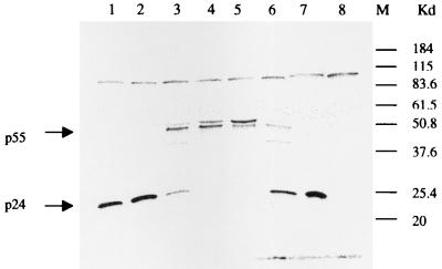
Atazanavir has an IC50 of approximately 47 nM for the proteolytic cleavage of the viral gag precursor p55 polyprotein in H9 cells infected with the virus. With an EC50 of 3.89 nM in RF/MT-2 strains, atazanavir demonstrates strong antiviral activity.[1] It is demonstrated that atazanavir inhibits bilirubin glucuronidation with an IC50 of 2.4 μM. Atazanavir has a Ki of 1.9 μM, which inhibits recombinant UGT1A1.[2] In U251, T98G, and LN229 glioblastoma cell lines, atazanavir inhibits cell growth with notably elevated levels of GRP78 and CHOP protein. In U251 glioblastoma cells, atazanavir induces a significant upregulation of polyubiquitinated proteins of diverse sizes.[3] With an IC50 of 26 μM, atazanavir also inhibits the human 20S proteasome. In HepG2 cells, atazanavir (30 μM) modifies the levels of ER stress and UPR gene expression.[4] In LS180V cells, atazanavir (30 mM) results in a 2.5-fold increase in immunoreactive P-gp expression and a decrease in intracellular Rh123.[5]
In vitro activity: Atazanavir inhibits the proteolytic cleavage of the viral gag precursor p55 polyprotein with IC50 of ~47 nM in virus-infected H9 cells. Atazanavir exhibits potent antiviral activity with EC50 of 3.89 nM in RF/MT-2 strains. Atazanavir is shown to be an inhibitor of bilirubin glucuronidation with IC50 of 2.4 μM. Atazanavir inhibits recombinant UGT1A1 with Ki of 1.9 μM. Atazanavir inhibits cell growth in U251, T98G, and LN229 glioblastoma cell lines, with strikingly increased GRP78 and CHOP protein levels. Atazanavir causes a prominent increase of polyubiquitinated proteins of various different sizes in U251 glioblastoma cells. Atazanavir also inhibits human 20S proteasome with IC50 of 26 μM. Atazanavir (30 μM) changes the magnitudes of ER stress and UPR gene expression in HepG2 cells. Atazanavir (30 mM) causes a 2.5-fold increase in immunoreactive P-gp expression with decreased intracellular Rh123 in LS180V cells. Kinase Assay: To determine the inhibition constants (Ki) for each Prt inhibitor, purified HIV-1 RF wild-type Prt (2.5 nM) is incubated at 37 ℃ with 1 μM to 15 μM fluorogenic substrate in reaction buffer (1 M NaCl, 1 mM EDTA, 0.1 M sodium acetate [pH 5.5], 0.1% polyethylene glycol 8000) in the presence or absence of Atazanavir. Cleavage of the substrate is quantified by measuring an increase in fluorescent emission at 490 nM after excitation at 340 nM using a Cytofluor 4000. Reactions are carried out using 1.36 μM, 1.66 μM, 2.1 μM, 3.0 μM, 5.0 μM, or 15 μM substrate in the presence of five concentrations of Atazanavir (1.25 nM to 25 nM). Substrate cleavage is monitored at 5-min intervals for 30 min. Cleavage rates are then determined for each sample at early time points in the reaction, and Ki values are determined from the slopes of the resulting Michaelis-Menten plots. Cell Assay: To determine cytotoxicity, host cells are incubated in the presence of serially diluted Atazanavir for 6 days and cell viability is quantitated using an XTT[2,3-bis(2-methoxy-4-nitro-5-sulfophenyl-2H-tetrazolium-5-carboxanilide] assay to calculate the 50% cytotoxic concentrations (CC50s). To assess the effect of human serum proteins on antiviral activity, the 10% fetal calf serum normally used for assays is replaced with 40% adult human serum or 1 mg of α1-acid glycoprotein/mL. Effects of Atazanavir on rCFs proliferation, collagen production and proteins expression [3] The rCFs were examined in the absence or the presence of CoCl2 to mimic a pro-fibrotic environment during hypoxic conditions. Following CoCl2 induced hypoxia, rCFs proliferation increased compared with the normal group (P < 0.01), as shown in Table 1, but was significantly inhibited in a concentration-dependent manner following Atazanavir sulfate treatment at concentrations between 1 and 10 μM compared with the CoCl2 group (P < 0.05). To further characterize this inhibitory effect, atazanavir sulfate treatment was combined with HCQ, a TLR 9 antagonist. However, it found no further decline in rCFs proliferation compared with the HCQ group (P > 0.05), as shown in Table 1. In addition, the content of collagen I and collagen III was measured in CoCl2 stimulated rCFs. The results showed the contents of collagen I and collagen III were increased compared with the normal group (P < 0.01). However, collagen I and collagen III levels were significantly reduced in a concentration-dependent manner following atazanavir sulfate treatment at concentrations between 1 and 10 μM compared with the CoCl2 group (P < 0.05)), as shown in Table 1. It found no further decline in collagen I and collagen III in atazanavir 3 μM plus HCQ 3 μM compared with the HCQ group (P > 0.05), as shown in Table 1. To further examine the mechanism of Atazanavirsulfate on reducing rCFs proliferation during hypoxia, we investigated the expression of HMGB1, p-NF-κB, p-IκBα and total NF-κB with or without atazanavir sulfate. Following CoCl2 induced hypoxia, HMGB1, p-NF-κB, p-IκBα and TLR 9 expression were increased compared with the normal group (P < 0.01), as shown in Fig. 1A and B, but HMGB1, p-IκBα and p-NF-κB expression were significantly inhibited following atazanavir sulfate treatment at 1–10 μM compared with the CoCl2 group (P < 0.05 or P < 0.01). Compared with the CoCl2 group, Atazanavir 3 μM group has no change in total NF-κB expression, and no decline in TLR 9 expression (P > 0.05), as shown in Fig. 1A and B. HCQ treatment reduced HMGB1, p-NF-κB and TLR 9 expression (P < 0.05 or P < 0.01). Atazanavir treatment was combined with HCQ has no further decline in HMGB1, TLR 9 and p-NF-κB expression (P > 0.05) compared with the HCQ group (P > 0.05), as shown in Fig. 1C and D. These findings suggest that atazanavir attenuates hypoxia induced rCFs proliferation by modulating the HMGB1/TLR 9 pathway. |
| ln Vivo |
Effects of Atazanavir sulfate on myocardial function [3]
We evaluated the effect of Atazanavir on LVSP and ± dp/dtmax of the left ventricle after MI 28 days. Compared with vehicle-treated animals, rats treated with Atazanavir had significantly improved LVSP, + dp/dtmax and − dp/dtmax 28 days after MI as shown in Table 2. In addition, we found no further change in SP, DP and HR compared with the HCQ group (P > 0.05). It is clear that continuous atazanavir treatment 28 days provided long-term benefits for the myocardial function recovery after MI. Effects of Atazanavir on cardiac collagen volume and myocytes hypertrophy after MI 28 days [3] To clarify the mechanism of long-term improved cardiac performance caused by atazanavir, we examined the effects of atazanavir treatment on mural hypertrophy and collagen volume in the non-infarcted region and infarct size. There was no difference in infarct size between the vehicle-treated group and atazanavir 30 mg/kg group (38.11 ± 4.15% and 38.80 ± 4.62%, respectively). The cross-sectional area and diameter of myocytes in the non-infarcted LV and hypertrophy of the myocytes significantly increased in vehicle-treated rats compared with Sham rats, while inhibited by atazanavir, as shown in Fig. 2A, C and D. Atazanavir significantly attenuated an increase in morphometrical collagen volume fraction in the border left ventricle, as shown in Fig. 2B and E. In agreement with the above results, the heart index (heart-weight to body-weight ratio) which was increased in the vehicle-treated rats compared with sham rats, was significantly (p < 0.05) lowered by continuous atazanavir treatment, as shown in Fig. 2F. Effects of Atazanavir on the expressions of α-SMA, HMGB1, p-NF-κB, TLR 9, collagen I, collagen III and the content of Hyp in vivo [3] Changes in the expressions of α-SMA, HMGB1, TLR 9, p-NF-κB, collagen I and collagen III were also examined by Western blot analysis, as shown in Figs. 3–5. In vehicle-treated rats, all of the examined protein expressional levels and the content of Hyp increased relative to the sham animals (P < 0.01), while those protein expressional levels and the content of Hyp decreased following Atazanavir treatment compared with the vehicle-treated rats (P < 0.01). The results of in vitro and in vivo investigations suggest that atazanavir can reduce fibroblast proliferation and collagen deposition by modulating the HMGB1/TLR 9 pathway. |
| Enzyme Assay |
Purified HIV-1 RF wild-type Prt (2.5 nM) is incubated at 37 °C with 1 μM to 15 μM fluorogenic substrate in reaction buffer (1 M NaCl, 1 mM EDTA, 0.1 M sodium acetate [pH 5.5], 0.1% polyethylene glycol 8000) with or without atazanavir in order to calculate the inhibition constants (Ki) for each Prt inhibitor. Using a Cytofluor 4000, cleavage of the substrate is measured as an increase in fluorescent emission at 490 nM following excitation at 340 nM. In five different concentrations of Atazanavir (1.25 nM to 25 nM), reactions are conducted with substrate that is 1.36 μM, 1.66 μM, 2.1 μM, 3.0 μM, 5.0 μM, or 15 μM. During a half-hour, the substrate cleavage is observed every five minutes. Then, at early stages of the reaction, cleavage rates are calculated for each sample, and Ki values are ascertained from the slopes of the ensuing Michaelis-Menten plots.
|
| Cell Assay |
In order to assess cytotoxicity, host cells are cultured for six days with serially diluted Atazanavir. Cell viability is then measured using the XTT[2,3-bis(2-methoxy-4-nitro-5-sulfophenyl-2H-tetrazolium-5-carboxanilide)] assay, which yields the 50% cytotoxic concentrations (CC50s). In order to evaluate how human serum proteins affect antiviral activity, 40% adult human serum or 1 mg of α1-acid glycoprotein/mL is added to the 10% fetal calf serum that is typically used for assays.
Cell culture and expressional analysis [3] Rat cardiac fibroblasts (rCFs) from newborn (1- to 2-day-old) Sprague-Dawley rats were isolated according to previous method (Villarreal et al., 1993). Cells were cultured in Dulbecco's Modified Eagle Medium (DMEM) with 10% fetal bovine serum, 100 kU/L penicillin and 100 mg/L streptomycin at 37 °C with 5% CO2 in a humidified incubator. The cells were cultured to approximately 70% confluency and starved in serum-free DMEM overnight prior to the treatment. The cells were then treated with 3 μM Atazanavir sulfate (purity > 99.0%; CAS No.: 229975-97-7) with or without cobalt chloride (CoCl2; 100 μM) for 72 h and thereafter proteins were extracted. rCFs proliferation assay and expressional assessment [3] To assess cellular proliferation, rCFs were maintained as described above. Cells were exposed to CoCl2 at 100 μM to mimic hypoxia and treated with varying concentrations of Atazanavir (0, 1, 3, 10 μM) with or without 3 μM hydroxychloroquine (HCQ), a TLR 9 antagonist, for 72 h. Cellular proliferation levels were determined via cell counting. To examine changes in expression, the cells were seeded into 6-well flat bottom plates and maintained as described above, with one well per plate maintained as an untreated control. Cells were treated with 3 μM Atazanavir sulfate with or without CoCl2 (100 μM) for 72 h and thereafter the supernatants were collected and the proteins were extracted. Collagen I and collagen III were examined by ELISA kits. The expression levels of TLR 9, HMGB1, p-NF-κB, p-IκBα and total NF-κB were examined by Western blot and normalized and displayed as described above. To investigate the possible mechanism of reduction in rCF proliferation, cells were treated with 3 μM atazanavir sulfate with or without 3 μM HCQ for 72 h, TLR 9 and expression levels of HMGB1 and p-NF-κB were examined using Western blot as described above. |
| Animal Protocol |
Induction of myocardial infarction (MI) model and experimental assessment [3]
Briefly, Rats were anesthetized with ketamine 100 mg/kg (i.m.) and xylazine 10 mg/kg (i.m.) and ventilated with room air using a rodent respirator. The chest was opened by middle thoracotomy and the left coronary artery was ligated at 2–3 mm from its origin between the left atrium and pulmonary artery conus using a 6-0 prolene suture. A successful operation was confirmed by the occurrence of ST-segment elevation in an electrocardiogram. This operation was performed by an experimenter who was blinded to the group assignments of the animals to avoid subjective bias of the experimenter on the outcome. The sham-operated group underwent thoracotomy and cardiac exposure without coronary ligation. Thirty rats were divided into three groups including (I) non-MI rats; (II) MI rats received saline alone; (III) MI rats received intragastric administration of Atazanavir sulfate (30 mg/kg) plus ritonavir (10 mg/kg). Atazanavir is a low oral bioavailability compound and, clinically, is generally coadministrated with Ritonavir, which boosts the oral bioavailability of atazanavir by inhibiting cytochrome P450 (CYP) 3A4, and P-glycoprotein via the same metabolic pathway (Le Tiec et al., 2005, 021567s026lbl). The rats were administered daily via intragastric administration of corresponding drug for continuous 28 days after MI 24 h. Treatment was orally administered on a daily basis for atazanavir-treated animals, while animals in the vehicle-treated and sham groups were given an equal volume of saline. At day 29, determine hemodynamics and analyze histopathological change. |
| ADME/Pharmacokinetics |
Absorption, Distribution and Excretion
Atazanavir is rapidly absorbed, with a time to peak concentration (Tmax) of approximately 2.5 hours. The pharmacokinetics of atazanavir are non-linear; within a once-daily dose range of 200 to 800 mg, both AUC and Cmax increase proportionally to the dose. Steady-state is reached between days 4 and 8, with a cumulative drug amount of approximately 2.3 times. Co-administration with food improves the bioavailability of atazanavir and reduces pharmacokinetic variability. A single dose of 400 mg atazanavir followed by a small amount of food (357 kcal, 8.2 g fat, 10.6 g protein) increased AUC and Cmax by 70% and 57%, respectively, compared to the fasting state. A single dose of 400 mg atazanavir followed by a high-fat meal (721 kcal, 37.3 g fat, 29.4 g protein) increased AUC by an average of 35%, while Cmax remained unchanged compared to the fasting state. Compared to the fasting state, the coefficients of variation of AUC and Cmax were reduced by approximately half when atazanavir was taken with either a light or high-fat meal. Following a single dose of 300 mg atazanavir and 100 mg ritonavir with a light meal (336 kcal, 5.1 g fat, 9.3 g protein), AUC increased by 33% and Cmax and 24-hour plasma concentration both increased by 40% compared to the fasting state. When taken with a high-fat meal (951 kcal, 54.7 g fat, 35.9 g protein), the AUC of atazanavir did not change significantly compared to the fasting state, and Cmax differed from the fasting value by less than 11%. 24-hour plasma concentration increased by approximately 33% due to delayed absorption after a high-fat meal; the median Tmax increased from 2.0 hours to 5.0 hours. Compared to the fasting state, the coefficients of variation of AUC and Cmax were reduced by approximately 25% when atazanavir was administered concurrently with ritonavir (regardless of whether a small or high-fat meal was consumed beforehand). Following a single dose of 400 mg of 14C-atazanavir, 79% and 13% of the total radioactive material, respectively, were excreted in feces and urine. Approximately 20% and 7% of the administered dose, respectively, were unmetabolized in feces and urine. The estimated volume of distribution of atazanavir in HIV-infected patients is 88.3 L. The estimated clearance of atazanavir in HIV-infected patients is 12.9 L/h. Atazanavir is rapidly absorbed, with a time to peak concentration (Tmax) of approximately 2.5 hours. The pharmacokinetics of atazanavir are non-linear, with both AUC and Cmax increasing dose-proportionally within the once-daily dose range of 200–800 mg. Steady-state plasma concentrations are reached between days 4 and 8, with a cumulative drug dose of approximately 2.3 times. Co-administration with food improves the bioavailability of atazanavir and reduces pharmacokinetic variability. After a single dose of atazanavir (with a bland diet (357 kcal, 8.2 g fat, 10.6 g protein), AUC increased by 70% and Cmax increased by 57% compared to the fasting state. After a single dose of atazanavir (with a high-fat diet (721 kcal, 37.3 g fat, 29.4 g protein), AUC increased by an average of 35% compared to the fasting state, while Cmax remained unchanged. Compared to the fasting state, atazanavir (with a bland diet) reduced the coefficients of variation of AUC and Cmax by approximately half, regardless of whether it was taken with a bland or high-fat diet. Peak plasma concentrations: In healthy subjects: after daily administration of 400 mg atazanavir with a bland diet on day 29, the peak plasma concentration was 5199 ng/mL. In HIV-infected patients: after daily administration of 400 mg with food, the plasma concentration on day 29 was 2298 ng/mL. ng/mL. Time to peak concentration: HIV-infected patients: 2 hours. For more complete data on absorption, distribution, and excretion of atazanavir (8 types), please visit the HSDB record page. Metabolism/Metabolites Atazanavir is extensively metabolized in the human body. The major biotransformation pathways of atazanavir in the human body include monooxygenation and dioxygenation. Other minor biotransformation pathways of atazanavir or its metabolites include glucuronidation, N-dealkylation, hydrolysis, and dehydrogenation. Two minor metabolites of atazanavir have been identified in plasma. Neither of these metabolites has shown antiviral activity in vitro. In vitro studies using human liver microsomes have shown that atazanavir... Atazanavir is primarily metabolized via CYP3A. Atazanavir is extensively metabolized in the human body. The major biotransformation pathways of atazanavir in the human body include monooxygenation and (atazanavir sulfate) dioxygenation. Other minor biotransformation pathways of atazanavir or its metabolites include glucuronidation, N-dealkylation, hydrolysis, and oxidative dehydrogenation. Two minor metabolites of atazanavir have been identified in plasma. Neither of these metabolites has shown in vitro antiviral activity. In vitro human liver microsomal studies have shown that atazanavir is metabolized by CYP3A. Biological half-life In healthy subjects (n=214) and HIV-1 infected adult subjects (n=13), daily administration of 400 mg/L... After taking 400 mg of atazanavir with food, the mean elimination half-life at steady state was approximately 7 hours. The elimination half-life in patients with impaired liver function was 12.1 hours (single dose of 400 mg). The mean half-life of atazanavir in subjects with impaired liver function was 12.1 hours, while the mean half-life in healthy volunteers was 6.4 hours. In healthy volunteers (n=214) and HIV-infected adults (n=13), the mean elimination half-life at steady state after taking 400 mg of atazanavir daily with a small amount of food was approximately 7 hours. Pharmacokinetics and Metabolism [2] Atazanavir is rapidly absorbed after oral administration (Tmax 2.5 hours) and exhibits nonlinear pharmacokinetic characteristics. Its bioavailability (AUC) is low in the daily dose range of 200–800 mg. The increase in Cmax was greater than the dose-proportional increase. Co-administration with food improved atazanavir's bioavailability and reduced pharmacokinetic variability. After absorption, atazanavir bound to plasma proteins α1-acid glycoprotein and albumin at similar rates (89% and 86%, respectively). Atazanavir primarily binds to hepatic cytochrome P450 (CYP). It is metabolized in the system, producing two major inactive metabolites, and is a substrate and inhibitor of CYP3A4 isoenzymes. In vitro studies have also shown that atazanavir is both an inhibitor and an inducer of the P-glycoprotein ATP-dependent efflux pump. This efflux pump has a wide cellular distribution and substrate specificity, which further increases the likelihood of drug interactions and pharmacokinetic variations in vivo [17]. Therefore, atazanavir should be used with caution in patients taking potent CYP3A4 inhibitors, intermediate or potent CYP3A4 inducers, and major CYP3A4 substrates. Concomitant use with CYP3A4 inducers such as rifampin may reduce plasma concentrations of atazanavir and weaken its clinical efficacy, while CYP3A4 inhibitors may increase plasma concentrations of atazanavir and increase its toxicity. Atazanavir 400 After being taken with food, the steady-state mean elimination half-life of atazanavir is approximately 7-8 hours, with 20% and 7% of the active drug excreted in feces and urine, respectively. In vitro studies have shown that free, non-protein-bound atazanavir directly inhibits UGT1A1-mediated bilirubin glucuronidation, providing a mechanistic explanation for dose-related hyperbilirubinemia. Indinavir may also inhibit UGT1A, therefore co-administration with atazanavir is not recommended. Population pharmacokinetic studies have shown significant inter- and intra-individual variability in atazanavir plasma concentrations, but currently, the dosage of atazanavir is the same regardless of differences in systemic blood and tissue distribution. The therapeutic range for atazanavir is 150 to 850 ng/ml [21,102]; however, it has been reported that in the absence of ritonavir, the plasma concentrations of both the patient and the drug are typically below the target trough concentration (Cmin) of 150. Significant individual variability in atazanavir exposure is considered an indication for twice-daily dosing or therapeutic monitoring. However, no significant correlation has been found between atazanavir plasma trough concentration (Cmin) and antiviral response in patients without protease inhibitor (PI) mutations. The wide variability in atazanavir exposure strongly supports the preferential use of ritonavir-enhanced atazanavir in individuals previously treated with protease inhibitors. |
| Toxicity/Toxicokinetics |
Effects During Pregnancy and Lactation
◉ Overview of Lactation Use Based on limited data, the levels of atazanavir in breast milk appear to be low. Combination therapy containing the CYP3A inhibitor cobicistat has not been studied during lactation, but the levels of atazanavir in breast milk are expected to be similar to or higher than those of single-agent formulations. Achieving and maintaining viral suppression through antiretroviral therapy can reduce the risk of breast milk transmission to below 1%, but not zero. For HIV-infected individuals receiving antiretroviral therapy with a persistently low viral load, breastfeeding should be supported if they choose to do so. If viral load is not suppressed, pasteurized donor breast milk or formula is recommended. ◉ Effects on Breastfed Infants As of the revision date, no relevant published information was found. ◉ Effects on Lactation and Breast Milk Gynecomastia has been reported in men receiving highly effective antiretroviral therapy. Gynecomastia initially presents unilaterally, but approximately half of cases progress to bilateral gynecomastia. No changes in serum prolactin levels have been observed, and it usually resolves spontaneously within one year even with continued treatment. Some case reports and in vitro studies suggest that protease inhibitors may cause hyperprolactinemia and galactorrhea in some male patients, but this remains controversial. The implications of these findings for lactating mothers are unclear. For mothers who have established lactation, prolactin levels may not affect their ability to breastfeed. Drug Interactions Pharmacological interactions exist with benzprodil (potentially leading to serious and/or life-threatening adverse reactions). Concomitant use of benzprodil and atazanavir is not recommended. Pharmacokinetic interactions exist with antiarrhythmic drugs (e.g., amiodarone, systemic lidocaine, quinidine). Potentially leading to serious and/or life-threatening adverse reactions. Plasma concentrations of these antiarrhythmic drugs should be monitored if used concomitantly with atazanavir. Potential pharmacokinetic interactions (elevated plasma concentrations of tricyclic antidepressants). It may cause serious and/or life-threatening adverse reactions. Plasma concentrations of these tricyclic antidepressants should be monitored if used concomitantly with atazanavir. There is a pharmacokinetic interaction with rifampin (significantly reducing peak plasma concentrations and area under the concentration-time curve (AUC) of HIV protease inhibitors by up to 90%). Concomitant use of atazanavir with rifampin is not recommended. For more complete data on interactions with atazanavir (34 in total), please visit the HSDB record page. Toxicity Overview There is currently no specific antidote for atazanavir toxicity. Patients should receive symptomatic and supportive care from healthcare professionals, with regular monitoring of vital signs and attention to signs of respiratory distress. ECG monitoring is recommended, as atazanavir may worsen atrioventricular block due to PR interval prolongation. If concomitant overdose of nucleoside reverse transcriptase inhibitors is suspected, clinicians should closely monitor patients for signs of lactic acidosis. Hepatotoxicity Atazanavir can cause various forms of liver injury, including transient elevations in serum enzymes, indirect hyperbilirubinemia, specific acute liver injury, and exacerbation of pre-existing chronic viral hepatitis. A significant proportion of patients taking antiretroviral regimens containing atazanavir will experience some degree of elevated serum transaminases. 3% to 10% of patients will experience moderate to severe elevations in serum transaminases (more than 5 times the upper limit of normal), and the incidence may be even higher in patients co-infected with HIV-HCV. These bilirubin elevations are usually asymptomatic and resolve spontaneously, returning to normal with continued use. Atazanavir treatment (similar to indinavir) can also lead to elevations in unconjugated bilirubin (indirect bilirubin) and total bilirubin, with up to 10% of patients developing jaundice. These elevations are due to inhibition of UDP-glucuronyltransferase, a liver enzyme responsible for bilirubin conjugation; patients with Gilbert's syndrome have insufficient UDP-glucuronyltransferase activity. Hyperbilirubinemia is usually mild, averaging 0.3-0.5 mg/dL, but can be more pronounced in patients with Gilbert's syndrome, with elevations reaching 1.5 mg/dL or more, accompanied by clinical jaundice. However, jaundice does not necessarily indicate liver damage. Clinically significant acute liver injury caused by atazanavir is rare, and its clinical manifestations, latency period, and recovery are not well understood. This type of liver injury is specific and rare, and may resemble liver injury caused by other HIV protease inhibitors. Liver injury typically appears 1 to 8 weeks after initiation of protease inhibitor use, with diverse elevations in liver enzymes, ranging from hepatocellular to cholestatic. Hypersensitivity reactions (fever, rash, eosinophilia) and autoantibody formation are rare. Acute liver injury is usually self-limiting and resolves within weeks after discontinuation of antiretroviral drugs (Case 1). Furthermore, in co-infected individuals, initiation of atazanavir-type antiretroviral therapy may lead to exacerbation of underlying chronic hepatitis B or C, typically occurring 2 to 12 months after treatment initiation, accompanied by elevated hepatocellular serum enzymes and elevated serum levels of hepatitis B virus (HBV) DNA or hepatitis C virus (HCV) RNA. Atazanavir treatment has not been clearly associated with lactic acidosis and acute fatty liver, adverse reactions commonly seen with various nucleoside analogue reverse transcriptase inhibitors. Effects during pregnancy and lactation ◉ Overview of medication use during lactation Based on limited data, the level of atazanavir in breast milk appears to be low. Combination therapy containing the CYP3A inhibitor cobicistat has not been studied during lactation, but the level of atazanavir in breast milk is expected to be similar to or higher than that of monotherapy. Achieving and maintaining viral suppression through antiretroviral therapy can reduce the risk of breast milk transmission to below 1%, but not zero. For HIV-infected individuals receiving antiretroviral therapy with a persistently low viral load, breastfeeding should be supported if they choose to do so. If viral load is not suppressed, pasteurized donor breast milk or formula is recommended. ◉ Effects on breastfed infants As of the revision date, no relevant published information was found. ◉ Effects on lactation and breast milk Gynecomastia has been reported in men receiving highly active antiretroviral therapy. Gynecomastia is initially unilateral, but about half of the cases develop into bilateral gynecomastia. No changes in serum prolactin levels have been observed, and it usually resolves spontaneously within one year even with continued treatment. Some case reports and in vitro studies suggest that protease inhibitors may cause hyperprolactinemia and galactorrhea in some male patients, but this remains controversial. The implications of these findings for lactating mothers are unclear. For mothers who have established lactation, prolactin levels may not affect their ability to breastfeed. Adverse Reactions Common adverse reactions of atazanavir include hyperbilirubinemia (35% to 49% in adults, 16% in children), rash (up to 21%), hypercholesterolemia (6% to 25%), hyperamylasemia (14% to 33%), jaundice (5% to 9% in adults, 13% to 15% in children), nausea (3% to 14%), cough (21% in children), and fever (2% in adults, 18% to 19% in children). Serious adverse reactions include Stevens-Johnson syndrome, toxic rash, erythema multiforme, angioedema, cholecystitis, pancreatitis, interstitial nephritis, diabetic ketoacidosis, and atrioventricular block. Other potential adverse reactions include kidney stones, gallstones, hyperlipidemia, hypertriglyceridemia, bleeding, pancreatitis, exacerbation of diabetes or hyperglycemia, and lactic acidosis when used in combination with nucleoside analogues. While immune reconstitution inflammatory syndrome (IRIS) is not a direct adverse reaction of atazanavir, it is noteworthy that a pathological inflammatory response may occur after initiation of antiretroviral therapy for HIV infection. There are reports of a mortality rate as high as 75% in IRIS cases associated with central nervous system tuberculosis. Although studies have shown that successful antiretroviral therapy can restore immune function, it may also exacerbate existing opportunistic infections (paradoxical immune reconstitution inflammatory syndrome, IRIS) or reveal previously undetected opportunistic infections (occult IRIS). Clinical symptoms may vary depending on the type of opportunistic infection, but common features include acute systemic or local inflammatory responses, such as fever or localized tissue edema. Therefore, the timing of initiating antiretroviral therapy is crucial for the prevention of IRIS. Drug Interactions Atazanavir is metabolized via the CYP3A4 pathway and inhibits CYP3A4, CYP1A2, and CYP2C9 enzymes. Therefore, patients taking medications that inhibit these enzymes or act as substrates for these enzymes, especially those with a narrow therapeutic index, should avoid using atazanavir. Significant drug interactions may occur with warfarin, irinotecan, diltiazem, simvastatin, lovastatin, phosphodiesterase inhibitors, St. John's wort, and tenofovir. Protein Binding Atazanavir binds to human serum proteins at a rate of 86%, and this binding is concentration-independent. Atazanavir binds similarly to α1-acid glycoprotein (AAG) and albumin (89% and 86%, respectively). |
| References | |
| Additional Infomation |
Atazanavir (brand name: Ritaz) is a prescription drug approved by the U.S. Food and Drug Administration (FDA) for the treatment of HIV infection in adults and children. Atazanavir is available in two different dosage forms: capsules and oral powder. Atazanavir capsules are approved for use in adults and children aged 6 years and older who weigh at least 15 kg (33 lbs). Atazanavir capsules can be used in combination with pharmacokinetic enhancers (synergists), such as ritonavir (brand name: Novell) or cobicistat (brand name: Tebost). (Fixed-dose combination tablets containing atazanavir and cobicistat [brand name: Evotaz] are also available.) Atazanavir oral powder is approved for use in children 3 months and older who weigh at least 5 kg (11 lbs) and must be used in combination with the synergist ritonavir. Atazanavir must be used in combination with other anti-HIV drugs.
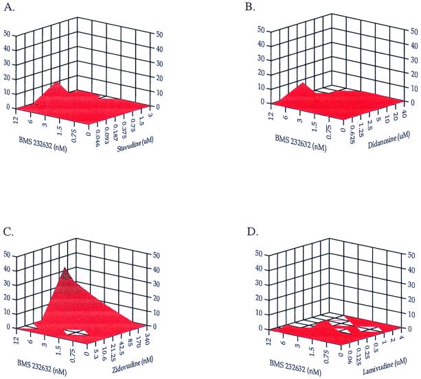
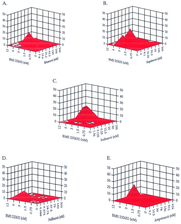
Atazanavir sulfate is the sulfate form of atazanavir, a nitrogen-based dipeptide analog with a (hydroxyethyl)hydrazine moiety containing a diaryl substituent. It is active against both wild-type and mutant HIV proteases. Atazanavir does not increase blood lipids, a common side effect of other protease inhibitors. Atazanavir is a nitrogen-based peptide HIV protease inhibitor used in combination with other anti-HIV drugs to treat HIV infection and AIDS. See also: Atazanavir (active ingredient); Atazanavir sulfate; Ritonavir (ingredient); Atazanavir sulfate; Cobicistat (ingredient)...See more... Drug Indications Ritaz capsules, used in combination with low-dose ritonavir, are indicated for the treatment of HIV-1 infection in adults and children aged 6 years and older, in combination with other antiretroviral drugs (see Section 4.2). Based on existing virological and clinical data in adult patients, no benefit is expected for patients carrying multiple protease inhibitor-resistant strains (mutations of 4 protease inhibitors). For previously treated adult and pediatric patients, selection of Reyataz should be based on individual viral resistance testing and the patient's treatment history (see Sections 4.4 and 5.1). Reyataz oral powder, in combination with low-dose ritonavir, is indicated for the treatment of HIV-1 infected pediatric patients at least 3 months of age and weighing at least 5 kg (see Section 4.2). Based on existing virological and clinical data from adult patients, patients carrying multiple protease inhibitor-resistant strains (≥4 protease inhibitor mutations) are not expected to benefit from Reyataz treatment. For previously treated adult and pediatric patients, selection of Reyataz should be based on individual viral resistance testing and the patient's treatment history (see Sections 4.4 and 5.1). Treatment of human immunodeficiency virus (HIV-1) infection. Therapeutic Use Atazanavir sulfate is indicated for the treatment of HIV-1 infection in combination with other antiretroviral agents. Atazanavir sulfate may be considered for adult patients with HIV infection who have previously received antiretroviral therapy and are genotyped and phenotypically identified as susceptible to atazanavir sulfate. /Included in the US product label/ Drug Warnings Patients receiving atazanavir in combination with nucleoside reverse transcriptase inhibitors (NRTIs) have been reported to have lactic acidosis syndrome (sometimes fatal) and symptomatic hyperlactatemia. Treatment with nucleoside reverse transcriptase inhibitors (NRTIs) is known to be associated with an increased risk of lactic acidosis syndrome; female sex and obesity are also known risk factors for this syndrome. Whether atazanavir increases the risk of lactic acidosis syndrome is yet to be determined. Patients receiving HIV protease inhibitors have been reported to have hyperglycemia (which may persist), new-onset diabetes, or exacerbation of pre-existing diabetes. Initiation of antidiabetic therapy (e.g., insulin, oral hypoglycemic agents) or adjustment of the dose of existing diabetes may be necessary; diabetic ketoacidosis may occur. Patients treated with atazanavir have experienced atrioventricular conduction abnormalities, including PR interval prolongation. Cardiac conduction abnormalities are usually limited to first-degree atrioventricular block; the QTc interval prolongation observed in HIV-infected patients treated with atazanavir has not been directly attributed to the drug. In clinical trials, asymptomatic first-degree atrioventricular block was observed in 5.9% of patients treated with regimens containing atazanavir or control antiretroviral agents (lopinavir/ritonavir, nelfinavir, efavirenz), and 3–10.4% of patients, respectively; second- or third-degree atrioventricular block was not observed. Due to a lack of clinical experience, atazanavir should be used with caution in patients with pre-existing cardiac conduction abnormalities (e.g., significant first-degree atrioventricular block; second- or third-degree atrioventricular block). Because atazanavir is a competitive inhibitor of uridine diphosphate glucuronyl transferase (UGT) 1A1 (an enzyme that catalyzes the glucuronidation of bilirubin), most patients treated with this drug experience reversible, asymptomatic elevations in indirect (unconjugated) bilirubin. In clinical trials, 35% to 47% of patients treated with this drug reported total bilirubin concentrations at least 2.6 times the upper limit of normal; long-term safety data are lacking for patients with persistent total bilirubin elevations exceeding 5 times the upper limit of normal. Elevated serum AST (SGOT) and/or ALT (SGPT) levels in patients with hyperbilirubinemia should be evaluated for other causes besides hyperbilirubinemia. If jaundice or scleral icterus caused by elevated bilirubin is cosmetically distressing, alternative antiretroviral therapies may be considered. Dosage reduction of atazanavir is not recommended (there are no efficacy data for dose reduction). For more drug warnings (full version) data on atazanavir (17 in total), please visit the HSDB record page. Pharmacodynamics Azanavir (ATV) is a nitrogen-containing peptide HIV-1 protease inhibitor (PI) active against human immunodeficiency virus type 1 (HIV-1). The HIV-1 protease is an enzyme responsible for hydrolyzing the viral polyprotein precursor protein into the various functional proteins of infectious HIV-1. Atazanavir binds to the active site of the protease and inhibits its activity. This inhibition prevents the cleavage of the viral polyprotein, thus avoiding the formation of immature, non-infectious viral particles. Protease inhibitors are almost always used in combination with at least two other anti-HIV drugs. Atazanavir is pharmacologically related to other protease inhibitors and currently available antiretroviral drugs, but structurally different. Atazanavir exhibits anti-HIV-1 activity against a variety of laboratory and clinically isolated HIV-1 strains with a mean half-maximal effective concentration (EC50) of 2 to 5 nM in the absence of human serum. These strains can be cultured in peripheral blood mononuclear cells, macrophages, CEM-SS cells, and MT-2 cells. Atazanavir is also active against HIV-1 M subtype A, B, C, D, AE, AG, F, G, and J isolates in cell culture. Atazanavir's activity against HIV-2 isolates varies (1.9–32 nM), with its EC50 value being higher than that of treatment-failed strains. Studies of the dual-drug antiviral activity of atazanavir showed that, in cell culture, atazanavir did not antagonize protease inhibitors (ampenavir, indinavir, lopinavir, nelfinavir, ritonavir, and saquinavir), non-nucleoside reverse transcriptase inhibitors (delavirin, efavirenz, and nevirapine), nucleoside reverse transcriptase inhibitors (abacavir, didanosine, emtricitabine, lamivudine, stavudine, tenofovir disoproxil fumarate, and zidovudine), the HIV-1 fusion inhibitor enfvirdi, or the two drugs used to treat viral hepatitis, adefovir and ribavirin, and did not enhance cytotoxicity. HIV-1 isolates with reduced sensitivity to atazanavir were screened in cell culture from patients who received atazanavir or atazanavir in combination with ritonavir. HIV-1 isolates with 93- to 183-fold reduced susceptibility to atazanavir were screened from three different viral strains and cultured in cell culture for 5 months. The amino acid substitutions in these HIV-1 viruses leading to atazanavir resistance included I50L, N88S, I84V, A71V, and M46I. Following drug screening, changes in protease cleavage sites also occurred. Recombinant viruses containing the I50L substitution but without other major protease inhibitor (PI) substitutions showed inhibited growth and increased susceptibility to other PIs (ampenavir, indinavir, lopinavir, nelfinavir, ritonavir, and saquinavir) in cell culture. The I50L and I50V substitutions resulted in selective resistance to atazanavir and ampravir, respectively, with no apparent cross-resistance. Concentration- and dose-dependent prolongation of the ECG PR interval was observed in healthy subjects taking atazanavir. In the placebo-controlled study AI424-076, the maximum mean (± standard deviation) change in the PR interval from the pre-dose value after oral administration of 400 mg atazanavir (n=65) was 24 (±15) ms, compared to 13 (±11) ms after placebo administration (n=67). The PR interval prolongation in this study was asymptomatic. Currently, information on pharmacodynamic interactions between atazanavir and other drugs that prolong the ECG PR interval in humans is limited. A clinical pharmacology study involving 72 healthy subjects determined the ECG effects of atazanavir. This study compared the efficacy of oral administration of 400 mg (maximum recommended dose) and 800 mg (twice the maximum recommended dose) of atazanavir with placebo; atazanavir had no concentration-dependent effect on the QTc interval (using Fridricia correction). In 1793 HIV-1 infected individuals receiving antiretroviral therapy, the degree of QTc interval prolongation was comparable between the atazanavir and control groups. In clinical trials, no healthy subjects or HIV-1 infected individuals receiving atazanavir experienced a QTc interval >500 ms. Atazanavir is a protease inhibitor (PI) approved for the treatment of HIV-1 infection. It is a substrate and inhibitor of cytochrome P450 isoenzyme 3A, and also an inhibitor and inducer of P-glycoprotein. In treatment-naïve patients, the virological efficacy of atazanavir is similar to that of efavirenz and lopinavir-enhanced ritonavir. Compared to other protease inhibitors, atazanavir has a smaller effect on blood lipids, making it suitable for patients who do not wish to develop hyperlipidemia. Ritonavir enhances the bioavailability of atazanavir but may cause elevated blood lipids; therefore, it is recommended for previously treated patients and those currently taking efavirenz or tenofovir. Atazanavir-enhanced ritonavir and lopinavir-enhanced ritonavir exhibit similar antiviral activity in both treatment-naïve and previously treated patients. Atazanavir can cause unconjugated bilirubinemia in more than 40% of patients, but the discontinuation rate is less than 2%. Atazanavir is approved for once-daily administration, and atazanavir/ritonavir and lopinavir/ritonavir are among the most commonly used protease inhibitors. [2] Atazanavir is a recently approved human immunodeficiency virus (HIV) protease inhibitor that plays an important role in the treatment of both treatment-naïve and treatment-experienced patients. Atazanavir (400 mg) is taken once daily in two capsules. Once-daily combination of atazanavir (300 mg) and ritonavir (100 mg) can safely increase drug exposure. Atazanavir does not cause an increase in serum total cholesterol, LDL cholesterol, or triglyceride levels, which may reduce the need for lipid-lowering drugs. Atazanavir causes an increase in unconjugated bilirubin levels, but this is generally not dose-limiting. In treatment-naïve patients, if virological rebound occurs after taking atazanavir, the I50L mutation of the HIV protease will occur, but this mutation will not lead to cross-resistance to other protease inhibitors. In patients who have previously received treatment and are highly resistant to other protease inhibitors, their sensitivity to atazanavir is usually reduced, and the best efficacy can be obtained when used in combination with ritonavir. Similar to other protease inhibitors, drug interactions must be considered when taking atazanavir and taking other drugs at the same time. [1] |
| Molecular Formula |
C38H54N6O11S
|
|---|---|
| Molecular Weight |
802.93
|
| Exact Mass |
802.357
|
| Elemental Analysis |
C, 56.84; H, 6.78; N, 10.47; O, 21.92; S, 3.99
|
| CAS # |
229975-97-7
|
| Related CAS # |
Atazanavir;198904-31-3
|
| PubChem CID |
158550
|
| Appearance |
White to light yellow solid powder
|
| Density |
1.164g/cm3
|
| Boiling Point |
995.5ºC at 760 mmHg
|
| Melting Point |
195.0°, or acetone; mp 198-199° (dec)
|
| Flash Point |
555.8ºC
|
| Vapour Pressure |
0mmHg at 25°C
|
| Source |
Endogenously produced metabolite
|
| LogP |
6.203
|
| Hydrogen Bond Donor Count |
7
|
| Hydrogen Bond Acceptor Count |
13
|
| Rotatable Bond Count |
18
|
| Heavy Atom Count |
56
|
| Complexity |
1190
|
| Defined Atom Stereocenter Count |
4
|
| SMILES |
S(=O)(=O)(O[H])O[H].O([H])[C@@]([H])(C([H])([H])N(C([H])([H])C1C([H])=C([H])C(C2=C([H])C([H])=C([H])C([H])=N2)=C([H])C=1[H])N([H])C([C@]([H])(C(C([H])([H])[H])(C([H])([H])[H])C([H])([H])[H])N([H])C(=O)OC([H])([H])[H])=O)[C@]([H])(C([H])([H])C1C([H])=C([H])C([H])=C([H])C=1[H])N([H])C([C@]([H])(C(C([H])([H])[H])(C([H])([H])[H])C([H])([H])[H])N([H])C(=O)OC([H])([H])[H])=O
|
| InChi Key |
DQSGVVGOPRWTKI-QVFAWCHISA-N
|
| InChi Code |
InChI=1S/C38H52N6O7.H2O4S/c1-37(2,3)31(41-35(48)50-7)33(46)40-29(22-25-14-10-9-11-15-25)30(45)24-44(43-34(47)32(38(4,5)6)42-36(49)51-8)23-26-17-19-27(20-18-26)28-16-12-13-21-39-28;1-5(2,3)4/h9-21,29-32,45H,22-24H2,1-8H3,(H,40,46)(H,41,48)(H,42,49)(H,43,47);(H2,1,2,3,4)/t29-,30-,31+,32+;/m0./s1
|
| Chemical Name |
methyl N-[(2S)-1-[2-[(2S,3S)-2-hydroxy-3-[[(2S)-2-(methoxycarbonylamino)-3,3-dimethylbutanoyl]amino]-4-phenylbutyl]-2-[(4-pyridin-2-ylphenyl)methyl]hydrazinyl]-3,3-dimethyl-1-oxobutan-2-yl]carbamate;sulfuric acid
|
| Synonyms |
Atazanavir sulfate; BMS-232632; 229975-97-7; Reyataz; Atazanavir sulphate; Atazanavir (sulfate); BMS 232632; BMS232632
|
| HS Tariff Code |
2934.99.03.00
|
| Storage |
Powder -20°C 3 years 4°C 2 years In solvent -80°C 6 months -20°C 1 month Note: Please store this product in a sealed and protected environment, avoid exposure to moisture. |
| Shipping Condition |
Room temperature (This product is stable at ambient temperature for a few days during ordinary shipping and time spent in Customs)
|
| Solubility (In Vitro) |
DMSO: ~104 mg/mL (~129.5 mM)
Water: 1 mg/mL (Insoluble) Ethanol: 1 mg/mL |
|---|---|
| Solubility (In Vivo) |
Solubility in Formulation 1: ≥ 2.5 mg/mL (3.11 mM) (saturation unknown) in 10% DMSO + 40% PEG300 + 5% Tween80 + 45% Saline (add these co-solvents sequentially from left to right, and one by one), clear solution.
For example, if 1 mL of working solution is to be prepared, you can add 100 μL of 25.0 mg/mL clear DMSO stock solution to 400 μL PEG300 and mix evenly; then add 50 μL Tween-80 to the above solution and mix evenly; then add 450 μL normal saline to adjust the volume to 1 mL. Preparation of saline: Dissolve 0.9 g of sodium chloride in 100 mL ddH₂ O to obtain a clear solution. Solubility in Formulation 2: ≥ 2.5 mg/mL (3.11 mM) (saturation unknown) in 10% DMSO + 90% (20% SBE-β-CD in Saline) (add these co-solvents sequentially from left to right, and one by one), clear solution. For example, if 1 mL of working solution is to be prepared, you can add 100 μL of 25.0 mg/mL clear DMSO stock solution to 900 μL of 20% SBE-β-CD physiological saline solution and mix evenly. Preparation of 20% SBE-β-CD in Saline (4°C,1 week): Dissolve 2 g SBE-β-CD in 10 mL saline to obtain a clear solution. View More
Solubility in Formulation 3: ≥ 2.5 mg/mL (3.11 mM) (saturation unknown) in 10% DMSO + 90% Corn Oil (add these co-solvents sequentially from left to right, and one by one), clear solution. Solubility in Formulation 4: ≥ 2.5 mg/mL (3.11 mM) (saturation unknown) in 5% DMSO + 40% PEG300 + 5% Tween80 + 50% Saline (add these co-solvents sequentially from left to right, and one by one), clear solution. Preparation of saline: Dissolve 0.9 g of sodium chloride in 100 mL ddH₂ O to obtain a clear solution. Solubility in Formulation 5: ≥ 2.5 mg/mL (3.11 mM) (saturation unknown) in 5% DMSO + 95% (20% SBE-β-CD in Saline) (add these co-solvents sequentially from left to right, and one by one), clear solution. Preparation of 20% SBE-β-CD in Saline (4°C,1 week): Dissolve 2 g SBE-β-CD in 10 mL saline to obtain a clear solution. Solubility in Formulation 6: 30% PEG400+0.5% Tween80+5% propylene glycol: 30 mg/mL |
| Preparing Stock Solutions | 1 mg | 5 mg | 10 mg | |
| 1 mM | 1.2454 mL | 6.2272 mL | 12.4544 mL | |
| 5 mM | 0.2491 mL | 1.2454 mL | 2.4909 mL | |
| 10 mM | 0.1245 mL | 0.6227 mL | 1.2454 mL |
*Note: Please select an appropriate solvent for the preparation of stock solution based on your experiment needs. For most products, DMSO can be used for preparing stock solutions (e.g. 5 mM, 10 mM, or 20 mM concentration); some products with high aqueous solubility may be dissolved in water directly. Solubility information is available at the above Solubility Data section. Once the stock solution is prepared, aliquot it to routine usage volumes and store at -20°C or -80°C. Avoid repeated freeze and thaw cycles.
Calculation results
Working concentration: mg/mL;
Method for preparing DMSO stock solution: mg drug pre-dissolved in μL DMSO (stock solution concentration mg/mL). Please contact us first if the concentration exceeds the DMSO solubility of the batch of drug.
Method for preparing in vivo formulation::Take μL DMSO stock solution, next add μL PEG300, mix and clarify, next addμL Tween 80, mix and clarify, next add μL ddH2O,mix and clarify.
(1) Please be sure that the solution is clear before the addition of next solvent. Dissolution methods like vortex, ultrasound or warming and heat may be used to aid dissolving.
(2) Be sure to add the solvent(s) in order.
| NCT Number | Recruitment | interventions | Conditions | Sponsor/Collaborators | Start Date | Phases |
| NCT04468087 | Active Recruiting |
Drug: Atazanavir Drug: Daclatasvir 60 mg |
COVID-19 | Hospital do Coracao | February 15, 2021 | Phase 2 Phase 3 |
| NCT04121195 | Active Recruiting |
Drug: Dose escalation | HIV/AIDS Tuberculosis |
University of Liverpool | October 30, 2020 | Phase 2 Phase 3 |
| NCT02016924 | Recruiting | Drug: ATV Drug: DRV |
Acquired Immune Deficiency Syndrome (AIDS) HIV Infections |
Gilead Sciences | January 16, 2014 | Phase 2 Phase 3 |
| NCT04452565 | Recruiting | Drug: Drug: NA-831 Combination Product: NA-831 and Atazanavir |
Coronavirus Infection Severe Acute Respiratory Infection |
NeuroActiva, Inc. | June 15, 2022 | Phase 2 Phase 3 |
| NCT01837719 | Completed | Drug: Atazanavir Drug: Cobicistat |
HIV-1 | Bristol-Myers Squibb | April 2013 | Phase 1 |
|
|---|
|
|
|
|---|
|
Products are for research use only; We do not sell to patients
Copyright 2020 InvivoChem LLC | All Rights Reserved


 COA
COA