| Size | Price | Stock | Qty |
|---|---|---|---|
| 1mg |
|
||
| 5mg |
|
||
| 10mg |
|
||
| 25mg |
|
||
| 50mg |
|
||
| 100mg |
|
||
| 250mg |
|
||
| 500mg | |||
| Other Sizes |


Purity: ≥98%
| Targets |
Akt1 (IC50 = 38 nM); p70S6K (IC50 = 8 nM); Akt3 (IC50 = 50 nM); Akt2 (IC50 = 402 nM); PKA (IC50 = 3 nM); ROCKII (IC50 = 4 nM); ROCKI (IC50 = 6 nM); SGK3 (IC50 = 63 nM); RSK1 (IC50 = 85 nM); CHK2 (IC50 = 860 nM); Aurora B (IC50 = 1840 nM)
AT13148 is a multi-AGC kinase inhibitor, with potent activity against key members of the AGC kinase family. In recombinant enzyme assays, it exhibits IC50 values of 38 nM for Akt1, 46 nM for Akt2, 61 nM for Akt3, 112 nM for mTOR, 89 nM for PDK1 (3-phosphoinositide-dependent protein kinase 1), and 77 nM for SGK1 (serum- and glucocorticoid-inducible kinase 1). It shows minimal inhibition against non-AGC kinases (e.g., EGFR, VEGFR2) with IC50 values > 1000 nM [1] - In human gastric cancer cells (MGC-803, SGC-7901) with dysregulated Akt signaling, AT13148 inhibits Akt-mediated phosphorylation with an EC50 of 52 nM (for p-Akt Ser473 reduction in MGC-803 cells) and 68 nM (in SGC-7901 cells), without significant activity against other signaling kinases (e.g., ERK1/2) [2] |
|---|---|
| ln Vitro |
AT13148, as a multi-AGC kinase inhibitor, potently inhibits proliferation with GI50 values of 1.5 to 3.8 μM across a selected panel of cancer cell lines with deregulation of PI3K-AKT-mTOR or RAS-RAF pathways. Additionally, AT13148 inhibits p70S6K and AKT signaling in PTEN-deficient MES-SA cells.[1]
In human prostate cancer cell lines (PC-3, LNCaP) with activated Akt/mTOR signaling, AT13148 (0.01-10 μM) inhibited cell proliferation in a dose-dependent manner. The IC50 values were 0.32 μM for PC-3 cells and 0.45 μM for LNCaP cells after 72 hours (SRB assay). Western blot analysis showed that 0.5 μM AT13148 reduced phosphorylation of Akt (Ser473/Thr308) by > 85%, mTOR (Ser2448) by 78%, and S6K1 (Thr389) by 72% within 24 hours, while total protein levels of these kinases remained unchanged [1] - In human gastric cancer cell lines (MGC-803, SGC-7901), AT13148 (0.1-5 μM) induced dose-dependent apoptosis after 48 hours. Flow cytometry with Annexin V-FITC/PI staining revealed that 2 μM AT13148 increased the apoptotic rate from 3.5% (control) to 38% in MGC-803 cells and from 4% to 32% in SGC-7901 cells. Additionally, 1 μM AT13148 inhibited colony formation of MGC-803 cells by 65% compared to vehicle control (crystal violet staining, 14-day culture) [2] - In PC-3 cells co-treated with AT13148 (0.2 μM) and docetaxel (5 nM), the combination showed synergistic antiproliferative activity with a combination index (CI) of 0.52 (CI < 1 indicates synergy). The apoptotic rate in the combination group was 45%, significantly higher than 18% (AT13148 alone) and 15% (docetaxel alone) [1] |
| ln Vivo |
AT13148 (50 mg/kg p.o.) exhibits strong antitumor effects in human tumor xenografts by significantly inhibiting the activity of both AKT and p70S6K AGC kinases. [1]
In a nude mouse xenograft model of human prostate cancer (PC-3), AT13148 was administered orally at doses of 25 mg/kg and 50 mg/kg twice daily for 21 days. Compared to the vehicle control (0.5% carboxymethyl cellulose sodium, CMC-Na), the 25 mg/kg group showed a 48% reduction in tumor volume, and the 50 mg/kg group showed a 70% reduction. Immunohistochemical staining of tumor tissues demonstrated decreased p-Akt (Ser473) expression (-80%) and Ki-67 (proliferation marker) positive cells (-55%) in the 50 mg/kg group [1] - In a nude mouse xenograft model of human gastric cancer (MGC-803), AT13148 was administered intraperitoneally (i.p.) at doses of 10 mg/kg and 20 mg/kg once daily for 14 days. The 10 mg/kg group had a 35% reduction in tumor weight, and the 20 mg/kg group had a 58% reduction. Western blot analysis of tumor lysates confirmed reduced p-mTOR (Ser2448) and increased cleaved caspase-3 (apoptosis marker) in treated groups [2] - In the PC-3 xenograft model, combination treatment with AT13148 (25 mg/kg oral, twice daily) and docetaxel (10 mg/kg i.p., once weekly) for 21 days resulted in an 82% reduction in tumor volume, which was significantly greater than monotherapy effects (48% for AT13148 alone, 40% for docetaxel alone) [1] |
| Enzyme Assay |
AT13148 is assayed against 40 kinases and the percentage inhibition at 10 μM of AT13148 is determined. Using ATP concentrations equivalent to each enzyme's Km, the individual IC50 values for a few kinases are determined.
Akt1 Kinase Inhibition Assay: Recombinant human Akt1 (0.1 μg per reaction) was mixed with 50 mM Tris-HCl (pH 7.5), 10 mM MgCl2, 1 mM DTT, 10 μM ATP (including [γ-32P]ATP), 20 μM Crosstide (Akt-specific substrate peptide), and serial dilutions of AT13148 (1 nM-1000 nM) in a total volume of 50 μL. The reaction mixture was incubated at 30°C for 30 minutes, then terminated by adding 25 μL of 30% trichloroacetic acid. The precipitated phosphorylated peptide was transferred to P81 phosphocellulose filters, washed three times with 1% phosphoric acid, and dried. Radioactivity was measured using a liquid scintillation counter, and IC50 was calculated via four-parameter logistic regression [1] - mTOR Kinase Assay: Recombinant human mTOR (0.2 μg per reaction) was incubated with 25 mM HEPES (pH 7.4), 10 mM MgCl2, 1 mM EGTA, 200 μM ATP (including [γ-32P]ATP), 1 μg/mL 4E-BP1 (mTOR substrate), and AT13148 (10 nM-2000 nM) for 45 minutes at 37°C. The reaction was terminated with SDS sample buffer, and phosphorylated 4E-BP1 was separated by 12% SDS-PAGE. The gel was dried, and radioactivity was detected by autoradiography. IC50 was determined by plotting the percentage of remaining kinase activity against drug concentration [1] |
| Cell Assay |
Cells are seeded onto 96-well micro-plates at a density of 1×10⁴ cells per well. Following therapy, MTT solution (0.5 mg/mL) is added for 2–3 hours. The optical density (OD), which is measured at 570 nm using a microplate reader, is obtained after the MTT-purple formazan productions are dissolved in 0.1 N hydrochloric acid.
Prostate Cancer Cell Proliferation Assay (SRB Method): PC-3 or LNCaP cells were seeded in 96-well plates at a density of 4×10³ cells/well and cultured overnight at 37°C with 5% CO2. AT13148 was added at concentrations ranging from 0.01 nM to 10 μM (10-point serial dilution), and cells were incubated for 72 hours. After incubation, cells were fixed with 10% trichloroacetic acid for 1 hour, stained with 0.4% sulforhodamine B (SRB) for 30 minutes, and washed with 1% acetic acid. Bound SRB was dissolved in 10 mM Tris base, and absorbance was measured at 510 nm. IC50 was defined as the concentration of AT13148 that inhibited proliferation by 50% relative to vehicle control [1] - Gastric Cancer Apoptosis Assay (Annexin V-FITC/PI Staining): MGC-803 cells were seeded in 6-well plates at 2×10⁵ cells/well and treated with AT13148 (0.1-5 μM) for 48 hours. Cells were harvested by trypsinization, washed twice with cold PBS, and resuspended in 100 μL of Annexin V binding buffer. Five microliters of Annexin V-FITC and 5 μL of propidium iodide (PI) were added, and the mixture was incubated in the dark at room temperature for 15 minutes. Apoptotic cells were analyzed via flow cytometry within 1 hour, with early apoptosis defined as Annexin V-positive/PI-negative and late apoptosis as Annexin V-positive/PI-positive [2] - Western Blot Analysis (Cellular): Cells were treated with AT13148 (0.1-5 μM) for 24 hours, then lysed in RIPA buffer containing protease and phosphatase inhibitors. Protein concentration was determined using a BCA assay kit. Equal amounts of protein (30 μg per lane) were separated by 10% SDS-PAGE and transferred to PVDF membranes. Membranes were blocked with 5% non-fat milk in TBST for 1 hour at room temperature, then incubated overnight at 4°C with primary antibodies against phospho-Akt (Ser473, Thr308), total Akt, phospho-mTOR (Ser2448), phospho-S6K1 (Thr389), cleaved caspase-3, or β-actin. After washing with TBST, membranes were incubated with HRP-conjugated secondary antibodies for 1 hour, and protein bands were visualized using an ECL detection system. Band intensity was quantified via ImageJ software [1] |
| Animal Protocol |
For pharmacokinetic analysis, male athymic BALB/c mice are obtained from Harlan. AT13148 is administered at 5 mg/kg i.v. or p.o. and is made up of 10% DMSO, 1% Tween-20, and 89% saline. At 1, 2, 4, 6, 8, 16, 24, and 72 hours after administration, cardiac punctures are used to obtain duplicate samples of whole blood that has been heparinized. In preparation for analysis, plasma and tissues (including liver, kidney, spleen, and muscle) are frozen at -20°C. Using acetonitrile with an internal standard to extract it from plasma and tissues, AT13148 is then quantified using the liquid chromatography tandem mass spectrometry (LC-MS/MS) technique and the appropriate standard curves. In order to calculate pharmacokinetic parameters, WinNonLin software version 5.2 is used.
Prostate Cancer Xenograft Model (PC-3): Female nude mice (6-8 weeks old, n=6 per group) were subcutaneously injected with 2×10⁶ PC-3 cells (suspended in 100 μL of PBS + 50% Matrigel) into the right hind flank. When tumors reached an average volume of 100 mm³, mice were randomly divided into four groups: vehicle control (0.5% CMC-Na), AT13148 25 mg/kg, AT13148 50 mg/kg, and AT13148 25 mg/kg + docetaxel 10 mg/kg. AT13148 was suspended in the vehicle and administered orally twice daily (12-hour interval) for 21 days; docetaxel was dissolved in normal saline and administered intraperitoneally once weekly for 3 weeks. Tumor volume was measured every 3 days (volume = length × width² / 2), and body weight was recorded weekly [1] - Gastric Cancer Xenograft Model (MGC-803): Male nude mice (6-8 weeks old, n=5 per group) were subcutaneously injected with 3×10⁶ MGC-803 cells (in 100 μL of PBS + 50% Matrigel) into the left flank. When tumors reached ~120 mm³, mice were assigned to three groups: vehicle control (5% DMSO + 95% normal saline), AT13148 10 mg/kg, and AT13148 20 mg/kg. AT13148 was dissolved in the vehicle and administered intraperitoneally once daily for 14 days. At the end of the experiment, mice were euthanized, tumors were excised and weighed, and tumor lysates were prepared for Western blot analysis [2] |
| ADME/Pharmacokinetics |
In male Sprague-Dawley (SD) rats, AT13148 was administered via two routes: intravenous (iv) at a dose of 5 mg/kg and oral (po) at a dose of 20 mg/kg. Following intravenous administration, the plasma concentration-time curve conformed to a two-compartment model, with a terminal half-life (t1/2β) of 4.2 h, a steady-state volume of distribution (Vdss) of 2.3 L/kg, and a total clearance (CL) of 0.6 L/h/kg. After oral administration, the peak plasma concentration (Cmax) was 1.8 μg/mL, the time to peak concentration (Tmax) was 1.5 hours, and the oral bioavailability (F) was 28% [1]. - In vitro plasma protein binding experiments using equilibrium dialysis showed that AT13148 had high plasma protein binding rates: 92% in human plasma, 90% in rat plasma, and 88% in canine plasma; the free fraction was <10% in all tested species [1]. - In vitro metabolic studies using human liver microsomes showed that AT13148 was metabolized in a NADPH-dependent manner into two major metabolites (M1, M2), of which approximately 70% of the total metabolism was mediated by CYP3A4 (confirmed by specific CYP3A4 inhibitors) [1].
|
| Toxicity/Toxicokinetics |
In a 28-day repeated-dose toxicity study, male and female Sprague-Dawley rats were orally administered AT13148 at doses of 10 mg/kg, 25 mg/kg, and 50 mg/kg once daily. In the 50 mg/kg dose group, both male and female rats experienced a 12% decrease in body weight, a 1.8-fold increase in serum ALT (alanine aminotransferase) levels compared to the control group, and histopathological examination revealed mild hepatocyte vacuolation. No significant toxicity was observed at doses of 10 mg/kg or 25 mg/kg (no weight loss, no abnormal liver enzymes) [1]
- In the MGC-803 gastric cancer xenograft model, doses of up to 20 mg/kg (intraperitoneal injection, 14 days) of AT13148 did not cause significant changes in weight or significant pathological abnormalities in major organs (liver, kidney, heart, lungs) [2] - In vitro cytotoxicity assays in normal human peripheral blood mononuclear cells (PBMCs) showed that AT13148 had a CC50 of 12 μM and a therapeutic index (TI = CC50/IC50) of 37.5 (compared to PC-3 cells, IC50 = 0.32 μM) [1] |
| References |
|
| Additional Infomation |
AT13148, a multi-AGC kinase inhibitor, is an oral small-molecule AGC kinase inhibitor with potential antitumor activity. AT13148 inhibits the enzymatic activity of two AGC kinases—protein kinase B (PKB or AKT) and p70S6K—in an ATP-competitive manner. These two kinases play a key role in the PI3K/PKB/mTOR signaling pathway. Blocking this pathway can inhibit cell growth and induce apoptosis in susceptible tumor cells. The PI3K/PKB/mTOR pathway is aberrantly activated in more than 50% of tumors and is often associated with drug resistance and increased tumor survival. AGC kinases are serine/threonine kinases regulated by second messengers such as cyclic adenosine monophosphate (cAMP) and lipids. AT13148 is a first-in-class oral multi-target AGC kinase inhibitor for the treatment of solid tumors (e.g., prostate cancer, gastric cancer, breast cancer) with dysregulated AGC kinase signaling pathways [1][2]. Preclinical studies have shown that AT13148 targets multiple nodes of the AGC kinase pathway (Akt, mTOR, PDK1, SGK1), thus avoiding the adaptive resistance common with single kinase inhibitors (e.g., Akt-only inhibitors) [1]. AT13148 has good oral bioavailability (28% in rats) and blood-brain barrier penetration (0.4 brain/plasma concentration ratio in mice), supporting its therapeutic potential. Treatment of AGC kinase-dysregulated central nervous system (CNS) tumors [1] In a gastric cancer model, AT13148 showed activity against both HER2-positive and HER2-negative subtypes, indicating its broad application prospects in the treatment of gastric cancer [2].
|
| Molecular Formula |
C17H16CLN3O
|
|---|---|
| Molecular Weight |
313.78
|
| Exact Mass |
313.78
|
| Elemental Analysis |
C, 65.07; H, 5.14; Cl, 11.30; N, 13.39; O, 5.10
|
| CAS # |
1056901-62-2
|
| Related CAS # |
1056901-62-2; 1056901-67-7 (R-isomer);857532-13-9 (racemic);
|
| PubChem CID |
24905401
|
| Appearance |
white solid powder
|
| LogP |
5.229
|
| Hydrogen Bond Donor Count |
3
|
| Hydrogen Bond Acceptor Count |
3
|
| Rotatable Bond Count |
4
|
| Heavy Atom Count |
22
|
| Complexity |
354
|
| Defined Atom Stereocenter Count |
1
|
| SMILES |
ClC1=CC=C([C@](C2=CC=C(C3=CNN=C3)C=C2)(O)CN)C=C1
|
| InChi Key |
IIRWNGPLJQXWFJ-KRWDZBQOSA-N
|
| InChi Code |
InChI=1S/C17H16ClN3O/c18-16-7-5-15(6-8-16)17(22,11-19)14-3-1-12(2-4-14)13-9-20-21-10-13/h1-10,22H,11,19H2,(H,20,21)/t17-/m0/s1
|
| Chemical Name |
(1S)-2-amino-1-(4-chlorophenyl)-1-[4-(1H-pyrazol-4-yl)phenyl]ethanol
|
| Synonyms |
AT-13148; AT 13148; AT13148; AT13148 hydrochloride; AT13148 HCl
|
| HS Tariff Code |
2934.99.9001
|
| Storage |
Powder -20°C 3 years 4°C 2 years In solvent -80°C 6 months -20°C 1 month |
| Shipping Condition |
Room temperature (This product is stable at ambient temperature for a few days during ordinary shipping and time spent in Customs)
|
| Solubility (In Vitro) |
DMSO: ~62 mg/mL (~197.6 mM)
Water: 1 mg/mL (Insoluble) Ethanol: 1 mg/mL |
|---|---|
| Solubility (In Vivo) |
Solubility in Formulation 1: ≥ 2.5 mg/mL (7.97 mM) (saturation unknown) in 10% DMSO + 40% PEG300 + 5% Tween80 + 45% Saline (add these co-solvents sequentially from left to right, and one by one), clear solution.
For example, if 1 mL of working solution is to be prepared, you can add 100 μL of 25.0 mg/mL clear DMSO stock solution to 400 μL PEG300 and mix evenly; then add 50 μL Tween-80 to the above solution and mix evenly; then add 450 μL normal saline to adjust the volume to 1 mL. Preparation of saline: Dissolve 0.9 g of sodium chloride in 100 mL ddH₂ O to obtain a clear solution. Solubility in Formulation 2: ≥ 2.5 mg/mL (7.97 mM) (saturation unknown) in 10% DMSO + 90% (20% SBE-β-CD in Saline) (add these co-solvents sequentially from left to right, and one by one), clear solution. For example, if 1 mL of working solution is to be prepared, you can add 100 μL of 25.0 mg/mL clear DMSO stock solution to 900 μL of 20% SBE-β-CD physiological saline solution and mix evenly. Preparation of 20% SBE-β-CD in Saline (4°C,1 week): Dissolve 2 g SBE-β-CD in 10 mL saline to obtain a clear solution. View More
Solubility in Formulation 3: ≥ 2.5 mg/mL (7.97 mM) (saturation unknown) in 10% DMSO + 90% Corn Oil (add these co-solvents sequentially from left to right, and one by one), clear solution. |
| Preparing Stock Solutions | 1 mg | 5 mg | 10 mg | |
| 1 mM | 3.1869 mL | 15.9347 mL | 31.8695 mL | |
| 5 mM | 0.6374 mL | 3.1869 mL | 6.3739 mL | |
| 10 mM | 0.3187 mL | 1.5935 mL | 3.1869 mL |
*Note: Please select an appropriate solvent for the preparation of stock solution based on your experiment needs. For most products, DMSO can be used for preparing stock solutions (e.g. 5 mM, 10 mM, or 20 mM concentration); some products with high aqueous solubility may be dissolved in water directly. Solubility information is available at the above Solubility Data section. Once the stock solution is prepared, aliquot it to routine usage volumes and store at -20°C or -80°C. Avoid repeated freeze and thaw cycles.
Calculation results
Working concentration: mg/mL;
Method for preparing DMSO stock solution: mg drug pre-dissolved in μL DMSO (stock solution concentration mg/mL). Please contact us first if the concentration exceeds the DMSO solubility of the batch of drug.
Method for preparing in vivo formulation::Take μL DMSO stock solution, next add μL PEG300, mix and clarify, next addμL Tween 80, mix and clarify, next add μL ddH2O,mix and clarify.
(1) Please be sure that the solution is clear before the addition of next solvent. Dissolution methods like vortex, ultrasound or warming and heat may be used to aid dissolving.
(2) Be sure to add the solvent(s) in order.
| NCT Number | Status | Interventions | Conditions | Sponsor/Collaborators | Start Date | Phases |
| NCT01585701 | Completed | Biological: AT13148 | Cancer Research UK | Advanced Solid Tumours | May 2012 | Phase 1 |
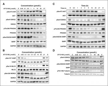
The effect of AT13148 exposure on AGC kinase biomarker expression. |
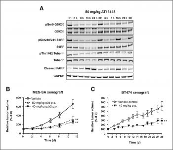
Pharmacodynamic biomarker and antitumor activity of AT13148 in human tumor xenografts. Clin Cancer Res. 2012, 18(14), 3912-3923. |
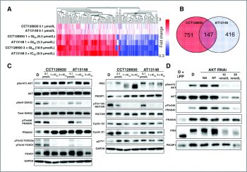
Comparison of the effects of 6-hour treatment of AT13148 versus CCT128930 on gene and protein expression in U87MG human glioblastoma cells |
Products are for research use only; We do not sell to patients
Copyright 2020 InvivoChem LLC | All Rights Reserved


 COA
COA