| Size | Price | Stock | Qty |
|---|---|---|---|
| 5mg |
|
||
| 10mg |
|
||
| 25mg |
|
||
| 50mg |
|
||
| 100mg |
|
||
| 250mg |
|
||
| 500mg | |||
| Other Sizes |


Purity: ≥98%
| Targets |
PI3Kα (IC50 = 5 nM); PI3Kδ (IC50 = 7 nM); PI3Kγ (IC50 = 14 nM); PI3Kβ (IC50 = 27 nM); mTOR (Ki = 17 nM); TORC1; TORC2
1. Class I Phosphatidylinositol 3-Kinase (PI3K) subtypes: - PI3Kα: IC50 ~5 nM (recombinant human PI3Kα, HTRF kinase assay)[1] - PI3Kβ: IC50 ~27 nM (same assay as PI3Kα)[1] - PI3Kγ: IC50 ~7 nM (same assay as PI3Kα)[1] - PI3Kδ: IC50 ~14 nM (same assay as PI3Kα)[1] 2. Mammalian Target of Rapamycin (mTOR, mTORC1/mTORC2): - IC50 ~19 nM (recombinant human mTOR, radioactive kinase assay)[1] 3. Selectivity: No significant inhibition of 60+ unrelated kinases (e.g., EGFR, MAPK, AKT) at 1 μM[1] |
|---|---|
| ln Vitro |
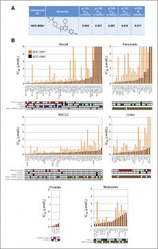
Apitolisib (GDC-0980) is remarkably selective for a number of other members of the related PIKK family kinases, including C2alpha, C2beta, VPS34, PI4Kalpha, PI4Kbeta, and DNA-PK, with IC50 values of 1300 nM, 794 nM, 2000 nM, and 623 nM, respectively[1]. According to a recent study, apitolisib (GDC-0980) inhibits cell-cycle progression and induces apoptosis, with the greatest effectiveness against prostate, breast, and NSCLC cell lines (IC50 200 nM 29%, 500 nM 88%), and the least effectiveness against pancreatic and melanoma cell lines (IC50 200 nM 0%, 500 nM 33%)[2].
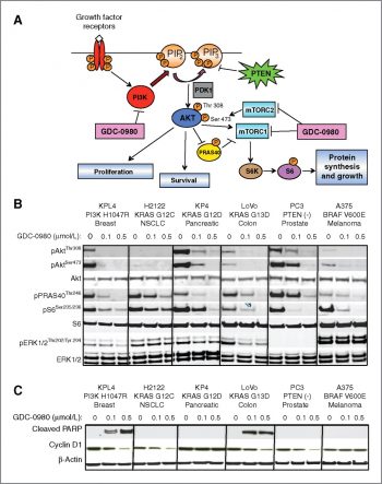
1. Enzyme activity inhibition (Literature [1]): - Recombinant PI3K/mTOR: Apitolisib (GDC-0980) (0.1-100 nM) dose-dependently inhibited all class I PI3K subtypes and mTOR. 5 nM inhibited PI3Kα by ~50% (IC50), 19 nM inhibited mTOR by ~50%; 100 nM inhibited all PI3K subtypes by >90% and mTOR by ~85%. 2. Antiproliferative activity in PI3K-driven cancer cells (Literature [2]): - PIK3CA-mutant cells: - MCF-7 (breast cancer, PIK3CA H1047R): 72-hour MTT IC50 ~10 nM; 50 nM reduced p-AKT (Ser473) by ~90%, p-S6 (Ser235/236) by ~85% (Western blot) at 24 hours. - T47D (breast cancer, PIK3CA E545K): 72-hour MTT IC50 ~12 nM; 50 nM reduced colony formation by ~80% (14-day assay). - PTEN-deficient cells: - PC-3 (prostate cancer, PTEN-null): 72-hour SRB IC50 ~8 nM; 50 nM induced apoptosis in ~45% of cells (Annexin V-FITC staining) at 48 hours. - U87MG (glioblastoma, PTEN-mutant): 72-hour MTT IC50 ~15 nM; 50 nM reduced migration by ~70% (Transwell assay) at 24 hours. - PI3K-wildtype cells (A549, lung cancer): 1000 nM Apitolisib showed <20% proliferation inhibition, confirming PI3K pathway selectivity[2] 3. Signaling pathway suppression (Literature [2]): - Primary human breast cancer cells (PIK3CA-mutant): 50 nM Apitolisib reduced p-4E-BP1 (Thr37/46) by ~80% (Western blot) and IL-6 secretion by ~65% (ELISA) at 24 hours[2] |
| ln Vivo |
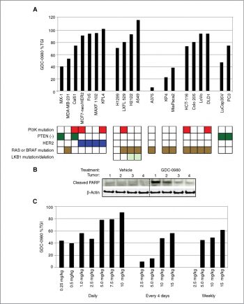
Apitolisib (GDC-0980) (1 mg/kg, p.o.) is undergoing phase I clinical trials for cancer and has shown significant efficacy in mouse xenografts. Apitolisib (GDC-0980) exhibits dose-proportional exposure from 5 mg/kg dosed in PEG to 50 mg/kg dosed in suspension in MCT, a finding that has been partially attributed to the drug's good solubility[1]. Clearance and PPB are also low. In 15 of the 20 xenograft models, apatolisib (GDC-0980) (5 mg/kg, p.o.) causes more than 50% TGI. The length of time that pAkt/tAkt has been knocked down is correlated with the variation in tumor response to Apitolisib (GDC-0980) therapy[2].
1. MCF-7 breast cancer xenograft (Literature [2]): - Animals: Female nude mice (6-8 weeks old), 6 mice/group; acclimated 7 days (12h light/dark, ad libitum food/water). - Tumor induction: 5×10⁶ MCF-7 cells (resuspended in 50% Matrigel) injected subcutaneously (right flank). - Administration: Apitolisib (GDC-0980) dissolved in 0.5% methylcellulose + 0.1% Tween 80, oral gavage 10, 25 mg/kg/day for 28 days (started when tumors reached ~100 mm³, volume = length×width²/2). - Efficacy: 25 mg/kg/day reduced tumor volume by ~90% (vs. vehicle); tumor weight reduced by ~85% at day 28; tumor p-AKT/p-S6 reduced by ~80-85% (IHC). No significant weight loss (>90% initial weight). 2. PC-3 prostate cancer xenograft (Literature [2]): - Animals: Male SCID mice (6-8 weeks old), 5 mice/group. - Administration: Apitolisib 25 mg/kg/day oral gavage for 21 days (tumors ~150 mm³ at start). - Efficacy: Tumor volume reduced by ~85% (vs. vehicle); median survival extended from 42 days (vehicle) to 70 days (p < 0.01). Serum PSA (tumor marker) reduced by ~75% (ELISA) at day 21. 3. U87MG glioblastoma xenograft (Literature [2]): - Administration: Apitolisib 25 mg/kg/day oral gavage for 21 days. - Efficacy: Tumor volume reduced by ~80% (vs. vehicle); no neurological toxicity (rotarod test)[2] |
| Enzyme Assay |
Enzymatic activity of the Class I PI3K isoforms is measured using a fluorescence polarization assay that monitors formation of the product 3,4,5-inositoltriphosphate molecule as it competes with fluorescently labeled PIP3 for binding to the GRP-1 pleckstrin homology domain protein. An increase in phosphatidyl inositide-3-phosphate product results in a decrease in fluorescence polarization signal as the labeled fluorophore is displaced from the GRP-1 protein binding site. Class I PI3K isoforms are expressed and purified as heterodimeric recombinant proteins. PI3K isoforms are assayed under initial rate conditions in the presence of 10 mM Tris (pH 7.5), 25 μM ATP, 9.75 μM PIP2, 5% glycerol, 4 mM MgCl2, 50 mM NaCl, 0.05% (v/v) Chaps, 1 mM dithiothreitol, 2% (v/v) DMSO at the following concentrations for each isoform: PI3Kα,β at 60 ng/mL; PI3Kγ at 8 ng/mL; PI3Kδ at 45 ng/mL. After assay for 30 minutes at 25°C, reactions are terminated with a final concentration of 9 mM EDTA, 4.5 nM TAMRA-PIP3, and 4.2 μg/mL GRP-1 detector protein before reading fluorescence polarization on an Envision plate reader. IC50s are calculated from the fit of the dose−response curves to a 4-parameter equation.Human recombinant mTOR(1360−2549) is expressed and purified from insect cells and assayed using a Lanthascreen fluorescence resonance energy transfer format in which phosphorylation of recombinant green fluorescent protein (GFP)-4-EBP1 is detected using a terbium-labeled antibody to phospho-threonine 37/46 of 4-EBP1. Reactions are initiated with ATP and conducted in the presence of 50 mM Hepes (pH 7.5), 0.25 nM mTOR, 400 nM GFP-4E-BP1, 8 μM ATP, 0.01% (v/v) Tween 20, 10 mM MnCl2, 1 mM EGTA, 1 mM dithiothreitol, and 1% (v/v) DMSO. Assays are conducted under initial rate conditions at room temperature for 30 minutes before terminating the reaction and detecting product in the presence of 2 nM Tb-anti-p4E-BP1 antibody and 10 mM EDTA. Dose−response curves are fit to an equation for competitive tight-binding inhibition and apparent Ki' s are calculated using the determined Km for ATP of 6.1 μM.
1. Class I PI3K kinase activity assay (HTRF-based): - Reagent preparation: Recombinant human PI3Kα/β/γ/δ (catalytic subunits + regulatory subunits p85α/p101) resuspended in assay buffer (50 mM Tris-HCl pH 7.5, 10 mM MgCl₂, 1 mM DTT, 0.01% Tween 20). Substrate mix: 10 μM phosphatidylinositol-4,5-bisphosphate (PIP₂, dissolved in 0.1% CHAPS) + 2 μM ATP + Eu³+-labeled ATP. - Reaction system: 50 μL mixture contained 5 nM PI3K (specific subtype), substrate mix, and serial Apitolisib (GDC-0980) (0.01-1000 nM). Vehicle control (0.1% DMSO) included. Incubated at 30℃ for 60 minutes. - Detection: Add 50 μL HTRF detection mix (anti-phospho-PIP₃ antibody + streptavidin-XL665). Incubate 30 minutes at RT. Measure fluorescence (excitation 337 nm, emission 620 nm/665 nm). Inhibition rate = (1 - (665/620 ratio)drug/(665/620 ratio)vehicle) × 100%. IC50 derived via nonlinear regression. 2. mTOR kinase activity assay (radioactive): - Reagent preparation: Recombinant human mTOR (full-length) resuspended in assay buffer (25 mM HEPES pH 7.4, 10 mM MgCl₂, 1 mM EGTA, 1 mM DTT). Substrate: 1 μg recombinant 4E-BP1. - Reaction system: 25 μL mixture contained 10 nM mTOR, 4E-BP1, 1 μCi [γ-³²P]-ATP, and serial Apitolisib (0.05-500 nM). Incubated at 37℃ for 45 minutes. - Detection: Reaction terminated by adding 5×SDS loading buffer. Proteins separated by SDS-PAGE, transferred to PVDF membrane. Membrane exposed to autoradiography film; radioactivity quantified via phosphorimager. IC50 calculated via dose-response curve[1] |
| Cell Assay |
Antiproliferative cellular assays are conducted using PC3 and MCF7.1 human tumor cell lines. MCF7.1 is an in vivo selected line and originally derived from the parental human MCF7 breast cancer cell line. Cell lines are cultured in RPMI supplemented with 10% fetal bovine serum, 100 units/mL penicillin, and 100 μg/mL streptomycin, 10 mM HEPES, and 2 mM glutamine at 3°C under 5% CO2. MCF7.1 cells or PC3 cells are seeded in 384-well plates in media at 1000 cells/well or 3000 cells/well, respectively, and incubated overnight prior to the addition of GDC-0980 to a final DMSO concentration of 0.5% v/v. MCF7.1 cells and PC3 cells are incubated for 3 days and 4 days, respectively, prior to the addition of CellTiter-Glo reagen and reading of luminescence using an Analyst plate reader. For antiproliferative assays, a cytostatic agent such as aphidicolin and a cytotoxic agent such as staurosporine are included as controls. Dose−response curves are fit to a 4-parameter equation and relative IC50s are calculated using Assay Explorer software.
1. Antiproliferation assay (MTT/SRB, Literature [2]): - MTT assay (MCF-7/T47D): - Cell culture: Cells maintained in RPMI 1640 + 10% FBS, seeded in 96-well plates (5×10³ cells/well) overnight. - Treatment: Incubated with Apitolisib (GDC-0980) (0.1-1000 nM) for 72 hours; vehicle (0.1% DMSO) as control. - Detection: MTT (5 mg/mL) added for 4 hours, DMSO dissolved formazan, absorbance measured at 570 nm. IC50 calculated via GraphPad Prism. - SRB assay (PC-3): - Cell culture: Cells seeded in 96-well plates (4×10³ cells/well) overnight. - Treatment: Incubated with Apitolisib (0.1-1000 nM) for 72 hours. - Detection: Cells fixed with 10% trichloroacetic acid, stained with 0.4% SRB. SRB dissolved in 10 mM Tris base; absorbance measured at 540 nm[2] 2. Western blot assay (Literature [2]): - Cell culture: MCF-7/PC-3 cells seeded in 6-well plates (2×10⁵ cells/well) overnight. - Treatment: Incubated with Apitolisib (10-500 nM) for 24 hours; MCF-7 cells stimulated with insulin (100 nM) for 30 minutes before lysis. - Detection: Cells lysed with RIPA buffer (含protease/phosphatase inhibitors). Proteins separated by SDS-PAGE, transferred to PVDF membrane, probed with antibodies against p-AKT (Ser473), p-S6 (Ser235/236), p-4E-BP1 (Thr37/46), and GAPDH (loading control). Band intensity quantified via ImageJ[2] 3. Apoptosis assay (Literature [2]): - Cell culture: PC-3 cells seeded in 24-well plates (1×10⁵ cells/well) overnight. - Treatment: Incubated with Apitolisib (10-500 nM) for 48 hours. - Detection: Cells harvested, stained with Annexin V-FITC/PI for 15 minutes at RT. Apoptosis rate analyzed via flow cytometry (FACS Calibur)[2] |
| Animal Protocol |
In Hank's Balanced Salt Solution, human prostate cancer PC3 cells are reconstituted, and 3 106 cells are implanted subcutaneously into the right hind flank of athymic nu/nu (nude) mice. Prior to starting treatment, tumors are watched until their mean tumor volume reaches 150–200 mm3. Athymic nu/nu (nude) mice with the right hind flank subcutaneously implanted with 5106 MCF7.1 cells that have been resuspended in a 1:1 mixture of Hank's Buffered Salt Solution and Matrigel Basement Membrane Matrix. Each naked mouse has a 0.36 mg/pellet of 17-estradiol (60-day release, no. SE-121) implanted beneath its dorsal shoulder blade before the cell inoculation. Tumors are watched after cell implantation until their mean tumor volume reaches 250–350 mm3 before dosing begins. 0.5% methylcellulose and 0.2% Tween-80 (MCT) are used to dissolve compound 2. Charles River Laboratories provides female nude (nu/nu) mice that are 20-30 g in weight, 6-8 weeks old, and in this condition. Depending on the xenograft model, tumor-bearing mice receive daily oral doses of 100 L of the test agent or vehicle (MCT) for 14–21 days.
1. MCF-7 xenograft protocol (Literature [2]): - Animals: Female nude mice (6-8 weeks old), 6 mice/group; acclimated to laboratory conditions for 7 days (12-hour light/dark cycle, free access to food and water). - Tumor induction: 5×10⁶ MCF-7 cells resuspended in 100 μL PBS + 50% Matrigel, injected subcutaneously into the right flank. - Drug preparation: Apitolisib (GDC-0980) dissolved in 0.5% methylcellulose + 0.1% Tween 80 (stirred at RT for 2 hours to ensure dissolution). Doses of 10 and 25 mg/kg were prepared by adjusting drug concentration. - Administration: When tumors reached ~100 mm³ (measured with calipers, volume = length×width²/2), oral gavage (10 μL/g body weight) was given once daily for 28 days. Vehicle control mice received the same volume of 0.5% methylcellulose + 0.1% Tween 80. - Assessment: Tumor volume and body weight measured twice weekly. At day 28, mice were euthanized; tumors excised, weighed, and fixed in 4% paraformaldehyde for p-AKT/p-S6 IHC staining. 2. Pharmacokinetic (PK) animal protocol (Literature [1]): - Animals: Male Sprague-Dawley rats (250-300 g) and female nude mice (20-25 g). - Drug preparation: Apitolisib dissolved in 10% DMSO + 90% PEG400 (rats, IV/oral) or 0.5% methylcellulose (mice, oral). - Administration: Rats received single IV dose (5 mg/kg) or oral dose (20 mg/kg); mice received single oral dose (20 mg/kg). - Assessment: Blood samples collected at 0.083, 0.25, 0.5, 1, 2, 4, 6, 8, 12, 24 hours post-dose. Plasma drug concentration measured via LC-MS/MS. PK parameters (AUC₀-∞, t₁/₂, Cmax) calculated via non-compartmental analysis[1] |
| ADME/Pharmacokinetics |
1. Oral bioavailability: - Rats: Comparison of a single oral dose (20 mg/kg) with an intravenous dose (5 mg/kg). Oral AUC₀-∞ was approximately 1,800 ng·h/mL, while intravenous AUC₀-∞ was approximately 6,000 ng·h/mL; oral bioavailability was approximately 30%. - Mice: Comparison of a single oral dose (20 mg/kg) with an intravenous dose (5 mg/kg). Oral AUC₀-∞ was approximately 1,200 ng·h/mL, while intravenous AUC₀-∞ was approximately 3,400 ng·h/mL; oral bioavailability was approximately 35%. 2. Half-life (t₁/₂): - Rats: Approximately 4.5 hours after oral administration, and approximately 3.8 hours after intravenous administration. - Mice: Approximately 3.2 hours after oral administration, and approximately 2.9 hours after intravenous administration. 3. Distribution: - Rat: Volume of distribution (Vd) is approximately 4.2 L/kg (intravenous injection), indicating good tissue penetration. - MCF-7 xenograft mice: Tumor/plasma concentration ratio is approximately 3.2 (24 hours after oral administration of 20 mg/kg). 4. Metabolism and excretion: - Rat: 72 hours after oral administration of 20 mg/kg, approximately 65% of the dose is excreted in feces (25% of which is the unchanged drug), and approximately 15% is excreted in urine (5% of which is the unchanged drug). - In vitro human liver microsomes: Apitolisib is mainly metabolized by CYP3A4; at a concentration of 10 μM, it has no significant inhibitory effect on CYP1A2, 2C9, 2C19, 2D6, and 3A4. 5. Plasma protein binding rate: - Human plasma: approximately 98% (ultrafiltration); Rat plasma: approximately 97%; Mouse plasma: ~96% [1]
|
| Toxicity/Toxicokinetics |
1. In vitro toxicity (Reference [2]): - PI3K-driven cancer cells (MCF-7, PC-3, U87MG) and PI3K wild-type cells (A549): Apitolisib (GDC-0980) at concentrations up to 1 μM did not show non-specific cytotoxicity (LDH release <10%); trypan blue exclusion assay showed that cell survival was >90% after 72 hours of exposure. 2. In vivo toxicity (Reference [2]): - Mice (oral administration of Apitolisib 10-25 mg/kg/day for 28 days): No deaths or abnormal behaviors (ataxia, lethargy); body weight was maintained at more than 90% of initial body weight. Serum ALT/AST (liver) and creatinine (kidney) were within the normal range. - Rats (oral administration of 20 mg/kg/day for 14 days): No hematological abnormalities (white blood cells, red blood cells, platelets) were observed. Histopathological examination of the liver, kidneys, and spleen revealed no drug-induced damage.[2]
|
| References |
|
| Additional Infomation |
Apittorib has been used in clinical trials for the treatment of various cancers, including solid tumors, breast cancer, prostate cancer, renal cell carcinoma, and endometrial cancer. Apittorib is an oral medication that targets phosphatidylinositol 3-kinase (PI3K) and mammalian target of rapamycin (mTOR) kinase in the PI3K/mTOR signaling pathway, exhibiting potential antitumor activity. Apittorib simultaneously inhibits both PI3K and mTOR kinases, leading to tumor cell apoptosis and inhibiting the growth of PI3K/mTOR-overexpressing cancer cells. Activation of the PI3K/mTOR pathway promotes cell growth and survival, and enhances resistance to chemotherapy and radiotherapy; mTOR is a serine/threonine kinase downstream of PI3K and can also be activated in a PI3K-independent manner.
1. Mechanism of Action: Apitolisib (GDC-0980) is a dual class I PI3K/mTOR inhibitor that binds to the ATP-binding pockets of PI3K (α/β/γ/δ) and mTOR (mTORC1/mTORC2). It blocks PI3K-mediated phosphorylation of PIP₂ to PIP₃, inhibiting the downstream AKT-S6/4E-BP1 signaling pathway—this inhibits the proliferation of PI3K-driven tumors (PIK3CA mutations/PTEN deficiencies) and induces their apoptosis. [1] 2. Preclinical significance: - Reference [1]: Apitolisib is confirmed to be an orally effective dual inhibitor with good ADME properties (reasonable bioavailability, tissue penetration and kinase selectivity). - Reference [2]: Significant efficacy was shown in various PI3K-driven tumor models (breast cancer, prostate cancer, glioblastoma), supporting its potential to treat cancers with PI3K pathway activation. 3. Limitations: - No clinical development data (e.g., FDA approval status) have been reported; preclinical studies show that the drug is only effective against PI3K-driven subtypes, limiting its therapeutic scope. - The oral bioavailability of the drug in rats/mice is moderate, and formulation optimization may be required for clinical use. [1] |
| Molecular Formula |
C23H30N8O3S
|
|---|---|
| Molecular Weight |
498.6011
|
| Exact Mass |
498.216
|
| Elemental Analysis |
C, 55.40; H, 6.06; N, 22.47; O, 9.63; S, 6.43
|
| CAS # |
1032754-93-0
|
| Related CAS # |
1032754-93-0
|
| PubChem CID |
25254071
|
| Appearance |
white solid powder
|
| Density |
1.4±0.1 g/cm3
|
| Boiling Point |
718.6±70.0 °C at 760 mmHg
|
| Flash Point |
388.4±35.7 °C
|
| Vapour Pressure |
0.0±2.4 mmHg at 25°C
|
| Index of Refraction |
1.677
|
| LogP |
0.45
|
| Hydrogen Bond Donor Count |
2
|
| Hydrogen Bond Acceptor Count |
11
|
| Rotatable Bond Count |
5
|
| Heavy Atom Count |
35
|
| Complexity |
715
|
| Defined Atom Stereocenter Count |
1
|
| SMILES |
S1C2C(=NC(C3=C([H])N=C(N([H])[H])N=C3[H])=NC=2C(C([H])([H])[H])=C1C([H])([H])N1C([H])([H])C([H])([H])N(C([C@]([H])(C([H])([H])[H])O[H])=O)C([H])([H])C1([H])[H])N1C([H])([H])C([H])([H])OC([H])([H])C1([H])[H]
|
| InChi Key |
YOVVNQKCSKSHKT-HNNXBMFYSA-N
|
| InChi Code |
InChI=1S/C23H30N8O3S/c1-14-17(13-29-3-5-31(6-4-29)22(33)15(2)32)35-19-18(14)27-20(16-11-25-23(24)26-12-16)28-21(19)30-7-9-34-10-8-30/h11-12,15,32H,3-10,13H2,1-2H3,(H2,24,25,26)/t15-/m0/s1
|
| Chemical Name |
(S)-1-(4-((2-(2-aminopyrimidin-5-yl)-7-methyl-4-morpholinothieno[3,2-d]pyrimidin-6-yl)methyl)piperazin-1-yl)-2-hydroxypropan-1-one.
|
| Synonyms |
GNE-390; GNE390; Apitolisib; GDC0980; GDC-0980; RG-7422; RG 7422; GNE 390; GDC 0980; RG7422
|
| HS Tariff Code |
2934.99.9001
|
| Storage |
Powder -20°C 3 years 4°C 2 years In solvent -80°C 6 months -20°C 1 month |
| Shipping Condition |
Room temperature (This product is stable at ambient temperature for a few days during ordinary shipping and time spent in Customs)
|
| Solubility (In Vitro) |
DMSO: ~20 mg/mL (40.1 mM)
Water: <1 mg/mL (Insoluble) Ethanol: <1 mg/mL |
|---|---|
| Solubility (In Vivo) |
Solubility in Formulation 1: ≥ 1.43 mg/mL (2.87 mM) (saturation unknown) in 10% DMSO + 40% PEG300 + 5% Tween80 + 45% Saline (add these co-solvents sequentially from left to right, and one by one), clear solution.
For example, if 1 mL of working solution is to be prepared, you can add 100 μL of 14.3 mg/mL clear DMSO stock solution to 400 μL PEG300 and mix evenly; then add 50 μL Tween-80 to the above solution and mix evenly; then add 450 μL normal saline to adjust the volume to 1 mL. Preparation of saline: Dissolve 0.9 g of sodium chloride in 100 mL ddH₂ O to obtain a clear solution. Solubility in Formulation 2: ≥ 1.43 mg/mL (2.87 mM) (saturation unknown) in 10% DMSO + 90% (20% SBE-β-CD in Saline) (add these co-solvents sequentially from left to right, and one by one), clear solution. For example, if 1 mL of working solution is to be prepared, you can add 100 μL of 14.3 mg/mL clear DMSO stock solution to 900 μL of 20% SBE-β-CD physiological saline solution and mix evenly. Preparation of 20% SBE-β-CD in Saline (4°C,1 week): Dissolve 2 g SBE-β-CD in 10 mL saline to obtain a clear solution. View More
Solubility in Formulation 3: ≥ 1.43 mg/mL (2.87 mM) (saturation unknown) in 10% DMSO + 90% Corn Oil (add these co-solvents sequentially from left to right, and one by one), clear solution. Solubility in Formulation 4: 0.5% methylcellulose+0.2%Tween 80: 30 mg/mL |
| Preparing Stock Solutions | 1 mg | 5 mg | 10 mg | |
| 1 mM | 2.0056 mL | 10.0281 mL | 20.0562 mL | |
| 5 mM | 0.4011 mL | 2.0056 mL | 4.0112 mL | |
| 10 mM | 0.2006 mL | 1.0028 mL | 2.0056 mL |
*Note: Please select an appropriate solvent for the preparation of stock solution based on your experiment needs. For most products, DMSO can be used for preparing stock solutions (e.g. 5 mM, 10 mM, or 20 mM concentration); some products with high aqueous solubility may be dissolved in water directly. Solubility information is available at the above Solubility Data section. Once the stock solution is prepared, aliquot it to routine usage volumes and store at -20°C or -80°C. Avoid repeated freeze and thaw cycles.
Calculation results
Working concentration: mg/mL;
Method for preparing DMSO stock solution: mg drug pre-dissolved in μL DMSO (stock solution concentration mg/mL). Please contact us first if the concentration exceeds the DMSO solubility of the batch of drug.
Method for preparing in vivo formulation::Take μL DMSO stock solution, next add μL PEG300, mix and clarify, next addμL Tween 80, mix and clarify, next add μL ddH2O,mix and clarify.
(1) Please be sure that the solution is clear before the addition of next solvent. Dissolution methods like vortex, ultrasound or warming and heat may be used to aid dissolving.
(2) Be sure to add the solvent(s) in order.
| NCT Number | Status | Interventions | Conditions | Sponsor/Collaborators | Start Date | Phases |
| NCT01485861 | Completed | Drug: Abiraterone Drug: Apitolisib |
Prostate Cancer | Genentech, Inc. | January 11, 2012 | Phase 1 Phase 2 |
|
Mol Cancer Ther, 2011, 10(12), 2426-2436. |
|
Products are for research use only; We do not sell to patients
Copyright 2020 InvivoChem LLC | All Rights Reserved


 COA
COA