| Size | Price | Stock | Qty |
|---|---|---|---|
| 50mg |
|
||
| 100mg |
|
||
| 250mg |
|
||
| 500mg |
|
||
| 1g |
|
||
| Other Sizes |
| Targets |
PTPMT1
Mitochondrial tyrosine phosphatase PTPMT1. [1] |
|---|---|
| ln Vitro |
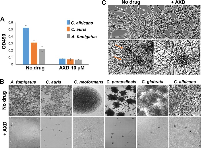
With the exception of Candida parapsilosis and Candida krusei, all isolates tested under planktonic conditions showed MIC values of ≤1.5 μg/mL. Alexidine dihydrochloride exhibits activity against most Candida spp. Fascinatingly, clinically significant fluconazole-resistant Candida isolates such as C. albicans (CA2, CA6, and CA10), C. glabrata (CG2 and CG5), C. parapsilosis (CP5), and C. auris (CAU-09 and CAU-03) are all significantly active against alexidine dihydrochloride[1].
When Alexidine dihydrochloride is used to inhibit planktonic growth, it completely stops the fungi being imaged from filamentating or proliferating. Alexidine dihydrochloride is able to decimate at low concentrations (1.5 to 6 μg/mL) mature biofilms of Candida, Cryptococcus, and Aspergillus spp. that are known to be resistant to almost all classes of antifungal drugs. In reality, alexidine dihydrochloride could prevent lateral yeast formation and biofilm dispersal in Candida albicans at concentrations of planktonic MICs that are 10-fold lower—150 ng/mL—than those of Candida albicans[1]. HUVECs and lung epithelial cells are 50% killed by alexidine dihydrochloride at concentrations five to ten times higher than the minimum inhibitory concentration needed to kill planktonically growing fungal pathogens[1]. AXD displayed broad-spectrum antifungal activity against planktonic cells of diverse fungi. MIC50 values for most Candida spp. were ≤1.5 μg/ml, except for C. parapsilosis and C. krusei. Against fluconazole-resistant clinical isolates (C. albicans CA2, CA6, CA10; C. glabrata CG2, CG5; C. parapsilosis CP5; C. auris CAU-09 and CAU-03), AXD MIC50 ranged from 0.73 to 1.5 μg/ml. For C. neoformans, MIC50 was 0.73–1.5 μg/ml. For filamentous fungi including Mucorales (R. delemar, R. oryzae, M. circinelloides, L. corymbifera, C. bertholletiae) and Aspergillus spp., AXD MIC50 values were 1.5–3 μg/ml. For S. apiospermum, MIC50 was 1.5 μg/ml. [1] AXD at 10 μM killed >80% of mature (48 h) biofilms of C. albicans, C. auris, and A. fumigatus. At as low as 150 ng/ml (0.15 μg/ml), AXD inhibited lateral yeast formation and biofilm dispersal in C. albicans. [1] In checkerboard synergy assays, AXD at 1.25 μg/ml reduced the MIC50 of fluconazole against mature C. albicans biofilms from >256 μg/ml to 1 μg/ml, with a fractional inhibitory concentration (FIC) index of 0.42, indicating synergistic interaction. [1] Phase-contrast microscopy showed that AXD at its respective MIC50 completely inhibited filamentation or proliferation of planktonic fungi. [1] |
| ln Vivo |
decided to concentrate on C. albicans biofilm formation because this fungus has a well-established murine biofilm model that is used to evaluate the efficacy of both new and old antifungal medications. When the drugs are seen under a microscope, the impact they have on the 24-hour-old biofilms that are developing in the mice's jugular vein catheters is evident. The biofilm density in the catheters treated with Alexidine dihydrochloride is considerably lower. In fact, compared to the untreated control biofilms, alexidine dihydrochloride inhibits 67% of the growth and viability of fungal biofilms, according to fungal CFU determination[1].
In a mouse central venous catheter model of C. albicans biofilm, intraluminal treatment with AXD at 3 μg/ml for 24 h (applied to 24-h preformed biofilms) reduced fungal biofilm viability by 67% compared to untreated controls, as determined by CFU enumeration. Microscopic visualization revealed significantly lower biofilm density in AXD-treated catheters. [1] |
| Cell Assay |
Cytotoxicity of AXD was evaluated in primary human umbilical vascular endothelial cells (HUVECs). Cells were grown in M-199 medium with supplements, seeded in 96-well plates, and treated with various concentrations of AXD (range 0.12–59 μg/ml) for 24 h at 37°C in 5% CO2. Cytotoxicity was quantified by chromium-51 release assay. The CC50 (concentration causing 50% killing) for HUVECs was >7.37 μg/ml. [1]
Cytotoxicity was also tested in human lung carcinoma A549 epithelial cells. A549 cells were grown in DMEM with 10% FBS, seeded at 1.5×10^5 cells/well in 96-well plates, incubated for 24 h, then treated with AXD in DMEM with 1% FBS for 24 h. Chromium-51 release assay showed a CC50 >7.37 μg/ml. [1] For immune cell cytotoxicity, primary bone marrow-derived macrophages from C57BL/6 mice were cultured in complete RPMI with M-CSF for 7 days, seeded at 1×10^5 cells/well, incubated overnight, then treated with various AXD concentrations for 24 h. Viability was assessed by DAPI staining using fluorescence microscopy. The CC50 for macrophages was >5 μg/ml. [1] Additionally, HL60 human promyelocytic cells were stained with CFSE (2 mM for 5 min), washed, seeded at 5×10^6 cells/ml in 96-well round-bottom plates, and treated with serially diluted AXD (0.004–10 μg/ml) for 48 h at 37°C. Cell proliferation was measured by flow cytometry (CFSE dilution). AXD at 5 μg/ml prevented cell division; at 10 μg/ml, cells did not divide. [1] |
| Animal Protocol |
In vivo biofilm drug susceptibility was assessed using a mouse central venous catheter infection model. Catheterized 8-week-old C57BL/6 male mice (jugular vein silastic catheter) were used. Catheters were instilled with 25 μl of C. albicans inoculum (5×10^6 cells/ml) and biofilms were allowed to develop for 24 h. Then, catheters were treated intraluminally with AXD at 3 μg/ml for 48 h. Control groups received fluconazole (250 μg/ml) or caspofungin (0.125 μg/ml). After treatment, catheters were cut laterally and imaged under phase-contrast microscopy. The distal 2 cm of catheters was cut into pieces, vortexed, sonicated, and plated on YPD agar for CFU enumeration. Each group had 6 mice. Percent biofilm reduction was calculated compared to untreated controls. [1]
|
| Toxicity/Toxicokinetics |
In vitro mammalian cell cytotoxicity: CC50 for HUVECs >7.37 μg/ml; for A549 lung epithelial cells >7.37 μg/ml; for bone marrow-derived macrophages >5 μg/ml. AXD at 5 μg/ml prevented proliferation of HL60 human promyelocytic cells. The concentrations causing host cell toxicity were at least 3- to 4-fold higher than AXD levels required to inhibit planktonic fungal cells (e.g., C. albicans MIC50 0.73–1.5 μg/ml). [1]
|
| References | |
| Additional Infomation |
AXD is a bis-biguanide where a 2-ethylhexyl chain is attached to each biguanide unit and the two units are linked by a 1,6-hexanediamine chain. It was originally identified for antibacterial properties and as an antiplaque agent/mouthwash, with potential for endodontic treatment to eliminate biofilms. AXD has been reported to have activity against C. neoformans by targeting phospholipases. It is an anticancer drug that induces mitochondrial apoptosis via PTPMT1 inhibition in mammalian cells. In this study, AXD was found to be fungicidal against common and emerging drug-resistant pathogens, including azole-resistant clinical isolates and Mucorales species (Rhizopus, L. corymbifera, S. apiospermum). The drug shows potential as a pan-antifungal antibiofilm agent, especially in combination with fluconazole to overcome biofilm resistance. [1]
|
| Molecular Formula |
C26H58CL2N10
|
|---|---|
| Molecular Weight |
581.71172
|
| Exact Mass |
580.422
|
| Elemental Analysis |
C, 53.68; H, 10.05; Cl, 12.19; N, 24.08
|
| CAS # |
1715-30-6
|
| Related CAS # |
Alexidine;22573-93-9
|
| PubChem CID |
102678
|
| Appearance |
White to light yellow solid powder
|
| Density |
1.1g/cm3
|
| Boiling Point |
658.2ºC at 760mmHg
|
| Melting Point |
220.6-223.4ºC
|
| Flash Point |
351.8ºC
|
| Vapour Pressure |
3.38E-17mmHg at 25°C
|
| LogP |
8.604
|
| Hydrogen Bond Donor Count |
8
|
| Hydrogen Bond Acceptor Count |
4
|
| Rotatable Bond Count |
23
|
| Heavy Atom Count |
38
|
| Complexity |
601
|
| Defined Atom Stereocenter Count |
0
|
| SMILES |
CCCCC(CC)CNC(=N)NC(=N)NCCCCCCNC(=N)NC(=N)NCC(CC)CCCC.Cl.Cl
|
| InChi Key |
BRJJFBHTDVWTCJ-UHFFFAOYSA-N
|
| InChi Code |
InChI=1S/C26H56N10.2ClH/c1-5-9-15-21(7-3)19-33-25(29)35-23(27)31-17-13-11-12-14-18-32-24(28)36-26(30)34-20-22(8-4)16-10-6-2;;/h21-22H,5-20H2,1-4H3,(H5,27,29,31,33,35)(H5,28,30,32,34,36);2*1H
|
| Chemical Name |
1-[N'-[6-[[amino-[[N'-(2-ethylhexyl)carbamimidoyl]amino]methylidene]amino]hexyl]carbamimidoyl]-2-(2-ethylhexyl)guanidine;dihydrochloride
|
| Synonyms |
Alexidine Dihydrochloride; Alexidine 2HCl;
|
| HS Tariff Code |
2934.99.9001
|
| Storage |
Powder -20°C 3 years 4°C 2 years In solvent -80°C 6 months -20°C 1 month Note: Please store this product in a sealed and protected environment, avoid exposure to moisture. |
| Shipping Condition |
Room temperature (This product is stable at ambient temperature for a few days during ordinary shipping and time spent in Customs)
|
| Solubility (In Vitro) |
DMSO :~125 mg/mL (~214.88 mM)
|
|---|---|
| Solubility (In Vivo) |
Solubility in Formulation 1: ≥ 2.08 mg/mL (3.58 mM) (saturation unknown) in 10% DMSO + 40% PEG300 + 5% Tween80 + 45% Saline (add these co-solvents sequentially from left to right, and one by one), clear solution.
For example, if 1 mL of working solution is to be prepared, you can add 100 μL of 20.8 mg/mL clear DMSO stock solution to 400 μL PEG300 and mix evenly; then add 50 μL Tween-80 to the above solution and mix evenly; then add 450 μL normal saline to adjust the volume to 1 mL. Preparation of saline: Dissolve 0.9 g of sodium chloride in 100 mL ddH₂ O to obtain a clear solution. Solubility in Formulation 2: ≥ 2.08 mg/mL (3.58 mM) (saturation unknown) in 10% DMSO + 90% (20% SBE-β-CD in Saline) (add these co-solvents sequentially from left to right, and one by one), clear solution. For example, if 1 mL of working solution is to be prepared, you can add 100 μL of 20.8 mg/mL clear DMSO stock solution to 900 μL of 20% SBE-β-CD physiological saline solution and mix evenly. Preparation of 20% SBE-β-CD in Saline (4°C,1 week): Dissolve 2 g SBE-β-CD in 10 mL saline to obtain a clear solution. View More
Solubility in Formulation 3: ≥ 2.08 mg/mL (3.58 mM) (saturation unknown) in 10% DMSO + 90% Corn Oil (add these co-solvents sequentially from left to right, and one by one), clear solution. Solubility in Formulation 4: 10% DMSO+40% PEG300+5% Tween-80+45% Saline: ≥ 2.08 mg/mL (3.58 mM) |
| Preparing Stock Solutions | 1 mg | 5 mg | 10 mg | |
| 1 mM | 1.7191 mL | 8.5953 mL | 17.1907 mL | |
| 5 mM | 0.3438 mL | 1.7191 mL | 3.4381 mL | |
| 10 mM | 0.1719 mL | 0.8595 mL | 1.7191 mL |
*Note: Please select an appropriate solvent for the preparation of stock solution based on your experiment needs. For most products, DMSO can be used for preparing stock solutions (e.g. 5 mM, 10 mM, or 20 mM concentration); some products with high aqueous solubility may be dissolved in water directly. Solubility information is available at the above Solubility Data section. Once the stock solution is prepared, aliquot it to routine usage volumes and store at -20°C or -80°C. Avoid repeated freeze and thaw cycles.
Calculation results
Working concentration: mg/mL;
Method for preparing DMSO stock solution: mg drug pre-dissolved in μL DMSO (stock solution concentration mg/mL). Please contact us first if the concentration exceeds the DMSO solubility of the batch of drug.
Method for preparing in vivo formulation::Take μL DMSO stock solution, next add μL PEG300, mix and clarify, next addμL Tween 80, mix and clarify, next add μL ddH2O,mix and clarify.
(1) Please be sure that the solution is clear before the addition of next solvent. Dissolution methods like vortex, ultrasound or warming and heat may be used to aid dissolving.
(2) Be sure to add the solvent(s) in order.
| NCT Number | Recruitment | interventions | Conditions | Sponsor/Collaborators | Start Date | Phases |
| NCT03140254 | Completed | Drug: Chlorhexidine gluconate Drug: N,N'-(1,10-decanediyldi-1(4H)-Pyridinyl-4- ylidene)-Bis-(1-octanamine) Dihydrochloride Drug: Placebo |
Preoperative Skin Preparation | CareFusion | 2016-09 | Phase 2 |
| NCT00515151 | Completed | Drug: 0.1% Octenidine with 30% 1-propanol and 45% 2-propanol Drug: 74% Ethanol with 10% 2-propanol |
Bacteremia Bacterial Infections Catheter-Associated Infections Catheterization, Central Venous |
University Hospital Freiburg | 2002-05 | Phase 4 |
| NCT02169167 | Completed | Device: Resin salve treatment Device: Octenidine treatment |
Diabetes Complications Diabetes Mellitus Diabetic Neuropathies Wound Infection |
Janne J. Jokinen | 2014-06 | Not Applicable |
|
Products are for research use only; We do not sell to patients
Copyright 2020 InvivoChem LLC | All Rights Reserved