| Size | Price | Stock | Qty |
|---|---|---|---|
| 10mg |
|
||
| 25mg |
|
||
| 50mg |
|
||
| 100mg |
|
||
| 250mg |
|
||
| 500mg |
|
||
| 1g |
|
||
| Other Sizes |


Purity: =100%
Alda-1 is a potent and selective Aldehyde Dehydrogenase-2 Agonist. Alda-1 reverses alcohol-induced hepatic steatosis and cell death in mice. Alda-1 inhibits atherosclerosis and attenuates hepatic steatosis in apolipoprotein E-knockout mice. Alda-1 reduces cerebral ischemia/reperfusion injury in rat through clearance of reactive aldehydes. Pharmacological activation of ALDH2 by Alda-1 reversed alcoholic steatosis and apoptosis through accelerating aldehyde clearance. This study indicates that ALDH2 is a promising molecular target and Alda-1 has therapeutic potential for treating alcoholic liver disease.
| Targets |
ALDH2
Mitochondrial aldehyde dehydrogenase (ALDH2). [1] |
|---|---|
| ln Vitro |
ROS was also higher in H/R cells than in control cells, which was aggravated upon treatment with 4-HNE, and reduced by Alda-1 treatment. Cell viability and mitochondrial membrane potential were inhibited in H/R cells, which was attenuated upon Alda-1 treatment. Activation of ALDH2 by Alda-1 attenuates hepatic I/R injury via clearance of cytotoxic aldehydes [6].
Alda-1 administration (1.5 mg/kg i.p. for 14 days) in prenatally stressed rats significantly decreased 4-HNE-protein content in the plasma (confirmed by immunoblotting, with albumin at 66 kDa as the main aldehyde protein). [2] Alda-1 treatment significantly increased PGC-1α mRNA expression in both the frontal cortex and hippocampus of prenatally stressed rats, and showed a trend toward increased PGC-1α protein expression in the frontal cortex (9.55 ± 3.00 ng/mg total protein in stress + Alda-1 group vs. stress group). [2] Alda-1 administration up-regulated Bcl-2 mRNA expression in the frontal cortex of prenatally stressed rats. [2] Alda-1 treatment significantly reduced TNF-α mRNA up-regulation in the frontal cortex and hippocampus, and tended to decrease IL-1β mRNA expression in the frontal cortex of prenatally stressed rats. [2] Proteomic analysis revealed that Alda-1 treatment resulted in a nearly 2-fold increase in the expression of Hsp60 (60 kDa heat shock protein) and up-regulation of mitochondrial carnitine O-palmitoyltransferase 2 (Cpt2) in the frontal cortex of prenatally stressed rats. In the hippocampus, Alda-1 treatment caused a 1.5-fold increase in dihydropyrimidine-related protein 2 (Drp2). [2] |
| ln Vivo |
Alda-1 therapy resulted in a considerable decrease in 4-HNE protein concentration in the plasma of apoE−/− animals. Alda-1 injection resulted in a small increase in the expression of genes associated to neurogenesis (Nog), mitochondrial biogenesis (CYTB, ND1), and apoptosis (Bax, Gsk3b) in Hp of apoE−/− mice. Alda-1 treatment resulted in the synthesis of 2 and 10 differently expressed proteins in FCx and Hp, respectively, of apoE−/− mice [1]. Alda-1 (1.5 mg/kg, bw, IP) injection significantly increased climbing time in prenatally stressed rats in the forced swim test, tending to decrease immobility time and increase swimming time. Furthermore, treatment of prenatally stressed rats with Alda-1 dramatically enhanced the number of entry into the open arms of the labyrinth and the duration spent in the maze, as determined by the elevated plus maze test [2]. After 24 hours of glucose administration, Alda-1 (8.5 mg/kg; intraperitoneal injection) and glucose dramatically reduced the number of 4-HNE and FJB-positive cells in the cerebral cortex of Alda-1-treated rats, which was significantly lower than that of DMSO-treated rats. [3]. Alda-1 (10 mg/kg per day) therapy avoids aldehyde excess, mitochondrial dysfunction and improves cardiac function in animals with post-MI cardiomyopathy [4].
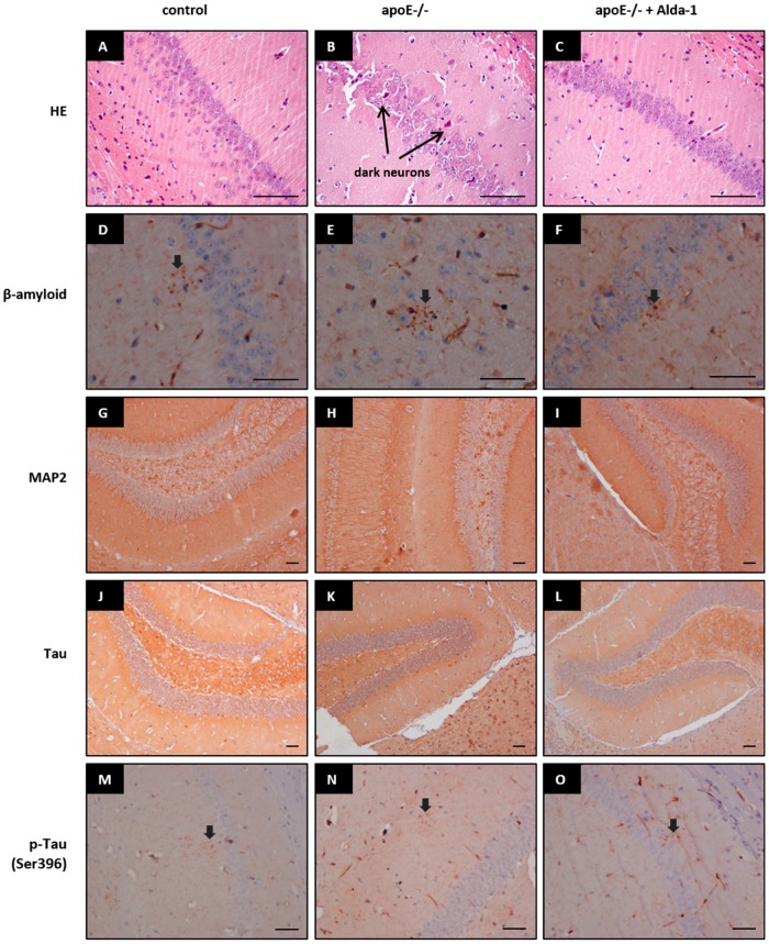
In apolipoprotein E knockout (apoE-/-) mice, prolonged treatment with Alda-1 (5 mg/kg/day mixed with chow diet for 4 months) resulted in a significant decrease of 4-HNE-protein adducts in the plasma (0.27 vs. 0.44 pmol; F(2,6)=97.78; p<0.05) and frontal cortex (11.91 vs. 19.41 pmol/g; F(2,6)=10.53; p<0.05) compared to untreated apoE-/- mice. [1] In the hippocampus of apoE-/- mice, Alda-1 administration led to a slight increase in gene expression related to neurogenesis (Nog, 1.20-fold, p=0.0268), mitochondrial biogenesis (CYTB, 1.33-fold, p=0.0258; ND1, 1.35-fold, p=0.0312), apoptosis (Bax, 1.22-fold, p=0.0303), and Gsk3b (1.24-fold, p=0.0058) as measured by real-time PCR. In the frontal cortex, Alda-1 treatment resulted in a reduction of Bmp4 mRNA expression (-1.28-fold, p=0.0366). [1] Proteomic analysis (iTRAQ) of mitochondria-enriched fractions from the frontal cortex of apoE-/- mice treated with Alda-1 showed upregulation of NADH dehydrogenase ubiquinone 1α subcomplex subunit 10 (1.27-fold) and excitatory amino acid transporter 2 (EAAT2, 1.12-fold) compared to untreated apoE-/- mice. [1] In the hippocampus, Alda-1 treatment upregulated myelin basic protein (1.37-fold), carbonic anhydrase 2 (1.27-fold), myelin proteolipid protein (1.25-fold), 2',3'-cyclic-nucleotide 3'-phosphodiesterase (CNP, 1.25-fold), sodium/potassium-transporting ATPase subunit α-2 (1.11-fold), and EAAT2 (1.09-fold), while downregulating spectrin β chain non-erythrocytic 1 (-1.06-fold), spectrin α chain non-erythrocytic 1 (-1.06-fold), actin aortic smooth muscle (-1.09-fold), and OCIA domain-containing protein 2 (-1.40-fold). [1] Histological examination (hematoxylin/eosin staining) showed that signs of mild tissue injury (shrunken and deeply stained "dark neurons") were visually slightly less pronounced in brains of Alda-1-treated apoE-/- mice compared to untreated apoE-/- mice. Immunohistochemistry for β-amyloid, MAP2, Tau, and phospho-Tau (Ser396) showed no differences between groups. [1] |
| Enzyme Assay |
Measurement of ALDH2 activity All procedures were performed in strict accordance with the manufacturer’s instructions. The principle behind this is that ALDH2 catalyzes the reaction between acetaldehyde and NAD+, and the change in absorbance of NADH at 340 nm can be used to calculate the activity of ALDH2 [5].
|
| Cell Assay |
In vitro model [5]
We used H9C2 cells within ten passages for cell culture. The medium was changed when the cells reached approximately 60% confluence. The cell models were divided into three plates: Alda-1 (CON, LPS, Alda-1, Alda-1 + LPS), daidzin (CON, LPS, daidzin (daidzin is an ALDH2 inhibitor), daidzin + LPS), cGAS-siRNA (CON, LPS, cGAS-siRNA + LPS, cGAS-siRNA + daidzin + LPS). The LPS treatment duration was 12 h, Alda-1 and daidzin pretreatment was performed for 2 h, and siRNA transfection was carried out for 48 h. The final concentrations of LPS, Alda-1 and daidzin were 10 μg/ml (Wang et al. 2019), 20 μM (Ji et al. 2016) and 48 μM (Ji et al. 2016), respectively.[5] siRNA transfection[5] Cell culture was performed in a 6-well plate per the manufacturer’s instructions. When the cells reached 30–40% confluence, the medium was changed to Opti-MEM™ I with reduced serum, and Lipofectamine 3000 was to transfect NC-small interfering RNA and cGAS-siRNA. The medium was replaced with complete culture medium after 6 h.[5] Spleen cells (4×10^6 cells/ml) were stimulated with concanavalin A (Con A; 2.5 μg/ml and 0.6 μg/ml) and lipopolysaccharide (LPS, 5 μg/ml) and incubated in 96-well plates at a final volume of 0.2 ml for 72 hours. Cell proliferation was determined by adding 0.5 μCi of [3H]-thymidine per well 16 hours before the end of incubation. The cultures were harvested with an automatic cell harvester, and [3H]thymidine incorporation was assessed using a liquid scintillation counter. [2] Production of IL-1β and TNF-α was examined after 72 hours of Con A (2.5 μg/ml) or LPS (5 μg/ml) stimulation. Cytokines were measured by enzyme-linked immunosorbent assay (ELISA) using commercially available kits. [2] |
| Animal Protocol |
We investigated the influence of Alda-1, a small-molecule activator of ALDH2, on depressive- and anxiety-like behaviors in an animal model of depression - the prenatally stressed rats - using behavioral, molecular and proteomic methods. Prolonged Alda-1 administration significantly increased the climbing time, tended to reduce the immobility time and increased the swimming time of the prenatally stressed rats in the forced swim test. Moreover, treatment of prenatally stressed rats with Alda-1 significantly increased number of entries into the open arms of the maze and the time spent therein, as assessed by elevated plus-maze test. Such actions were associated with reduction of plasma 4-HNE-protein content, decrease of TNF-α mRNA and increase of PGC-1α (regulator of mitochondrial biogenesis) mRNA level in the frontal cortex and hippocampus of the prenatally stressed rats as well as with normalization of peripheral immune parameters and significant changes in expression of 6 and 4 proteins related to mitochondrial functions in the frontal cortex and hippocampus, respectively. Collectively, ALDH2 activation by Alda-1 led to a significant attenuation of depressive- and anxiety-like behaviors in the prenatally stressed rats. The pattern of changes suggested mitoprotective effect of Alda-1, however the exact functional consequences of the revealed alterations require further investigation [2].
Three groups of animals were studied: control C57BL/6J mice (chow diet, n=10), untreated apoE-/- mice (Taconic, chow diet, n=10), and apoE-/- mice treated with Alda-1 (n=10). At eight weeks of age, Alda-1 was administered to mice at a dose of 5 mg per kg of body weight per day, mixed without heating with chow diet, for four months. At six months of age, mice were euthanized 5 minutes after injection of fraxiparine (1000 UI), and blood from the right ventricle was collected. Entire brains or dissected hippocampi and frontal cortices were collected on ice-cold glass plates. [1] For biochemical measurement, blood was centrifuged at 1000g-force at 4°C for 10 minutes, and plasma was stored at -80°C. Frontal cortex tissues were submerged in PBS buffer and homogenized using high-speed shaking (120 seconds) in plastic tubes with stainless steel beads. [1] For histology and immunohistochemistry, brain tissue samples were formalin fixed, embedded in paraffin, and 2 μm paraffin sections were stained with hematoxylin-eosin or used for immunohistochemistry with antibodies against MAP2, β-amyloid, Tau, and phospho-Tau (Ser396). [1] For real-time PCR, total RNA was isolated from homogenized frontal cortex and hippocampus using a kit. Relative gene expression analysis was carried out in triplicate with internal reference genes. [1] For iTRAQ proteomics, mitochondria-enriched fractions were isolated from freshly harvested hippocampus and frontal cortex at 4°C. Mitochondria-enriched pellets were resuspended in lysis buffer (7 M urea, 2 M thiourea, 4% CHAPS, 1% DTT, protease inhibitors), incubated at 25°C for 30 minutes, then centrifuged at 12,000×g for 15 minutes. Protein concentration was determined by Bradford method. 100 μg of protein from each sample was precipitated with ice-cold acetone (1:6 v/v) overnight, centrifuged at 12,000×g for 10 minutes at 4°C, air-dried, then dissolved, reduced, alkylated, and digested with trypsin overnight at 37°C (1:50 w/w ratio). Samples were labeled with iTRAQ reagents, combined, dried, dissolved in 5% acetonitrile/0.1% TFA, purified, and subjected to strong cation exchange fractionation with 12 fractions collected by centrifugation. [1] |
| References |
[1]. Stachowicz A, et al. Proteomic Analysis of Mitochondria-Enriched Fraction Isolated from the Frontal Cortex and Hippocampus of Apolipoprotein E Knockout Mice Treated with Alda-1, an Activator of Mitochondrial Aldehyde Dehydrogenase (ALDH2). Int J Mol Sci. 2017 Feb 17;18(2):435.
[2]. Stachowicz A, et al. The impact of mitochondrial aldehyde dehydrogenase (ALDH2) activation by Alda-1 on the behavioral and biochemical disturbances in animal model of depression. Brain Behav Immun. 2016 Jan;51:144-53. [3]. Ikeda T, et al. Effects of Alda-1, an Aldehyde Dehydrogenase-2 Agonist, on Hypoglycemic Neuronal Death. PLoS One. 2015 Jun 17;10(6):e0128844. [4]. Gomes KM, et al. Aldehydic load and aldehyde dehydrogenase 2 profile during the progression of post-myocardial infarction cardiomyopathy: benefits of Alda-1. Int J Cardiol. 2015 Jan 20;179:129-138. [5]. ALDH2 mitigates LPS-induced cardiac dysfunction, inflammation, and apoptosis through the cGAS/STING pathway. Mol Med. 2023; 29: 171. [6]. Free Radic Res. 2018 Jun;52(6):629-638. doi: 10.1080/10715762.2018.1459042. |
| Additional Infomation |
N-(1,3-benzodioxane-5-ylmethyl)-2,6-dichlorobenzamide is a carbonyl compound and an organohalide compound.
Alda-1 is an activator of mitochondrial aldehyde dehydrogenase (ALDH2), an enzyme responsible for detoxification of toxic aldehydes such as 4-hydroxynonenal (4-HNE) to non-toxic acids. In this study, Alda-1 treatment decreased 4-HNE-protein adducts in plasma and frontal cortex of apoE-/- mice, indicating ALDH2 activation. The pattern of gene and protein expression changes implies a mitoprotective action of Alda-1, though accurate functional consequences require further research. [1] |
| Molecular Formula |
C15H11CL2NO3
|
|---|---|
| Molecular Weight |
324.16
|
| Exact Mass |
323.012
|
| Elemental Analysis |
C, 55.58; H, 3.42; Cl, 21.87; N, 4.32; O, 14.81
|
| CAS # |
349438-38-6
|
| Related CAS # |
349438-38-6;
|
| PubChem CID |
831629
|
| Appearance |
White to off-white solid
|
| LogP |
4.226
|
| Hydrogen Bond Donor Count |
1
|
| Hydrogen Bond Acceptor Count |
3
|
| Rotatable Bond Count |
3
|
| Heavy Atom Count |
21
|
| Complexity |
372
|
| Defined Atom Stereocenter Count |
0
|
| SMILES |
ClC1C([H])=C([H])C([H])=C(C=1C(N([H])C([H])([H])C1C([H])=C([H])C2=C(C=1[H])OC([H])([H])O2)=O)Cl
|
| InChi Key |
NMKJFZCBCIUYHI-UHFFFAOYSA-N
|
| InChi Code |
InChI=1S/C15H11Cl2NO3/c16-10-2-1-3-11(17)14(10)15(19)18-7-9-4-5-12-13(6-9)21-8-20-12/h1-6H,7-8H2,(H,18,19)
|
| Chemical Name |
N-(1,3-Benzodioxol-5-ylmethyl)-2,6-dichlorobenzamide
|
| Synonyms |
Alda-1 Alda1 Alda 1.
|
| HS Tariff Code |
2934.99.9001
|
| Storage |
Powder -20°C 3 years 4°C 2 years In solvent -80°C 6 months -20°C 1 month |
| Shipping Condition |
Room temperature (This product is stable at ambient temperature for a few days during ordinary shipping and time spent in Customs)
|
| Solubility (In Vitro) |
DMSO : ≥ 51 mg/mL (~157.33 mM)
|
|---|---|
| Solubility (In Vivo) |
Solubility in Formulation 1: ≥ 2.5 mg/mL (7.71 mM) (saturation unknown) in 10% DMSO + 90% Corn Oil (add these co-solvents sequentially from left to right, and one by one), clear solution.
For example, if 1 mL of working solution is to be prepared, you can add 100 μL of 25.0 mg/mL clear DMSO stock solution to 900 μL of corn oil and mix evenly. Solubility in Formulation 2: 24 mg/mL (74.04 mM) in 0.5% CMC-Na/saline water (add these co-solvents sequentially from left to right, and one by one), suspension solution; with ultrasonication. Preparation of saline: Dissolve 0.9 g of sodium chloride in 100 mL ddH₂ O to obtain a clear solution. (Please use freshly prepared in vivo formulations for optimal results.) |
| Preparing Stock Solutions | 1 mg | 5 mg | 10 mg | |
| 1 mM | 3.0849 mL | 15.4245 mL | 30.8490 mL | |
| 5 mM | 0.6170 mL | 3.0849 mL | 6.1698 mL | |
| 10 mM | 0.3085 mL | 1.5424 mL | 3.0849 mL |
*Note: Please select an appropriate solvent for the preparation of stock solution based on your experiment needs. For most products, DMSO can be used for preparing stock solutions (e.g. 5 mM, 10 mM, or 20 mM concentration); some products with high aqueous solubility may be dissolved in water directly. Solubility information is available at the above Solubility Data section. Once the stock solution is prepared, aliquot it to routine usage volumes and store at -20°C or -80°C. Avoid repeated freeze and thaw cycles.
Calculation results
Working concentration: mg/mL;
Method for preparing DMSO stock solution: mg drug pre-dissolved in μL DMSO (stock solution concentration mg/mL). Please contact us first if the concentration exceeds the DMSO solubility of the batch of drug.
Method for preparing in vivo formulation::Take μL DMSO stock solution, next add μL PEG300, mix and clarify, next addμL Tween 80, mix and clarify, next add μL ddH2O,mix and clarify.
(1) Please be sure that the solution is clear before the addition of next solvent. Dissolution methods like vortex, ultrasound or warming and heat may be used to aid dissolving.
(2) Be sure to add the solvent(s) in order.
|
|
Products are for research use only; We do not sell to patients
Copyright 2020 InvivoChem LLC | All Rights Reserved


 COA
COA