| Size | Price | Stock | Qty |
|---|---|---|---|
| 5mg |
|
||
| 10mg |
|
||
| 25mg |
|
||
| 50mg |
|
||
| 100mg |
|
||
| 250mg |
|
||
| 500mg | |||
| Other Sizes |


Purity: ≥98%
| Targets |
In both wild-type mouse hippocampus slice cultures and native N2a neuroblastoma cells, AK-7 (10 μM) lowers cholesterol levels. Neuroprotective effects of AK-7 (1 μM) on striatal neurons involved in Huntington's disease (HD) have been reported [1]. In primary midbrain cultures, the fraction of DA neurons is decreased by AK-7 (12.5 μM) [3].
|
|---|---|
| ln Vitro |
In both wild-type mouse hippocampus slice cultures and native N2a neuroblastoma cells, AK-7 (10 μM) lowers cholesterol levels. Neuroprotective effects of AK-7 (1 μM) on striatal neurons involved in Huntington's disease (HD) have been reported [1]. In primary midbrain cultures, the fraction of DA neurons is decreased by AK-7 (12.5 μM) [3].
AK-7 was identified as the most potent and selective SIRT2 inhibitor from a screen of a brain-permeable compound library. It demonstrated dose-dependent inhibition of recombinant SIRT2 deacetylase activity in biochemical assays.[1] In a striatal neuronal model of Huntington's disease (HD), AK-7 (1 µM) significantly rescued neuronal cell death induced by expression of a mutant huntingtin fragment (N171-82Q). This neuroprotective effect was achieved at a concentration lower than its biochemical IC₅₀.[1] AK-7 (1 µM) decreased the number of polyglutamine inclusions per neuron in the same HD neuronal model.[1] AK-7 treatment (10 µM for 24 hours) did not alter the expression level of the mutant huntingtin transgene in primary striatal neurons, as assessed by Western blot.[1] Consistent with the mechanism of SIRT2 inhibition, AK-7 (10 µM) reduced the nuclear localization of the transcription factor SREBP-2 in primary striatal neurons, leading to transcriptional downregulation of key cholesterol biosynthetic genes (FDFT1, HMGCR, HMGCS1). This resulted in a significant reduction of total cholesterol levels in primary striatal neuron cultures, N2a neuroblastoma cells, and hippocampal brain slice cultures from wild-type mice. The cholesterol-lowering effect was observed at 10 µM but not at a lower concentration (1 µM) in N2a cells and hippocampal slices.[1] |
| ln Vivo |
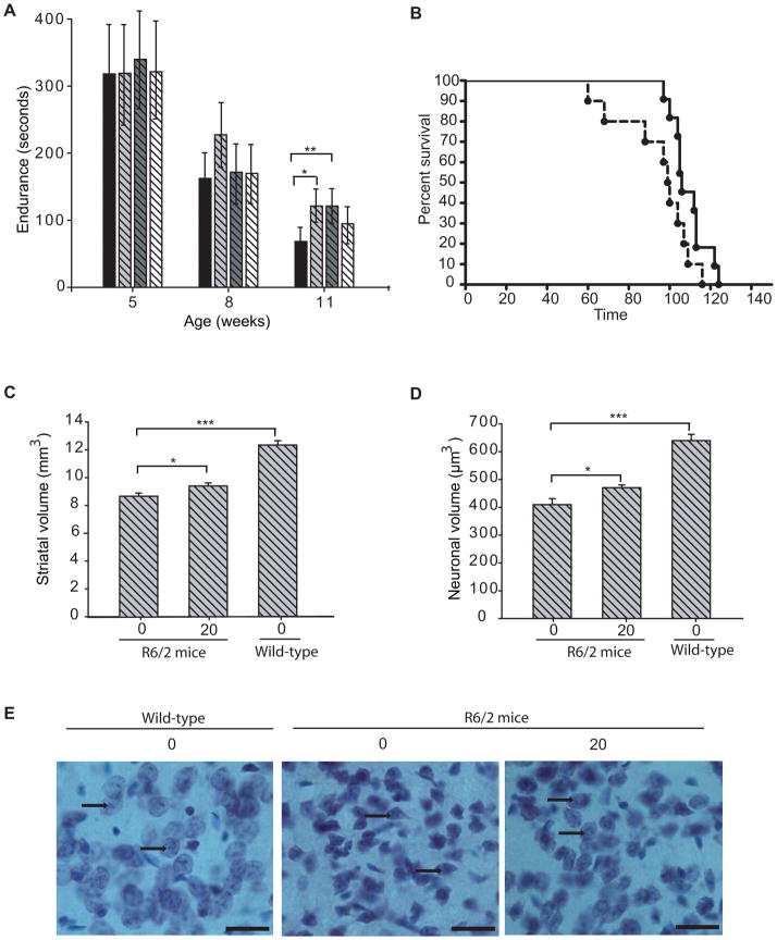
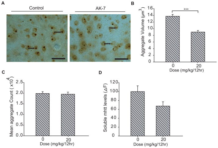
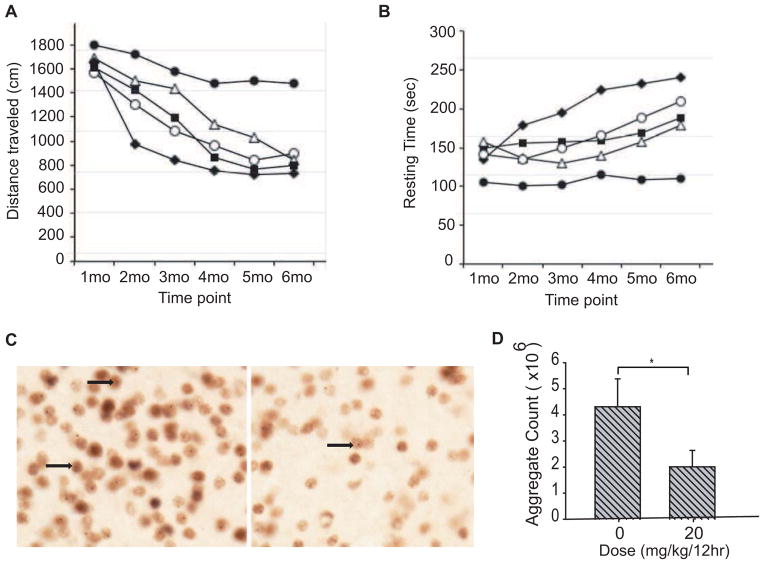
In both wild-type and HD mice, AK-7 (15 mg/kg/dose, intraperitoneal) penetrates the brain [1]. In R6/2 HD mice, AK-7 (10, 20 mg/kg, intraperitoneally) prolongs survival and improves behavioral and neuropathological characteristics. In R6/2 mice, AK-7 (20 mg/kg) ameliorates HD neuropathology. In R6/2 brains, AK-7 also lessens polyglutamine aggregation. Additionally, the locomotor performance of 140CAG mice treated with AK-7 changed in a way that was comparable to that of untreated wild-type mice [2]. The most effective dose was 20 mg/kg, which also differed considerably from untreated 140CAG mice.
Chronic treatment with the brain-permeable SIRT2 inhibitor AK-7 (10, 20, 30 mg/kg, i.p., twice daily) demonstrated significant efficacy in two genetic mouse models of Huntington’s disease (HD). In the R6/2 transgenic mouse model, AK-7 treatment (10 and 20 mg/kg) significantly improved motor performance on the accelerating rotarod at 11 weeks of age, increasing latency to fall by 44%. Treatment at 10 mg/kg extended mean survival by 13.2%. Neuropathological analysis at 12 weeks showed that treatment (20 mg/kg) increased total striatal volume by 9% and striatal neuronal cell body volume by 15% compared to vehicle-treated controls. In the 140CAG full-length Htt knock-in mouse model, AK-7 treatment (20 mg/kg, most effective dose) significantly improved locomotor activity in open field tests, normalizing distance traveled and resting time from 2 to 5 months of age compared to untreated HD mice.[2] |
| Enzyme Assay |
Sirtuin deacetylase activity was assessed using a fluorometric assay. Recombinant active enzymes (SIRT1, SIRT2, or the catalytically active fragment of SIRT3) were incubated with a fluorogenic deacetylase substrate and NAD⁺ in the supplied reaction buffer. Test compounds or a DMSO control were added to the reaction mixture. Enzyme activity, measured as the rate of deacetylation, was quantified by fluorescence (excitation 355 nm, emission 460 nm) using a plate reader. Activity was normalized to defined units per reaction well. Each compound concentration was tested in triplicate, and autofluorescent background signals were subtracted.[1]
|
| Cell Assay |
Primary striatal neurons were derived from embryonic rat ganglionic eminences. An HD model was established by lentiviral transduction to express mutant huntingtin fragments (N171-82Q or control N171-18Q). Neurons were treated with AK-7 (1 µM for neuroprotection/inclusion count; 10 µM for other assays) or DMSO control for specified durations. Neuronal survival was assessed by counting NeuN-positive cells. Polyglutamine inclusions were quantified by immunolabeling with an anti-huntingtin antibody and automated imaging/analysis.[1]
For analysis of SREBP-2 compartmentalization, primary neurons on coverslips were immunolabeled for endogenous SREBP-2. Images were acquired, and the percentage of SREBP-2 signal localized to the nucleus (traced using Hoechst staining) relative to the soma was quantified using image analysis software.[1] For cholesterol assays, N2a neuroblastoma cells were treated with AK-7 or DMSO for 48 hours. Hippocampal slice cultures from wild-type mice were treated for 48 hours. Total cholesterol levels were measured using enzymatic assay kits and normalized to protein content.[1] Gene expression analysis was performed by extracting RNA from treated primary neurons, reverse transcribing to cDNA, and quantifying mRNA levels of cholesterol biosynthesis genes using real-time PCR.[1] |
| Animal Protocol |
Pharmacokinetic studies were performed in wild-type and R6/2 transgenic HD model mice. AK-7 was formulated at 1.5 mg/mL in a vehicle of 25% Cremophor EL, 10% DMSO in water. The compound was administered via a single intraperitoneal injection at a dose of 15 mg/kg to 11-week-old mice. Groups of mice (n=3 per genotype per time point) were sacrificed at 15, 30, 60, 120, 240, and 480 minutes post-injection. Blood was collected for serum separation, and brains were harvested. Tissues were immediately frozen for subsequent analysis of AK-7 concentrations.[1]
|
| ADME/Pharmacokinetics |
AK-7 can cross the blood-brain barrier. After a single intraperitoneal injection (15 mg/kg), AK-7 is rapidly cleared from the serum, reaching peak concentrations 15 to 30 minutes after administration and becoming almost undetectable after 1 hour. The peak concentration of AK-7 in the brain of wild-type mice is approximately 3 µM, and in R6/2 HD mice it is approximately 4 µM. The brain concentration drops to approximately 2 µM 2 hours after injection and to 0.2 µM 4 hours after injection, indicating that its metabolic stability in the brain is limited (half-life is approximately 2 hours). The brain osmotic kinetics of wild-type mice and R6/2 mice are similar. [1]
|
| Toxicity/Toxicokinetics |
In acute toxicity studies, AK-7 was safe at doses up to 2500 mg/kg, with no deaths observed at this dose. In chronic treatment of HD in a mouse model, a dose of 30 mg/kg (twice daily) did not improve survival in R6/2 mice, likely due to systemic toxicity at this high dose. [2]
|
| References |
|
| Additional Infomation |
AK-7 (3-(1-azacycloheptaylsulfonyl)-N-(3-bromophenyl)benzamide) is the first reported SIRT2 deacetylase inhibitor that can cross the blood-brain barrier. It is a sulfobenzoic acid derivative, and its scaffold has attracted much attention due to its good drug-like properties for targets in the central nervous system. In a Huntington's disease model, its neuroprotective mechanism is related to SIRT2 inhibition, which leads to the retention of SREBP-2 in the cytoplasm, downregulation of cholesterol biosynthesis, and thus a reduction in neuronal cholesterol levels. It also reduces the accumulation of mutant huntingtin protein. Although it only reaches low micromolar concentrations in the brain (below its in vitro IC₅₀), it has shown efficacy in neuronal models, providing proof of concept for targeting SIRT2. This study highlights the need for further optimization of the scaffold to improve its potency and metabolic stability. [1]
|
| Molecular Formula |
C19H21BRN2O3S
|
|
|---|---|---|
| Molecular Weight |
437.35
|
|
| Exact Mass |
436.045
|
|
| CAS # |
420831-40-9
|
|
| Related CAS # |
|
|
| PubChem CID |
1328033
|
|
| Appearance |
White to off-white solid powder
|
|
| Density |
1.5±0.1 g/cm3
|
|
| Index of Refraction |
1.631
|
|
| LogP |
4.93
|
|
| Hydrogen Bond Donor Count |
1
|
|
| Hydrogen Bond Acceptor Count |
4
|
|
| Rotatable Bond Count |
4
|
|
| Heavy Atom Count |
26
|
|
| Complexity |
570
|
|
| Defined Atom Stereocenter Count |
0
|
|
| SMILES |
O=S(N1CCCCCC1)(C2=CC(C(NC3=CC(Br)=CC=C3)=O)=CC=C2)=O
|
|
| InChi Key |
IYAYHZZWYNXHEQ-UHFFFAOYSA-N
|
|
| InChi Code |
InChI=1S/C19H21BrN2O3S/c20-16-8-6-9-17(14-16)21-19(23)15-7-5-10-18(13-15)26(24,25)22-11-3-1-2-4-12-22/h5-10,13-14H,1-4,11-12H2,(H,21,23)
|
|
| Chemical Name |
|
|
| Synonyms |
|
|
| HS Tariff Code |
2934.99.9001
|
|
| Storage |
Powder -20°C 3 years 4°C 2 years In solvent -80°C 6 months -20°C 1 month |
|
| Shipping Condition |
Room temperature (This product is stable at ambient temperature for a few days during ordinary shipping and time spent in Customs)
|
| Solubility (In Vitro) |
|
|||
|---|---|---|---|---|
| Solubility (In Vivo) |
Solubility in Formulation 1: ≥ 2.5 mg/mL (5.72 mM) (saturation unknown) in 10% DMSO + 40% PEG300 + 5% Tween80 + 45% Saline (add these co-solvents sequentially from left to right, and one by one), clear solution.
For example, if 1 mL of working solution is to be prepared, you can add 100 μL of 25.0 mg/mL clear DMSO stock solution to 400 μL PEG300 and mix evenly; then add 50 μL Tween-80 to the above solution and mix evenly; then add 450 μL normal saline to adjust the volume to 1 mL. Preparation of saline: Dissolve 0.9 g of sodium chloride in 100 mL ddH₂ O to obtain a clear solution. Solubility in Formulation 2: ≥ 2.5 mg/mL (5.72 mM) (saturation unknown) in 10% DMSO + 90% Corn Oil (add these co-solvents sequentially from left to right, and one by one), clear solution. For example, if 1 mL of working solution is to be prepared, you can add 100 μL of 25.0 mg/mL clear DMSO stock solution to 900 μL of corn oil and mix evenly. (Please use freshly prepared in vivo formulations for optimal results.) |
| Preparing Stock Solutions | 1 mg | 5 mg | 10 mg | |
| 1 mM | 2.2865 mL | 11.4325 mL | 22.8650 mL | |
| 5 mM | 0.4573 mL | 2.2865 mL | 4.5730 mL | |
| 10 mM | 0.2286 mL | 1.1432 mL | 2.2865 mL |
*Note: Please select an appropriate solvent for the preparation of stock solution based on your experiment needs. For most products, DMSO can be used for preparing stock solutions (e.g. 5 mM, 10 mM, or 20 mM concentration); some products with high aqueous solubility may be dissolved in water directly. Solubility information is available at the above Solubility Data section. Once the stock solution is prepared, aliquot it to routine usage volumes and store at -20°C or -80°C. Avoid repeated freeze and thaw cycles.
Calculation results
Working concentration: mg/mL;
Method for preparing DMSO stock solution: mg drug pre-dissolved in μL DMSO (stock solution concentration mg/mL). Please contact us first if the concentration exceeds the DMSO solubility of the batch of drug.
Method for preparing in vivo formulation::Take μL DMSO stock solution, next add μL PEG300, mix and clarify, next addμL Tween 80, mix and clarify, next add μL ddH2O,mix and clarify.
(1) Please be sure that the solution is clear before the addition of next solvent. Dissolution methods like vortex, ultrasound or warming and heat may be used to aid dissolving.
(2) Be sure to add the solvent(s) in order.
Products are for research use only; We do not sell to patients
Copyright 2020 InvivoChem LLC | All Rights Reserved


 COA
COA