| Size | Price | Stock | Qty |
|---|---|---|---|
| 10mg |
|
||
| 50mg |
|
||
| 100mg |
|
||
| 250mg |
|
||
| 500mg |
|
||
| 1g |
|
||
| Other Sizes |


Purity: ≥98%
| Targets |
AGI-5198 specifically targets mutant isocitrate dehydrogenase 1 (IDH1), with the highest selectivity for the IDH1-R132H mutation (the most common mutant form in gliomas). The IC50 value for inhibiting recombinant IDH1-R132H enzyme activity is approximately 100 nM [1];
AGI-5198 also targets other IDH1 mutant variants (e.g., IDH1-R132C) found in cancer cells. It does not inhibit wild-type IDH1 at concentrations up to 10 μM [2] |
|---|---|
| ln Vitro |
The amount of R-2HG in TS603 glioma cell pellets was measured, and the results showed that AGI-5198 inhibited the mutant IDH1 enzyme in a dose-dependent manner. In two patient-derived glioma cell lines (TS676 and TS516) that express exclusively wild-type IDH1 alleles, AGI-5198 has no effect on colony formation [1]. When exposed to ionizing radiation (IR), cancer cells heterozygous for the IDH1 (R132H) mutation show reduced IDH-mediated NADPH generation and consequently increased amounts of reactive oxygen species, or DNA, compared to IDH1 wild-type cells. Double-strand breaks and cell death are also observed. These effects are reversed by AGI-5198, an IDH1 (R132H) inhibitor [2].
1. Treatment of glioma cell lines harboring IDH1-R132H (e.g., U87-IDH1-R132H, HT1080-IDH1-R132H) with AGI-5198 resulted in concentration-dependent inhibition of 2-hydroxyglutarate (2-HG) production (a carcinogenic oncometabolite generated by mutant IDH1). The IC50 for 2-HG inhibition in U87-IDH1-R132H cells was ~100 nM, consistent with its enzyme inhibitory activity. After 72 hours of treatment with 3-10 μM AGI-5198, cell proliferation was significantly suppressed (by ~50-70% compared to vehicle control) as measured by MTT assay. Additionally, immunoblotting showed increased expression of glial fibrillary acidic protein (GFAP, a marker of astrocyte differentiation) and βIII-tubulin (a marker of neuronal differentiation), indicating promotion of glioma cell differentiation [1]; 2. In IDH1-R132H-mutated glioma cells (U87-IDH1-R132H) and IDH1-R132C-mutated cholangiocarcinoma cells (HCCC-9810-IDH1-R132C), pre-treatment with 1 μM AGI-5198 for 24 hours reduced ionizing radiation (IR)-induced DNA damage. Immunofluorescence staining for γH2AX (a marker of DNA double-strand breaks) revealed a ~40-50% decrease in γH2AX foci 24 hours post-IR (2-4 Gy) compared to vehicle-treated irradiated cells. Clone formation assays showed that AGI-5198 treatment increased the survival fraction of irradiated mutant IDH1 cells: at 4 Gy IR, the survival fraction of U87-IDH1-R132H cells treated with 1 μM AGI-5198 was ~0.35, compared to ~0.15 in vehicle controls. This radioprotective effect was not observed in wild-type IDH1 cells (e.g., parental U87 cells) [2] |
| ln Vivo |
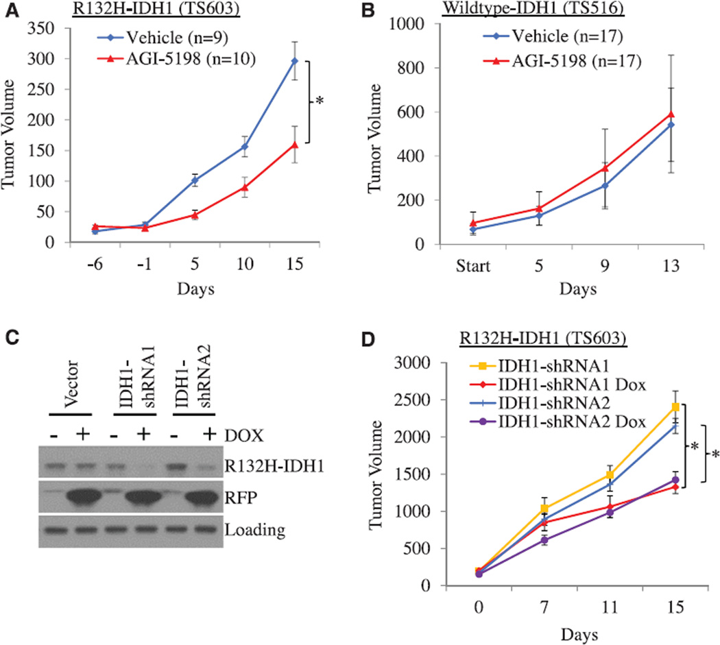
In human glioma xenografts, AGI-5198 (450 mg/kg, po) inhibits tumor growth by 50% to 60%. Mice treated with AGI-5198 had tumors that stained less strongly with anti-Ki-67 antibodies. Glioma xenografts of the IDH1 wild-type grow unaffected by AGI-5198 [1].
1. Nude mice (female, 6-8 weeks old) were subcutaneously implanted with U87-IDH1-R132H glioma cells (5×10⁶ cells/mouse). When tumors reached a volume of ~100 mm³, mice were randomly divided into two groups (n=6 per group): vehicle control (oral gavage of 0.5% methylcellulose + 0.1% Tween 80) and AGI-5198-treated (50 mg/kg, oral gavage, twice daily [bid] for 21 consecutive days). Tumor volume was measured every 3 days: at day 21, the mean tumor volume in the AGI-5198 group was ~350 mm³, compared to ~800 mm³ in the control group (a ~56% reduction). Liquid chromatography-tandem mass spectrometry (LC-MS/MS) analysis of tumor homogenates showed a ~90% decrease in 2-HG levels in AGI-5198-treated tumors. Histopathological examination revealed increased GFAP expression in treated tumors, confirming in vivo promotion of differentiation [1]; 2. Nude mice (male, 6-8 weeks old) were orthotopically implanted with U87-IDH1-R132H glioma cells (2×10⁵ cells/mouse, injected into the right striatum). Seven days post-implantation, mice were assigned to four groups (n=5 per group): vehicle control (intraperitoneal [i.p.] injection of DMSO + normal saline), AGI-5198 alone (25 mg/kg, i.p., once daily [qd] for 5 days), IR alone (4 Gy, single dose to the brain on day 3 of treatment), and AGI-5198 + IR. At day 28 post-implantation, magnetic resonance imaging (MRI) showed that the AGI-5198 + IR group had a mean tumor volume of ~220 mm³, which was significantly larger than the IR alone group (~120 mm³) but smaller than the vehicle control group (~450 mm³). This confirmed that AGI-5198 attenuated the anti-tumor efficacy of IR in mutant IDH1 gliomas (radioprotective effect) [2] |
| Enzyme Assay |
1. Recombinant IDH1-R132H enzyme activity assay: The reaction mixture (100 μL total volume) contained 50 mM Tris-HCl buffer (pH 7.5), 2 mM NADP⁺, 10 mM α-ketoglutarate (α-KG, substrate), 5 mM MgCl₂, 1 μg recombinant IDH1-R132H protein, and serial concentrations of AGI-5198 (0.01-10 μM). The reaction was initiated by adding α-KG and incubated at 37°C for 30 minutes. The production of NADPH (a byproduct of mutant IDH1-catalyzed 2-HG synthesis) was measured spectrophotometrically at 340 nm every 5 minutes for 30 minutes. Enzyme activity was calculated as the rate of NADPH formation, and the IC50 was determined by fitting the inhibition curve using nonlinear regression [1];
2. Mutant IDH1 enzyme inhibition validation assay: For IDH1-R132C, the assay protocol was similar to that for IDH1-R132H, with minor adjustments: the reaction buffer included 20 mM HEPES (pH 7.2) instead of Tris-HCl, and the incubation time was extended to 45 minutes. AGI-5198 was tested at concentrations of 0.1-5 μM, and enzyme activity was quantified by measuring NADPH fluorescence (excitation 340 nm, emission 460 nm) to enhance sensitivity. No inhibition of wild-type IDH1 was observed even at the highest AGI-5198 concentration (10 μM) [2] |
| Cell Assay |
1. Glioma cell proliferation and differentiation assay: U87-IDH1-R132H cells were seeded in 96-well plates at 5×10³ cells/well and allowed to adhere overnight. Cells were treated with AGI-5198 (0.1-10 μM) or vehicle (0.1% DMSO) for 72 hours. For proliferation assessment, 10 μL MTT reagent (5 mg/mL) was added to each well, followed by 4 hours of incubation at 37°C. The supernatant was removed, 100 μL DMSO was added to dissolve formazan crystals, and absorbance was measured at 570 nm. For differentiation analysis, cells were seeded in 6-well plates at 2×10⁵ cells/well, treated with 5 μM AGI-5198 for 5 days, then lysed with RIPA buffer. Equal amounts of protein (40 μg) were separated by 12% SDS-PAGE, transferred to PVDF membranes, and probed with primary antibodies against GFAP and βIII-tubulin (1:1000 dilution). HRP-conjugated secondary antibodies (1:5000 dilution) and ECL reagent were used for band detection [1];
2. γH2AX immunofluorescence and clone formation assay: For γH2AX staining, U87-IDH1-R132H cells were seeded on coverslips in 24-well plates (1×10⁴ cells/well), treated with 1 μM AGI-5198 for 24 hours, then irradiated with 2 Gy IR. At 24 hours post-IR, cells were fixed with 4% paraformaldehyde, permeabilized with 0.2% Triton X-100, blocked with 5% BSA, and incubated with anti-γH2AX antibody (1:500 dilution) overnight at 4°C. Alexa Fluor 488-conjugated secondary antibody (1:1000 dilution) and DAPI (for nuclear staining) were added, and images were captured using a fluorescence microscope. γH2AX foci per cell were counted in 50 random cells per group. For clone formation, cells were seeded in 6-well plates at 200 cells/well, treated with 1 μM AGI-5198 for 24 hours, irradiated with 0-6 Gy IR, and cultured for 14 days. Colonies (>50 cells) were stained with crystal violet and counted, and survival fractions were calculated [2] |
| Animal Protocol |
Dissolved in 0.5% MC and 0.2% Tween 80; 150 mg/kg, 450 mg/kg per day; oral gavage
IDH1 mutant glioma xenografts 1. Subcutaneous xenograft model for anti-tumor efficacy: Female nude mice (6-8 weeks old) were acclimated for 1 week before experimentation. U87-IDH1-R132H cells (5×10⁶ cells in 100 μL PBS + 50% Matrigel) were subcutaneously injected into the right flank of each mouse. When tumors reached ~100 mm³ (day 0), mice were divided into two groups (n=6): vehicle group (oral gavage of 0.5% methylcellulose + 0.1% Tween 80, 100 μL/mouse, bid) and AGI-5198 group (50 mg/kg in the same vehicle, oral gavage, 100 μL/mouse, bid). Tumor volume was calculated every 3 days using the formula: Volume = (length × width²)/2. After 21 days of treatment, mice were euthanized, tumors were excised, weighed, and homogenized for 2-HG measurement by LC-MS/MS [1]; 2. Orthotopic xenograft model for radioprotection: Male nude mice (6-8 weeks old) were anesthetized with isoflurane. U87-IDH1-R132H cells (2×10⁵ cells in 2 μL PBS) were injected into the right striatum using a stereotaxic frame (coordinates: 0.5 mm anterior, 2 mm lateral, 3 mm depth from bregma). Seven days post-injection, mice were assigned to four groups (n=5): (1) Vehicle: i.p. injection of 100 μL DMSO + normal saline (1:10 v/v), qd for 5 days; (2) AGI-5198 alone: 25 mg/kg AGI-5198 (dissolved in DMSO + normal saline, 1:10 v/v), i.p., qd for 5 days; (3) IR alone: single 4 Gy brain IR on day 3 (using a linear accelerator, field size 5×5 mm); (4) AGI-5198 + IR: combination of AGI-5198 treatment and IR. At day 28 post-implantation, mice underwent MRI to measure tumor volume, then were euthanized for histopathological analysis [2] |
| Toxicity/Toxicokinetics |
1. In the subcutaneous xenograft study (nude mice, 50 mg/kg AGI-5198, orally twice daily for 21 days), no significant changes in body weight (mean weight loss <5% from baseline) or clinical signs of toxicity (e.g., lethargy, diarrhea, alopecia) were observed. Serum alanine aminotransferase (ALT) and aspartate aminotransferase (AST) levels measured at euthanasia were within the normal range, indicating no significant hepatotoxicity [1]; 2. In the orthotopic xenograft study (nude mice, 25 mg/kg AGI-5198, intraperitoneally once daily for 5 days), no deaths or serious toxicities were reported. Throughout the experiment, the mean body weight of mice in the AGI-5198 treatment group was comparable to that in the vector control group [2].
|
| References | |
| Additional Infomation |
AGI-5198 (IDH-C35) is a potent and selective inhibitor of the IDH1 R132H mutant. N-cyclohexyl-2-(N-(3-fluorophenyl)-2-(2-methyl-1H-imidazol-1-yl)acetamido)-2-(o-tolyl)acetamide has been reported in Aspergillus flavus, and related data have been reported. 1. AGI-5198 is the first small-molecule inhibitor with high selectivity against the IDH1 mutant. Its antitumor mechanism in glioma primarily involves inhibiting the IDH1 mutant to reduce the production of 2-HG—2-HG accumulates in IDH1 mutant cells, disrupting histone and DNA methylation, leading to impaired cell differentiation and uncontrolled proliferation. By reducing 2-HG levels, AGI-5198 can restore normal epigenetic regulation and promote the differentiation of glioma cells into mature glial cells or neuronal lineages [1]; 2. The radioprotective effect of AGI-5198 on IDH1 mutant cancer cells is a key consideration for its clinical application. Literature suggests that this effect may be related to the reduction of 2-HG levels induced by AGI-5198, thereby indirectly enhancing the DNA damage repair capacity (manifested as a reduction in γH2AX foci after IR). This finding means that the combined use of AGI-5198 and radiotherapy may reduce the treatment effect in patients with IDH1 mutant cancer, so further research is needed to optimize the treatment strategy [2]; 3. AGI-5198 has no activity against wild-type IDH1, which minimizes the off-target effects on normal cells (because wild-type IDH1 is essential for cell metabolism, such as the tricarboxylic acid cycle). This selectivity makes it an ideal candidate for targeted therapy against cancers driven by mutant IDH1, such as gliomas and cholangiocarcinomas [1, 2].
|
| Molecular Formula |
C27H31FN4O2
|
|
|---|---|---|
| Molecular Weight |
462.56
|
|
| Exact Mass |
462.243
|
|
| CAS # |
1355326-35-0
|
|
| Related CAS # |
|
|
| PubChem CID |
56645356
|
|
| Appearance |
White to off-white solid powder
|
|
| Density |
1.2±0.1 g/cm3
|
|
| Boiling Point |
707.6±60.0 °C at 760 mmHg
|
|
| Flash Point |
381.7±32.9 °C
|
|
| Vapour Pressure |
0.0±2.3 mmHg at 25°C
|
|
| Index of Refraction |
1.613
|
|
| LogP |
4.05
|
|
| Hydrogen Bond Donor Count |
1
|
|
| Hydrogen Bond Acceptor Count |
4
|
|
| Rotatable Bond Count |
7
|
|
| Heavy Atom Count |
34
|
|
| Complexity |
686
|
|
| Defined Atom Stereocenter Count |
0
|
|
| SMILES |
FC1=CC=CC(N(C(C2=CC=CC=C2C)C(NC3CCCCC3)=O)C(CN4C=CN=C4C)=O)=C1
|
|
| InChi Key |
FNYGWXSATBUBER-UHFFFAOYSA-N
|
|
| InChi Code |
InChI=1S/C27H31FN4O2/c1-19-9-6-7-14-24(19)26(27(34)30-22-11-4-3-5-12-22)32(23-13-8-10-21(28)17-23)25(33)18-31-16-15-29-20(31)2/h6-10,13-17,22,26H,3-5,11-12,18H2,1-2H3,(H,30,34)
|
|
| Chemical Name |
N-cyclohexyl-2-(N-(3-fluorophenyl)-2-(2-methyl-1H-imidazol-1-yl)acetamido)-2-(o-tolyl)acetamide
|
|
| Synonyms |
IDH C35; AGI5198; IDH-C35; IDHC35; AGI-5198; AGI 5198;
|
|
| HS Tariff Code |
2934.99.9001
|
|
| Storage |
Powder -20°C 3 years 4°C 2 years In solvent -80°C 6 months -20°C 1 month |
|
| Shipping Condition |
Room temperature (This product is stable at ambient temperature for a few days during ordinary shipping and time spent in Customs)
|
| Solubility (In Vitro) |
DMSO: 24 mg/mL (51.9 mM)
Water: 1 mg/mL (Insoluble) Ethanol: 14 mg/mL (30.3 mM) |
|---|---|
| Solubility (In Vivo) |
Solubility in Formulation 1: ≥ 2.08 mg/mL (4.50 mM) (saturation unknown) in 10% DMSO + 40% PEG300 + 5% Tween80 + 45% Saline (add these co-solvents sequentially from left to right, and one by one), clear solution.
For example, if 1 mL of working solution is to be prepared, you can add 100 μL of 20.8 mg/mL clear DMSO stock solution to 400 μL PEG300 and mix evenly; then add 50 μL Tween-80 to the above solution and mix evenly; then add 450 μL normal saline to adjust the volume to 1 mL. Preparation of saline: Dissolve 0.9 g of sodium chloride in 100 mL ddH₂ O to obtain a clear solution. Solubility in Formulation 2: 2.08 mg/mL (4.50 mM) in 10% DMSO + 90% (20% SBE-β-CD in Saline) (add these co-solvents sequentially from left to right, and one by one), suspension solution; with ultrasonication. For example, if 1 mL of working solution is to be prepared, you can add 100 μL of 20.8 mg/mL clear DMSO stock solution to 900 μL of 20% SBE-β-CD physiological saline solution and mix evenly. Preparation of 20% SBE-β-CD in Saline (4°C,1 week): Dissolve 2 g SBE-β-CD in 10 mL saline to obtain a clear solution. View More
Solubility in Formulation 3: ≥ 2.08 mg/mL (4.50 mM) (saturation unknown) in 10% DMSO + 90% Corn Oil (add these co-solvents sequentially from left to right, and one by one), clear solution. Solubility in Formulation 4: 0.5% methylcellulose+0.2% Tween 80: 30mg/mL |
| Preparing Stock Solutions | 1 mg | 5 mg | 10 mg | |
| 1 mM | 2.1619 mL | 10.8094 mL | 21.6188 mL | |
| 5 mM | 0.4324 mL | 2.1619 mL | 4.3238 mL | |
| 10 mM | 0.2162 mL | 1.0809 mL | 2.1619 mL |
*Note: Please select an appropriate solvent for the preparation of stock solution based on your experiment needs. For most products, DMSO can be used for preparing stock solutions (e.g. 5 mM, 10 mM, or 20 mM concentration); some products with high aqueous solubility may be dissolved in water directly. Solubility information is available at the above Solubility Data section. Once the stock solution is prepared, aliquot it to routine usage volumes and store at -20°C or -80°C. Avoid repeated freeze and thaw cycles.
Calculation results
Working concentration: mg/mL;
Method for preparing DMSO stock solution: mg drug pre-dissolved in μL DMSO (stock solution concentration mg/mL). Please contact us first if the concentration exceeds the DMSO solubility of the batch of drug.
Method for preparing in vivo formulation::Take μL DMSO stock solution, next add μL PEG300, mix and clarify, next addμL Tween 80, mix and clarify, next add μL ddH2O,mix and clarify.
(1) Please be sure that the solution is clear before the addition of next solvent. Dissolution methods like vortex, ultrasound or warming and heat may be used to aid dissolving.
(2) Be sure to add the solvent(s) in order.
|
|---|
|
|
Products are for research use only; We do not sell to patients
Copyright 2020 InvivoChem LLC | All Rights Reserved


 COA
COA