| Size | Price | Stock | Qty |
|---|---|---|---|
| 5mg |
|
||
| 10mg |
|
||
| 25mg |
|
||
| 50mg |
|
||
| 100mg |
|
||
| 250mg |
|
||
| 500mg | |||
| Other Sizes |


Purity: ≥98%
| Targets |
PARP-1 ( Ki = 0.5 nM )
AG-14361 is a potent and selective inhibitor of poly(ADP-ribose) polymerase 1 (PARP1), with an IC50 of 1.5 nM in recombinant human PARP1 enzyme assays. It exhibits minimal activity against PARP2 (IC50 = 45 nM) and no significant inhibition of other DNA repair enzymes (e.g., DNA-PK, ATM) at concentrations up to 10 μM [1] |
|---|---|
| ln Vitro |
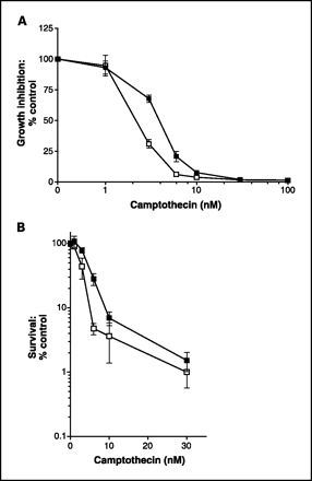
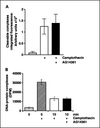
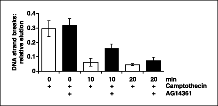
AG14361 is a minimum of 1000-fold more potent than benzamides. In permeabilized SW620 cells, the IC50 for AG14361 is 29 nM, whereas in intact SW620 cells, it is 14 nM. The tricyclic ring system of AG14361, as revealed by crystallographic analysis of the compound bound to the chicken PARP-1 catalytic domain, is situated in a pocket made up of amino acid residues Trp861, His862, Gly863, Tyr896, Phe897, Ala898, Lys903, Ser904, Tyr907, and Glu988. AG14361 establishes significant hydrogen bonds with Gly863, Ser904, and Glu988, as well as a water-mediated hydrogen bond. Since maximal PARP-1 inhibition is observed at much lower concentrations (≤1 μM) than the GI50, AG14361-induced growth inhibition cannot be attributed to PARP-1-related effects. At 0.4 μM, AG14361 has no effect on the growth or gene expression of cancer cells. However, it increases the antiproliferative activity of topotecan and temozolomide and inhibits 73% of LoVo cells' ability to recover from potentially fatal γ-radiation damage. In addition, microarray analysis demonstrates that 0.4 μM AG14361 does not significantly change gene expression. The expression of the 6800 genes remains unchanged in A549 cells exposed to 0.4 μM AG14361 for a duration of 17 hours. Thus, it can be concluded that the cellular effects of this low concentration of AG14361 are specific to PARP-1 inhibition because, despite inhibiting cellular PARP-1 activity by more than 85%, 0.4 μM AG14361 essentially does not change gene expression or cell proliferation. Gene expression is impacted by higher, growth-inhibitory concentrations of AG14361; however, since PARP-/- and PARP-1+/+ cells are equally affected in terms of cell proliferation, these effects are unlikely to be related to PARP-1 inhibition. Lower concentrations of AG14361 are found in the brain, but it is quickly absorbed into the bloodstream and distributed to the tumor and liver. In both xenograft models, the tissue-to-plasma concentration ratio shows that AG14361 is sustained in tumor tissue over time, with tumor concentrations (≥15 μM for 2 hours) higher than those needed to inhibit PARP-1 activity in vitro.[1] AG14361 overcomes temozolomide resistance by increasing temozolomide activity in all MMR-proficient cells (1.5–3.3-fold), but it is more successful in MMR-deficient cells (3.7–5.2-fold potentiation). On the other hand, benzylguanine is ineffective in MMR-deficient cells and only boosts the effectiveness of temozolomide in MMR-proficient cells.[2] AG14361 amplifies the cytotoxic and growth-inhibitory properties of topoisomerase I poisons. The DNA single-strand breaks caused by camptothecin are more persistent when AG14361 is present.[3]
Antiproliferative activity in HR-deficient cells: AG-14361 shows preferential cytotoxicity to homologous recombination (HR)-deficient cancer cells. IC50 values (72 h, MTT assay): BRCA2-mutant Capan-1 (pancreatic cancer, 0.8 μM), BRCA1-mutant MDA-MB-436 (breast cancer, 1.1 μM); vs. HR-proficient MCF-7 (breast cancer, IC50 = 16 μM), normal human foreskin fibroblasts (HFF, IC50 >50 μM) [1] - PARP inhibition and DNA damage accumulation: In Capan-1 cells, AG-14361 (0.1–2 μM) dose-dependently reduced poly(ADP-ribose) (PAR) polymer levels: 1 μM reduced PAR by 90% vs. control (western blot). It also increased γ-H2AX foci (DNA double-strand break marker) by 4.5-fold (immunofluorescence) and induced G2/M cell cycle arrest (40% G2/M cells vs. 18% control) at 0.5 μM [1] - Synergy with DNA-damaging agents: Combination of AG-14361 (0.2 μM) with carboplatin (0.5 μg/mL) in BRCA1-mutant MDA-MB-436 cells enhanced cytotoxicity: cell viability reduced to 20% (combination) vs. 65% (AG-14361 alone) and 70% (carboplatin alone), with a combination index (CI) of 0.4. Similar synergy was observed with temozolomide (0.1 μg/mL, CI = 0.38) [2] - Apoptosis induction: In Capan-1 cells treated with AG-14361 (1 μM) for 48 h, the percentage of apoptotic cells (Annexin V-positive/PI-positive) increased from 3% (control) to 32%, accompanied by a 3.0-fold increase in cleaved caspase-3 protein levels (western blot) [1] |
| ln Vivo |
The administration of AG14361 prior to radiation therapy has a statistically significant effect on the sensitivity of mice harboring LoVo xenografts to radiation therapy. In xenografts, AG14361 statistically significantly increases blood flow, which may enhance medication delivery to tumor xenografts. Nontoxic dosages of AG14361 cause a 2- to 3-fold delay in LoVo xenograft growth when induced by irinotecan, x-irradiation, or temozolomide in vivo. The tumor growth delay was increased from 3 days to 9 days and from 10 days to 15 days by AG14361 at 5 mg/kg and 15 mg/kg, respectively, when coadministration of AG14361 with temozolomide was observed. This increase in temozolomide activity against LoVo xenografts was statistically significant. AG14361 and temozolomide together result in the full remission of SW620 xenograft tumors. Following intraperitoneal injection of AG14361 (10 mg/kg), PARP-1 activity in SW620 xenografts is inhibited by more than 75% for at least 4 hours, as determined by pharmacodynamic assay. This is consistent with the concentration of AG14361 remaining in the tumor.[1]
BRCA-mutant pancreatic cancer xenografts: Female nude mice (6–8 weeks old) bearing subcutaneous BRCA2-mutant Capan-1 tumors were treated with AG-14361 (10 mg/kg, oral, daily) for 28 days. Tumor growth inhibition (TGI) was 80% (treated volume: 250 mm³ vs. vehicle: 1250 mm³, P<0.001). Combination with carboplatin (5 mg/kg, intraperitoneal, weekly) increased TGI to 92% [1] - Ovarian cancer xenograft model: Female BALB/c nude mice (7 weeks old) with BRCA1-mutant OVCAR-8 tumors were grouped (n=5/group): vehicle, AG-14361 (15 mg/kg, oral, daily), cisplatin (3 mg/kg, intraperitoneal, weekly), combination. The combination group had a tumor weight of 0.22 g vs. 1.05 g (vehicle), 0.48 g (AG-14361 alone), and 0.52 g (cisplatin alone), with a local control rate of 60% [3] - Pharmacodynamic validation in vivo: In Capan-1 xenografts, AG-14361 (10 mg/kg, oral) reduced tumor PAR levels by 75% (western blot) and increased γ-H2AX expression by 3.8-fold (immunohistochemistry) vs. vehicle, confirming in vivo PARP inhibition and DNA damage [1] |
| Enzyme Assay |
Full-length recombinant human PARP-1 activity is assessed in a reaction mixture containing activated calf thymus DNA (10 μg/mL) at 25 °C, 500 μM NAD+ plus [32P]NAD+ (0.1–0.3 μCi per reaction mixture), and 20 nM PARP-1. The reaction is stopped after 4 minutes by adding ice-cold 10% (wt/vol) trichloroacetic acid. A PhosphorImager is used to quantify the reaction product [32P]ADP-ribose that is integrated into acid-insoluble material after it has been deposited onto Whatman GF/C glass fiber filters using a Bio-Dot microfiltration device. Inhibition of PARP-1 activity by AG14361 at 0–600 nM is measured, and the Ki for AG14361 is calculated by nonlinear regression analysis.
Recombinant PARP1 activity assay: Purified recombinant human PARP1 was incubated with a biotinylated double-stranded DNA (dsDNA) activator and NAD⁺ (substrate) in assay buffer (50 mM Tris-HCl pH 8.0, 10 mM MgCl₂, 1 mM DTT) at 37°C for 20 min. Serial concentrations of AG-14361 (0.1–50 nM) were added, and incubation continued for 30 min. The reaction was terminated by adding 10% trichloroacetic acid (TCA). PAR polymer was detected via a streptavidin-horseradish peroxidase (HRP) conjugate and chemiluminescence. IC50 values were calculated by fitting the percentage of remaining PARP activity (vs. vehicle) to a four-parameter logistic model [1] |
| Cell Assay |
The luciferase-coupled ATP quantization assay of metabolically active cells in a 96-well plate is used in the cell viability assay along with MTT. One well of a 24-well plate is plated with one to two × 104 cells for MTT. The target medications (AG14361) are dissolved in DMSO at different concentrations and subsequently added to the cells in Dulbecco's modified Eagle's medium (DMEM) containing 10% fetal bovine serum. Additionally, each well receives an addition of the IC50 concentration of AG14361. The addition to medium results in a final DMSO concentration of 0.1%. The medium is removed after 48 hours, and each well receives 0.3 mL of 0.1% MTT in phosphate-buffered saline (PBS). The MTT solution is removed and 0.8 mL of 2-propanol is added after 30 minutes of incubation in a 37°C CO2 incubator. A plate reader is used to measure the OD560 following 30 minutes of shaking. Every time point is platented three times.
MTT antiproliferation assay: HR-deficient (Capan-1, MDA-MB-436) or HR-proficient (MCF-7, HFF) cells were seeded in 96-well plates at a density of 5×10³ cells/well and incubated overnight (37°C, 5% CO₂). AG-14361 (0.01–100 μM) was added, and cells were cultured for 72 h. MTT reagent (5 mg/mL, 10 μL/well) was added, incubation continued for 4 h, and formazan crystals were dissolved in DMSO. Absorbance at 570 nm was measured, and IC50 values were calculated using GraphPad Prism software [1] - γ-H2AX immunofluorescence assay: Capan-1 cells were treated with AG-14361 (0.1–2 μM) for 24 h, fixed with 4% paraformaldehyde, and permeabilized with 0.2% Triton X-100. Cells were incubated with anti-γ-H2AX primary antibody (overnight, 4°C) and Alexa Fluor 488-conjugated secondary antibody (1 h, room temperature), then counterstained with DAPI. γ-H2AX foci per cell were counted using fluorescence microscopy (≥100 cells per group) [1] - Annexin V-FITC/PI apoptosis assay: Capan-1 cells were treated with AG-14361 (0.5–2 μM) for 48 h, harvested by trypsinization, washed with cold PBS, and resuspended in Annexin V binding buffer. Annexin V-FITC (5 μL) and propidium iodide (PI, 10 μL) were added, and cells were incubated in the dark (15 min, room temperature). Apoptotic cells were analyzed via flow cytometry [1] - Western blot for PAR and cleaved caspase-3: Cells treated with AG-14361 were lysed in RIPA buffer (with protease inhibitors), and 30 μg of protein was separated by 12% SDS-PAGE. Proteins were transferred to PVDF membranes, blocked with 5% non-fat milk (1 h, room temperature), and incubated with anti-PAR or anti-cleaved caspase-3 primary antibodies (overnight, 4°C). HRP-conjugated secondary antibodies were added (1 h, room temperature), and bands were visualized via enhanced chemiluminescence (ECL) [1] |
| Animal Protocol |
CD-1 nude mice with palpable, subcutaneous SW620 or LoVo xenografts are given intraperitoneally AG14361 (at 5 or 15 mg/kg) or normal saline (control animals) once a day for five days (five mice per group). In cases where drugs are combined, AG14361 is injected intraperitoneally once a day for five days.This is done either right before the cytotoxic medication (NSC 362856 at 68 mg/kg or CPT-11 at 2.5 mg/kg) is administered or half an hour before applying 2 Gy of x-irradiation locally to the tumor once a day for five days. The tumor volumes are expressed as median relative tumor volume (RTV), which are calculated using the formula a2 × b/2, where a is the tumor's width and b its length. These volumes are obtained from two-dimensional caliper measurements. In other words, RTV1 represents the tumor volume on day 0 of treatment, and RTV4 represents the tumor volume four times that on day 0 of treatment. The term "tumor growth delay" refers to the difference between the time to RTV4 in mice receiving medication or radiation therapy and that of control mice (vehicle alone).
Pancreatic cancer xenograft protocol: Female nude mice (6–8 weeks old) were subcutaneously injected with 5×10⁶ Capan-1 cells (suspended in 100 μL of a 1:1 mixture of PBS and matrigel) into the right flank. When tumors reached an average volume of ~100 mm³, mice were randomly divided into four groups (n=6/group): (1) Vehicle: 0.5% methylcellulose in PBS, oral gavage, daily; (2) AG-14361: 10 mg/kg, dissolved in 0.5% methylcellulose, oral gavage, daily; (3) Carboplatin: 5 mg/kg, intraperitoneal injection, weekly; (4) Combination: AG-14361 + carboplatin. Treatment lasted 28 days. Tumor volume (length × width² / 2) was measured every 3 days, and body weight was recorded weekly [1] - Ovarian cancer xenograft protocol: Female BALB/c nude mice (7 weeks old) were subcutaneously injected with 4×10⁶ OVCAR-8 cells (100 μL PBS/matrigel, 1:1). When tumors reached ~120 mm³, mice were grouped (n=5/group): vehicle (0.5% methylcellulose, oral, daily), AG-14361 (15 mg/kg, oral, daily), cisplatin (3 mg/kg, intraperitoneal, weekly), combination. Treatment continued for 21 days. At euthanasia, tumors were excised and weighed, and tumor tissues were collected for immunohistochemical analysis of γ-H2AX [3] |
| ADME/Pharmacokinetics |
Oral bioavailability in rats: Male Sprague-Dawley rats (250-300 g) were administered AG-14361 via oral gavage (10 mg/kg) or intravenous injection (2 mg/kg). The oral bioavailability was 65%. After oral administration: peak plasma concentration (Cmax) = 2.8 μg/mL (time to peak, Tmax = 1.2 h), terminal half-life (t1/2) = 4.2 h, area under the concentration-time curve (AUC0-24h) = 16.5 μg·h/mL. Intravenous administration: Cmax = 7.2 μg/mL, t1/2 = 3.8 h, AUC0-∞ = 19.8 μg·h/mL [1]
- Plasma protein binding rate: In human plasma, the protein binding rate of AG-14361 was 92%, mainly bound to albumin (as determined by 37°C equilibrium dialysis method) [1] - Tissue distribution in mice: After oral administration of 10 mg/kg AG-14361 to Capan-1 xenograft mice, the highest tissue concentrations were found in the liver (4.5 μg/g at 2 hours) and tumor (3.2 μg/g at 2 hours), followed by plasma (2.8 μg/mL at 1.2 hours). The brain tissue concentration was <0.4 μg/g, indicating that its blood-brain barrier penetration was extremely low [1] |
| Toxicity/Toxicokinetics |
Repeated-dose toxicity in rats: Male/female Sprague-Dawley rats (n=4 per sex per group) were orally administered AG-14361 (5, 20, 100 mg/kg) daily for 28 consecutive days. No deaths were observed. The no-observed-adverse-effect level (NOAEL) was 20 mg/kg. Mild anemia (18% decrease in hemoglobin compared to the control group) and elevated serum aspartate aminotransferase (AST) (1.6-fold increase compared to the control group) were observed in the 100 mg/kg group, but no histopathological changes were observed in the liver and kidneys [1]. Xenograft toxicity in mice: In all tumor models, daily oral administration of AG-14361 (10-15 mg/kg) resulted in a weight loss of ≤5%, with no obvious toxic reactions (e.g., somnolence, diarrhea). Combination therapy with platinum-based drugs (carboplatin, cisplatin) did not increase toxicity compared to monotherapy [1,3]
- In vitro safety in normal cells: In human HFF cells and peripheral blood mononuclear cells (PBMCs), treatment with AG-14361 (≤20 μM) for 72 hours had no significant effect on cell viability (MTT assay, viability >85% vs. control group) [1] |
| References | |
| Additional Infomation |
LSM-1988 belongs to the benzimidazole class of compounds. Mechanism of action: AG-14361 inhibits PARP1, a key enzyme in DNA single-strand break base excision repair (BER). In homologous recombination (HR)-deficient cells (e.g., BRCA1/2 mutant cells), BER inhibition leads to unrepaired DNA damage, accumulation of double-strand breaks, and ultimately, synthetic lethality. This mechanism explains its selectivity for HR-deficient cancers and its synergistic effect with DNA damaging agents [1,2]. Preclinical development focus: AG-14361 is a pioneering PARP inhibitor that has been preclinically evaluated for the treatment of HR-deficient solid tumors, including BRCA-mutated pancreatic, breast, and ovarian cancers. It provides foundational data for the development of subsequent clinically approved PARP inhibitors (such as olaparib and rucaparib) [1,3] - Limitations in clinical translation: Despite the strong efficacy shown in preclinical studies, AG-14361 failed to enter clinical trials due to challenges in optimizing its pharmacokinetic properties (e.g., moderate oral bioavailability) and potential drug interactions (which have been identified in in vitro metabolism studies) [1].
|
| Molecular Formula |
C19H20N4O
|
|
|---|---|---|
| Molecular Weight |
320.39
|
|
| Exact Mass |
320.163
|
|
| Elemental Analysis |
C, 71.23; H, 6.29; N, 17.49; O, 4.99
|
|
| CAS # |
328543-09-5
|
|
| Related CAS # |
|
|
| PubChem CID |
9840076
|
|
| Appearance |
White to off-white solid powder
|
|
| Density |
1.3±0.1 g/cm3
|
|
| Index of Refraction |
1.676
|
|
| LogP |
1.92
|
|
| Hydrogen Bond Donor Count |
1
|
|
| Hydrogen Bond Acceptor Count |
3
|
|
| Rotatable Bond Count |
3
|
|
| Heavy Atom Count |
24
|
|
| Complexity |
460
|
|
| Defined Atom Stereocenter Count |
0
|
|
| SMILES |
O=C1NCCN2C3=C1C=CC=C3N=C2C4=CC=C(C=C4)CN(C)C
|
|
| InChi Key |
SEKJSSBJKFLZIT-UHFFFAOYSA-N
|
|
| InChi Code |
InChI=1S/C19H20N4O/c1-22(2)12-13-6-8-14(9-7-13)18-21-16-5-3-4-15-17(16)23(18)11-10-20-19(15)24/h3-9H,10-12H2,1-2H3,(H,20,24)
|
|
| Chemical Name |
2-[4-[(dimethylamino)methyl]phenyl]-1,3,10-triazatricyclo[6.4.1.04,13]trideca-2,4,6,8(13)-tetraen-9-one
|
|
| Synonyms |
AG 14361; AG14361; AG-14361
|
|
| HS Tariff Code |
2934.99.9001
|
|
| Storage |
Powder -20°C 3 years 4°C 2 years In solvent -80°C 6 months -20°C 1 month |
|
| Shipping Condition |
Room temperature (This product is stable at ambient temperature for a few days during ordinary shipping and time spent in Customs)
|
| Solubility (In Vitro) |
DMSO: 10~12 mg/mL (31.2~37.5 mM)
Water: 1 mg/mL (Insoluble) Ethanol: 1 mg/mL |
|---|---|
| Solubility (In Vivo) |
Solubility in Formulation 1: ≥ 1 mg/mL (3.12 mM) (saturation unknown) in 10% DMSO + 40% PEG300 + 5% Tween80 + 45% Saline (add these co-solvents sequentially from left to right, and one by one), clear solution.
For example, if 1 mL of working solution is to be prepared, you can add 100 μL of 10.0 mg/mL clear DMSO stock solution to 400 μL of PEG300 and mix evenly; then add 50 μL of Tween-80 to the above solution and mix evenly; then add 450 μL of normal saline to adjust the volume to 1 mL. Preparation of saline: Dissolve 0.9 g of sodium chloride in 100 mL ddH₂ O to obtain a clear solution. Solubility in Formulation 2: ≥ 1 mg/mL (3.12 mM) (saturation unknown) in 10% DMSO + 90% (20% SBE-β-CD in Saline) (add these co-solvents sequentially from left to right, and one by one), clear solution. For example, if 1 mL of working solution is to be prepared, you can add 100 μL of 10.0 mg/mL clear DMSO stock solution to 900 μL of 20% SBE-β-CD physiological saline solution and mix evenly. Preparation of 20% SBE-β-CD in Saline (4°C,1 week): Dissolve 2 g SBE-β-CD in 10 mL saline to obtain a clear solution. View More
Solubility in Formulation 3: ≥ 1 mg/mL (3.12 mM) (saturation unknown) in 10% DMSO + 90% Corn Oil (add these co-solvents sequentially from left to right, and one by one), clear solution. Solubility in Formulation 4: 4% DMSO+ddH2O: 2mg/mL |
| Preparing Stock Solutions | 1 mg | 5 mg | 10 mg | |
| 1 mM | 3.1212 mL | 15.6060 mL | 31.2120 mL | |
| 5 mM | 0.6242 mL | 3.1212 mL | 6.2424 mL | |
| 10 mM | 0.3121 mL | 1.5606 mL | 3.1212 mL |
*Note: Please select an appropriate solvent for the preparation of stock solution based on your experiment needs. For most products, DMSO can be used for preparing stock solutions (e.g. 5 mM, 10 mM, or 20 mM concentration); some products with high aqueous solubility may be dissolved in water directly. Solubility information is available at the above Solubility Data section. Once the stock solution is prepared, aliquot it to routine usage volumes and store at -20°C or -80°C. Avoid repeated freeze and thaw cycles.
Calculation results
Working concentration: mg/mL;
Method for preparing DMSO stock solution: mg drug pre-dissolved in μL DMSO (stock solution concentration mg/mL). Please contact us first if the concentration exceeds the DMSO solubility of the batch of drug.
Method for preparing in vivo formulation::Take μL DMSO stock solution, next add μL PEG300, mix and clarify, next addμL Tween 80, mix and clarify, next add μL ddH2O,mix and clarify.
(1) Please be sure that the solution is clear before the addition of next solvent. Dissolution methods like vortex, ultrasound or warming and heat may be used to aid dissolving.
(2) Be sure to add the solvent(s) in order.
Effect of AG14361 on topo I poison-induced growth inhibition and cytotoxicity in PARP-1+/+, PARP-1−/−, and human leukemia cell lines.Clin Cancer Res. 2005 Dec 1;11(23):8449-57. |
|---|
Effect of AG14361 on camptothecin-stabilized cleavable complex induction and removal.Clin Cancer Res. 2005 Dec 1;11(23):8449-57. |
Repair of camptothecin-induced DNA single-strand breaks in K562 cells in the presence or absence of AG14361.K562 cells were exposed to 30 nmol/L camptothecin for 30 minutes followed by repair in drug-free medium (white columns) or medium containing 0.4 μmol/L AG14361 (black columns) for 0, 10, or 20 minutes. DNA strand breaks were measured by alkaline elution. Relative elution was calculated by comparison with DMSO or 0.4 μmol/L AG14361 alone control as appropriate. Columns, mean of three independent experiments; bars, SE.Clin Cancer Res. 2005 Dec 1;11(23):8449-57. |
Products are for research use only; We do not sell to patients
Copyright 2020 InvivoChem LLC | All Rights Reserved


 COA
COA