| Size | Price | Stock | Qty |
|---|---|---|---|
| 25mg |
|
||
| 50mg |
|
||
| 100mg |
|
||
| 250mg |
|
||
| 500mg | |||
| 1g |
|
||
| Other Sizes |


Purity: ≥98%
ADT-OH is a analogue of anethole dithiolethione (ADT) and synthetic hydrogen sulfide (H2S) donor. ADT-OH significantly reduced tPA-enhanced Akt activation and VEGF expression in brain microvascular endothelial cells in the in vitro glucose-oxygen deprivation (OGD) model. Last but not least, ADT-OH enhanced functional outcomes in mice given MCAO and tPA infusion. H2S donors may have prevented the Akt-VEGF-MMP9 cascade from increasing tPA-induced cerebral hemorrhage. Giving out H2S donors could be a novel way to increase the safety of tPA after a stroke.
| Targets |
IκBα (inhibits degradation, leading to reduced NF-κB activation) [1]
Fas-associated protein with death domain (FADD) (increases protein level by inhibiting its ubiquitin-mediated degradation) [1] Makorin ring finger protein 1 (MKRN1) (downregulates expression) [1] XIAP (downregulates expression) [1] Bcl-2 (downregulates expression) [1] |
|---|---|
| ln Vitro |
ADT-OH releases H₂S in a concentration- and time-dependent manner in MEF and B16F10 cells. [1]
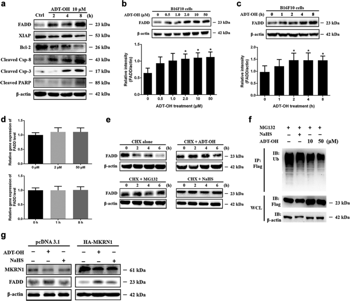
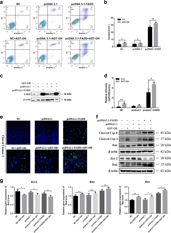
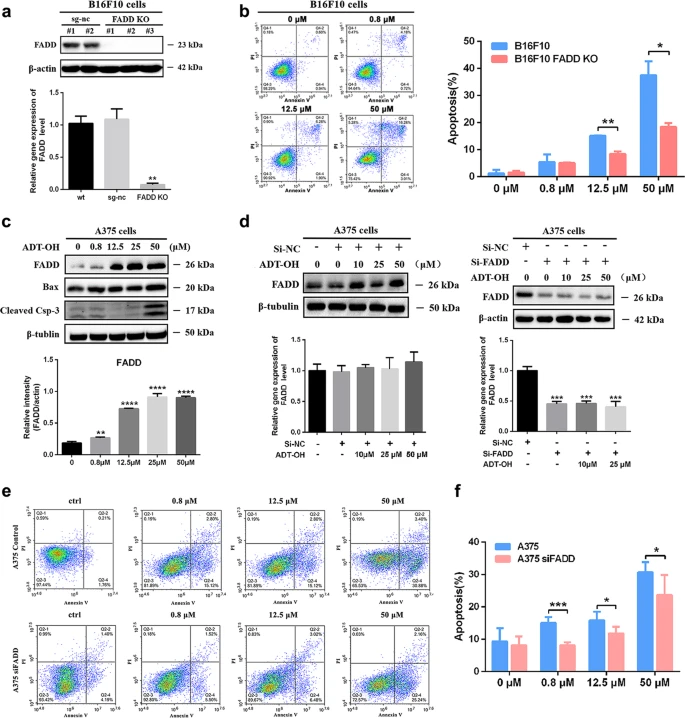
ADT-OH (0.8-100 μM) significantly inhibits the proliferation of various cancer cell lines, including B16F10 melanoma cells, as measured by CCK-8 assay. B16F10 cells were the most sensitive, with 12.5 μM ADT-OH reducing proliferation by 55.74% after 24h. It had a lesser effect on normal MEF cells (27.64% reduction at 12.5 μM). [1] ADT-OH (0.8-50 μM) induces apoptosis in B16F10 cells in a dose-dependent manner, with 12.5 μM and 25 μM inducing 15.02% and 41.95% apoptosis, respectively, after 24h. It had a minimal pro-apoptotic effect on normal MEF, HaCaT, and HK2 cells. [1] ADT-OH (10 μM) inhibits IκBα degradation, leading to reduced NF-κB activation and subsequent downregulation of anti-apoptotic proteins XIAP and Bcl-2 in B16F10 cells. It also increases the protein levels of cleaved caspase-8, cleaved caspase-3, and cleaved PARP, indicating induction of both intrinsic and extrinsic apoptosis pathways. [1] ADT-OH (10 μM) significantly increases the protein level of FADD in a time- and dose-dependent manner in B16F10 cells, without affecting its mRNA level. This effect was also observed in 4T1, LLC, A549, and HepG2 cell lines. [1] ADT-OH (10 μM) inhibits the ubiquitin-mediated degradation of FADD by decreasing the protein stability of its E3 ubiquitin ligase, MKRN1. The half-life of FADD is extended from ~4h to >6h with ADT-OH treatment. [1] In FADD-knockout B16F10 cells and FADD-knockdown A375 cells, the pro-apoptotic effect of ADT-OH (50 μM) is significantly blunted compared to control cells (22.26% vs. 41.95% apoptosis in B16F10 cells). [1] Combining FADD overexpression with low-dose ADT-OH (2 μM) treatment in B16F10 cells further increases apoptosis (44%) compared to FADD overexpression (27%) or ADT-OH alone. This combination also enhances the activation of caspase-3 and modulates the expression of Bcl-2 family proteins. [1] |
| ln Vivo |
In a B16F10 mouse xenograft model, oral administration of ADT-OH (37.5 mg/kg, every other day) significantly inhibits tumour growth and prolongs survival of tumour-bearing mice compared to vehicle control. [1]
Combining ADT-OH (37.5 mg/kg, oral, every other day) with tumour-targeted delivery of FADD via a recombinant Salmonella strain (VNP-FADD, 1x10⁵ cfu/mouse, i.p.) results in the most significant tumour growth inhibition, smallest tumour volume, longest tumour doubling time (29.63 days vs. 13.65 days for PBS), and most prolonged survival compared to either treatment alone. [1] Immunofluorescence and Western blot analysis of tumour tissues from mice treated with the combination show increased FADD protein levels and cleaved caspase-3, indicating enhanced apoptosis. [1] H&E staining and TUNEL assays of tumour sections reveal that the combination of ADT-OH and VNP-FADD induces the largest necrotic areas and the highest number of apoptotic cells compared to other treatment groups. [1] In a xenograft model using FADD-knockout B16F10 cells, ADT-OH (37.5 mg/kg, oral, every other day) has no significant therapeutic effect, with tumours growing similarly to untreated controls, demonstrating that FADD is essential for the anti-tumour activity of ADT-OH in vivo. [1] |
| Cell Assay |
Cell Proliferation (CCK-8): Cells (5x10³ cells/well in 96-well plates) were treated with ADT-OH (0.8-100 μM) for 24, 48, or 72h. CCK-8 reagent (10 μl) was added and incubated for 1h. Absorbance was measured at 450 nm. [1]
Apoptosis (Flow Cytometry): B16F10 or other cells were treated with ADT-OH (0.8-50 μM) for 24h. Cells were harvested and stained with Annexin V-EGFP and propidium iodide (PI) according to the manufacturer's instructions, then analyzed by flow cytometry. [1] H₂S Measurement: Cells in 96-well plates were cultured with 10 mM Cys, 10 μM PLP, and ADT-OH. Lead acetate paper was placed over the plate for 2-24h. For quantification, a H₂S detection kit was used on cell supernatants after treatment with ADT-OH according to the manufacturer's instructions. [1] Western Blotting: Cells were lysed in buffer with protease inhibitors. Proteins (40 μg) were separated by SDS-PAGE and transferred to membranes. Membranes were probed with primary antibodies (e.g., anti-FADD, anti-MKRN1, anti-cleaved caspase-3) and then with HRP-conjugated secondary antibodies. Proteins were detected using an ECL system. [1] Ubiquitination Assay: B16F10 cells were co-transfected with FADD-Flag and HA-Ubiquitin plasmids. After treatment with MG132 (10 μM for 6h) and/or ADT-OH (10 μM for 6h), cells were lysed in 1% SDS buffer. FADD-Flag was immunoprecipitated using an anti-FLAG antibody, followed by Western blot analysis using an anti-HA antibody to detect ubiquitinated FADD. [1] |
| Animal Protocol |
Efficacy Study (B16F10 Xenograft):** C57BL/6 mice were subcutaneously injected with B16F10 cells (2x10⁵ cells/mouse). Starting on day 1, mice (n=8/group) were treated orally with ADT-OH (37.5 mg/kg in 100 μl of 0.5% carboxymethylcellulose/PBS) or vehicle every other day. Tumour volume was measured periodically, and survival was monitored. [1]
* **Combination Study (ADT-OH + VNP-FADD):** B16F10 tumour-bearing mice were established as above. When tumours reached ~100-150 mm³, mice (n≥8/group) received an intraperitoneal injection of PBS, VNP (1x10⁵ cfu), or VNP-FADD (1x10⁵ cfu). The combination groups also received oral ADT-OH (37.5 mg/kg) every other day starting from this time. Tumour volume, doubling time, and survival were monitored. At day 15, some mice were sacrificed for tissue collection (tumour, liver, spleen) for histological and biochemical analysis. [1] * **FADD-KO Study:** C57BL/6 mice were subcutaneously injected with either B16F10 control cells or B16F10 FADD-knockout cells. Mice (n=8/group) were treated orally with vehicle or ADT-OH (37.5 mg/kg) every other day. Tumour growth was monitored. [1] Efficacy Study (B16F10 Xenograft): C57BL/6 mice were subcutaneously injected with B16F10 cells (2x10⁵ cells/mouse). Starting on day 1, mice (n=8/group) were treated orally with ADT-OH (37.5 mg/kg in 100 μl of 0.5% carboxymethylcellulose/PBS) or vehicle every other day. Tumour volume was measured periodically, and survival was monitored. [1] Combination Study (ADT-OH + VNP-FADD): B16F10 tumour-bearing mice were established as above. When tumours reached ~100-150 mm³, mice (n≥8/group) received an intraperitoneal injection of PBS, VNP (1x10⁵ cfu), or VNP-FADD (1x10⁵ cfu). The combination groups also received oral ADT-OH (37.5 mg/kg) every other day starting from this time. Tumour volume, doubling time, and survival were monitored. At day 15, some mice were sacrificed for tissue collection (tumour, liver, spleen) for histological and biochemical analysis. [1] FADD-KO Study: C57BL/6 mice were subcutaneously injected with either B16F10 control cells or B16F10 FADD-knockout cells. Mice (n=8/group) were treated orally with vehicle or ADT-OH (37.5 mg/kg) every other day. Tumour growth was monitored. [1] |
| Toxicity/Toxicokinetics |
ADT-OH showed a greater inhibitory effect on the proliferation of tumour cells (e.g., B16F10) than on normal cells (e.g., MEFs, HaCaT, HK2) in vitro. [1]
In vivo, the combination of low-dose ADT-OH (37.5 mg/kg) with VNP-FADD exhibited excellent tumour suppression with little apparent side effects, although specific toxicity parameters (e.g., body weight, organ histology) were not detailed. [1] |
| References | |
| Additional Infomation |
ADT-OH [5-(4-hydroxyphenyl)-3H-1,2-dithiole-3-thione] is a slow-releasing hydrogen sulfide (H₂S) donor. Its dithiolethione moiety is widely used for synthesising slow-releasing organic H₂S donors and is known for its chemopreventive and cytoprotective properties. [1]
The study identifies a novel mechanism of action for ADT-OH: it downregulates the E3 ubiquitin ligase MKRN1, which in turn stabilizes the FADD protein by preventing its ubiquitin-mediated degradation. This leads to enhanced extrinsic apoptosis signalling. This is in addition to its known effect on the NF-κB pathway and intrinsic apoptosis. [1] FADD is identified as a critical mediator of ADT-OH's anti-melanoma effects. The pro-apoptotic and anti-tumour activity of ADT-OH is significantly reduced in FADD-deficient cells and tumours. [1] The combination of ADT-OH with tumour-targeted FADD delivery (via VNP-FADD) shows strong synergistic anti-tumour effects in a mouse melanoma model, suggesting a promising new therapeutic strategy. [1] |
| Molecular Formula |
C9H6OS3
|
|---|---|
| Molecular Weight |
226.326
|
| Exact Mass |
225.958
|
| Elemental Analysis |
C, 47.76; H, 2.67; O, 7.07; S, 42.50
|
| CAS # |
18274-81-2
|
| Related CAS # |
18274-81-2
|
| PubChem CID |
3082127
|
| Appearance |
Yellow solid powder
|
| Density |
1.48g/cm3
|
| Boiling Point |
322.2ºC at 760mmHg
|
| Melting Point |
191-192 ºC
|
| Flash Point |
148.6ºC
|
| Vapour Pressure |
0.000284mmHg at 25°C
|
| Index of Refraction |
1.757
|
| LogP |
3.101
|
| Hydrogen Bond Donor Count |
1
|
| Hydrogen Bond Acceptor Count |
4
|
| Rotatable Bond Count |
1
|
| Heavy Atom Count |
13
|
| Complexity |
241
|
| Defined Atom Stereocenter Count |
0
|
| SMILES |
S1C(=C([H])C(=S)S1)C1C([H])=C([H])C(=C([H])C=1[H])O[H]
|
| InChi Key |
IWBBKLMHAILHAR-UHFFFAOYSA-N
|
| InChi Code |
InChI=1S/C9H6OS3/c10-7-3-1-6(2-4-7)8-5-9(11)13-12-8/h1-5,10H
|
| Chemical Name |
5-(4-hydroxyphenyl)dithiole-3-thione
|
| Synonyms |
Desmethylanethol trithione ACS1; ADT-OH
|
| HS Tariff Code |
2934.99.9001
|
| Storage |
Powder -20°C 3 years 4°C 2 years In solvent -80°C 6 months -20°C 1 month |
| Shipping Condition |
Room temperature (This product is stable at ambient temperature for a few days during ordinary shipping and time spent in Customs)
|
| Solubility (In Vitro) |
DMSO: 45~125 mg/mL (198.8~552.3 mM)
Ethanol: ~12 mg/mL (~53.0 mM) |
|---|---|
| Solubility (In Vivo) |
Solubility in Formulation 1: ≥ 2.08 mg/mL (9.19 mM) (saturation unknown) in 10% DMSO + 40% PEG300 + 5% Tween80 + 45% Saline (add these co-solvents sequentially from left to right, and one by one), clear solution.
For example, if 1 mL of working solution is to be prepared, you can add 100 μL of 20.8 mg/mL clear DMSO stock solution to 400 μL PEG300 and mix evenly; then add 50 μL Tween-80 to the above solution and mix evenly; then add 450 μL normal saline to adjust the volume to 1 mL. Preparation of saline: Dissolve 0.9 g of sodium chloride in 100 mL ddH₂ O to obtain a clear solution. Solubility in Formulation 2: ≥ 2.08 mg/mL (9.19 mM) (saturation unknown) in 10% DMSO + 90% (20% SBE-β-CD in Saline) (add these co-solvents sequentially from left to right, and one by one), clear solution. For example, if 1 mL of working solution is to be prepared, you can add 100 μL of 20.8 mg/mL clear DMSO stock solution to 900 μL of 20% SBE-β-CD physiological saline solution and mix evenly. Preparation of 20% SBE-β-CD in Saline (4°C,1 week): Dissolve 2 g SBE-β-CD in 10 mL saline to obtain a clear solution. (Please use freshly prepared in vivo formulations for optimal results.) |
| Preparing Stock Solutions | 1 mg | 5 mg | 10 mg | |
| 1 mM | 4.4183 mL | 22.0916 mL | 44.1833 mL | |
| 5 mM | 0.8837 mL | 4.4183 mL | 8.8367 mL | |
| 10 mM | 0.4418 mL | 2.2092 mL | 4.4183 mL |
*Note: Please select an appropriate solvent for the preparation of stock solution based on your experiment needs. For most products, DMSO can be used for preparing stock solutions (e.g. 5 mM, 10 mM, or 20 mM concentration); some products with high aqueous solubility may be dissolved in water directly. Solubility information is available at the above Solubility Data section. Once the stock solution is prepared, aliquot it to routine usage volumes and store at -20°C or -80°C. Avoid repeated freeze and thaw cycles.
Calculation results
Working concentration: mg/mL;
Method for preparing DMSO stock solution: mg drug pre-dissolved in μL DMSO (stock solution concentration mg/mL). Please contact us first if the concentration exceeds the DMSO solubility of the batch of drug.
Method for preparing in vivo formulation::Take μL DMSO stock solution, next add μL PEG300, mix and clarify, next addμL Tween 80, mix and clarify, next add μL ddH2O,mix and clarify.
(1) Please be sure that the solution is clear before the addition of next solvent. Dissolution methods like vortex, ultrasound or warming and heat may be used to aid dissolving.
(2) Be sure to add the solvent(s) in order.
|
|
|
Products are for research use only; We do not sell to patients
Copyright 2020 InvivoChem LLC | All Rights Reserved


 COA
COA