| Size | Price | Stock | Qty |
|---|---|---|---|
| 5mg |
|
||
| 10mg |
|
||
| 25mg |
|
||
| 50mg |
|
||
| 100mg |
|
||
| 250mg | |||
| Other Sizes |
Purity: =99.77%
| Targets |
OXPHOS/oxidative phosphorylation; VLX600 targets mitochondrial oxidative phosphorylation (OXPHOS), specifically inhibiting complexes I, II, and IV of the electron transport chain [2]. It reduces oxygen consumption rate and induces mitochondrial dysfunction [2]. The compound also inhibits mTOR signaling as a downstream effect [2].
|
|---|---|
| ln Vitro |
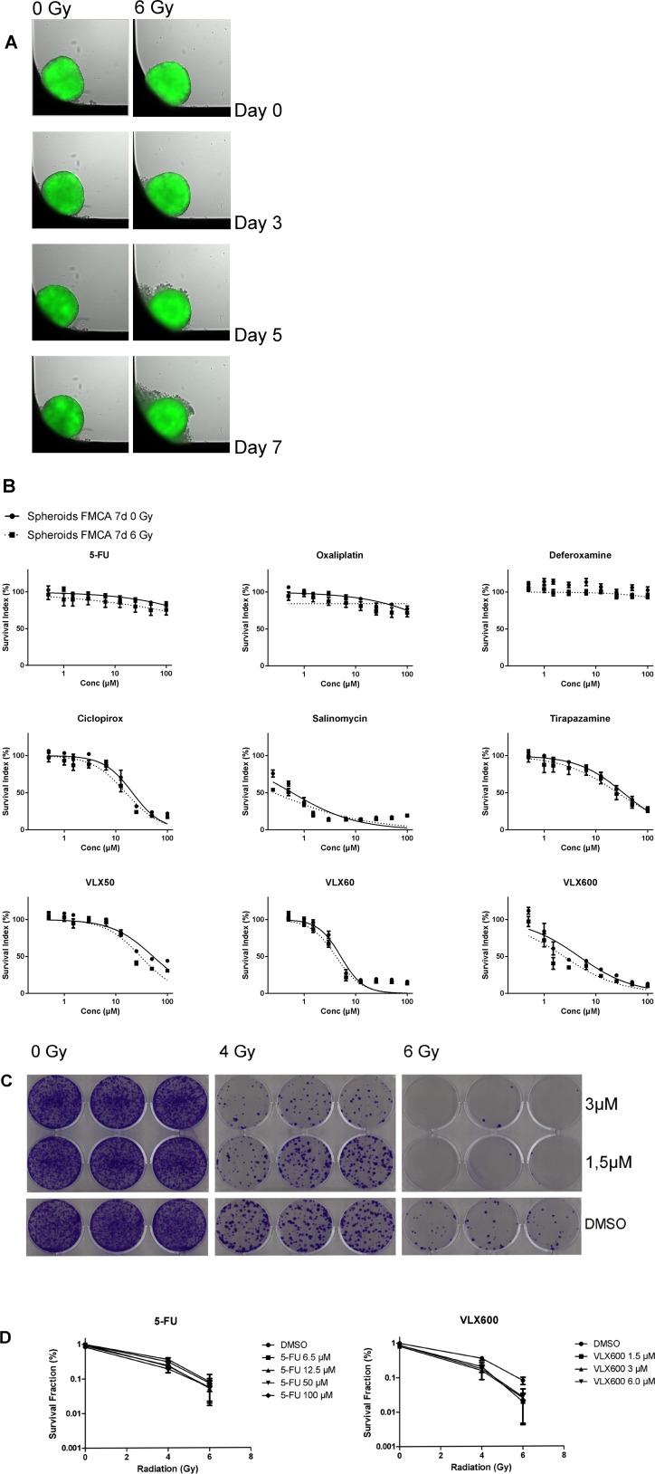
- VLX600 reduced viability of HCT116 multicellular spheroids after 6-hour exposure followed by 72-hour drug-free incubation, with central necrotic areas appearing after 72-96 hours [2].
- VLX600 strongly decreased clonogenicity of dispersed HCT116 spheroid cells [2]. - VLX600 inhibited proliferation of HCT116 monolayer cells with growth arrest at 48 hours followed by decreased cell numbers indicating cell death; IC50 values for colon cancer cells ranged from approximately 0.5-2 μM, while immortalized cells showed higher IC50 values (approximately 5-10 μM) [2]. - VLX600 showed selective cytotoxicity against primary colon cancer patient cells (n=22) with sensitivity below plasma concentrations achievable in vivo [2]. - VLX600 induced HIF-1α-dependent glycolytic response, upregulating genes associated with hypoxia and glycolysis [2]. - VLX600 induced autophagy as evidenced by LC3-II induction, autophagosome formation (Lysotracker and LC3 staining), and increased autophagic flux (further increased by chloroquine co-treatment) [2]. - VLX600 decreased cellular ATP levels in colon cancer cells (HCT116, DLD, HT29) but not in immortalized hTERT-RPE1 cells [2]. - VLX600 induced AMPK phosphorylation in colon cancer cells but not in immortalized cells [2]. - VLX600 (1-6.5 μM) synergistically sensitized HCT116 spheroid cells to radiation (4-6 Gy) in clonogenic assays; survival fraction at 6 Gy alone was 8.13%, and combination with VLX600 reduced survival further (interaction ratio 0.30-0.36) [1]. - VLX600 (1.5-3 μM) increased gamma-H2AX expression (DNA double-strand break marker) in HCT116 spheroids, with combination of 3 μM VLX600 + 6 Gy showing highest expression in both center and margin [1]. - VLX600 showed radiosensitizing effect specific to spheroids; no significant synergy observed in monolayer cultures [1]. - VLX600 in combination with irinotecan showed synergistic effects in colon cancer cells [2]. - VLX600 (6 μM) inhibited oxygen consumption rate in HCT116 cells, with transient increase followed by ~50% decrease over 10 hours [2]. - VLX600 reduced spheroid hypoxic fraction (pimonidazole staining) from 63±13% to 32±4% at 3 hours [2]. - VLX600 decreased COX-1 (mitochondrial complex IV subunit) expression in monolayer and spheroid cultures [2]. - VLX600 induced cell death with features of apoptosis (caspase activation) but overall survival was not affected by caspase inhibitors, suggesting apoptosis-independent mechanism [2]. - VLX600 cytotoxicity was potentiated by glucose starvation [2]. VLX600 (6 μM; 72 hours) promotes autophagy response [2]. VLX600 is cytotoxic to HCT116 spheroids. VLX600 induces HIF-1α-dependent glycolysis. VLX600 inhibits oxygen consumption in HCT116 cells. VLX600 suppresses the phosphorylation of mTOR downstream effectors 4EBP1 and p70-S6K through an HIF-1α-independent mechanism. VLX600 preferentially causes a decrease in ATP levels in cancer cells but not in normal cells [2]. |
| ln Vivo |
- VLX600 administered intravenously at maximally tolerated dose (16 mg/kg) showed antitumor activity in both HCT116 and HT29 colon cancer xenograft models [2].
- In HCT116 xenografts, VLX600 treatment resulted in decreased Ki67 labeling index consistent with growth arrest, and large cytoplasmic vesicles (autolysosomes) were observed in tumor sections [2]. - In an orthotopic colon cancer model (HT29-LUC cells injected into cecal wall), combination treatment with VLX600 (8 mg/kg, five cycles every second day) and irinotecan (45 mg/kg, two cycles 4 days apart) reduced median bioluminescent signal to approximately 10% of vehicle controls (P < 0.05) [2]. - VLX600 treatment showed minimal systemic toxicity with no loss of body mass and no or minor changes in plasma parameters (liver alanine aminotransferase, blood glucose, total protein) [2]. In human tumor xenografts, VLX600 (16 mg/kg; intravenously; every three days for 16 days) demonstrated anticancer efficacy [2]. |
| Enzyme Assay |
- Oxygen consumption rate measurement using Clark-type electrode: Cells were trypsinized, resuspended in medium, and placed in a respiratory chamber. Basal respiration was monitored for 3-4 minutes. State 4 respiration was measured in the presence of oligomycin (1 μg/mL) which blocks mitochondrial ATP synthase. Uncoupled respiration was assessed in the presence of carbonyl cyanide m-chlorophenylhydrazone (3 μM) [2].
- Seahorse XF analyzer measurement: Cells were plated in culture medium in XF24-well cell plates. Before measurements, medium was replaced with Seahorse assay media containing 1 mM pyruvate and 25 mM glucose at 37°C without CO2 for 1 hour. Oxygen consumption rate values were measured using the XF24 extracellular flux analyzer [2]. - Electron flow analysis in permeabilized cells: Cells were permeabilized with digitonin (6 mg per 10⁶ cells) in buffer containing 140 mM KCl, 5 mM KH₂PO₄, 1 mM MgCl₂, 1 mM ADP, and 5 mM Tris (pH 7.2) at 37°C. Substrates and inhibitors were used at the following concentrations: malate, pyruvate, and succinate all at 5 mM; rotenone 1 μM; malonate 10 mM; antimycin A 1 μM; ascorbate 5 mM; and N,N,N′,N′-tetramethyl-1,4-phenylene diamine at 0.5 mM [2]. - ATP assay: Cells (10,000 per well) were plated in 96-well plates and exposed to VLX600 for 24 or 48 hours. ATP was measured using an ATP assay kit [2]. - Lactate production assay: Cells (300,000) were plated in 6-well plates and exposed to VLX600 for 24 hours. Supernatant was collected and lactate concentration was measured using a lactate assay kit [2]. - Glucose determination in spheroids: Spheroids were trypsinized, washed in PBS, and cells were counted. Cells were lysed by three freeze-thaw cycles, and supernatant was used for glucose concentration measurement using a glucose assay kit [2]. |
| Cell Assay |
- Multicellular spheroid viability assay (acid phosphatase method): Spheroids were prepared by seeding 10,000 cells per well in poly-HEMA-coated 96-well plates with overfilled medium (170 μL) to create a convex surface, plates inverted for 24 hours, then returned to normal and incubated for 4 days. Spheroids were exposed to drugs for 6 hours, washed, and incubated for an additional 66 hours. Viability was determined by lysing spheroids in 100 μL of 0.1 M sodium acetate with 0.1% Triton X-100 and p-nitrophenyl phosphate, incubating at 37°C for 2 hours, adding NaOH, and measuring absorbance at 405 nm [2].
- Fluorometric microculture cytotoxicity assay (FMCA) for monolayer cells: Cells (1,000 per well in 384-well plates) were pre-incubated overnight, then drugs were added using an acoustic liquid handler. After 7 days of incubation, medium was removed, cells washed with PBS, fluorescein diacetate buffer and solution added, incubated 50-70 min at 37°C, and fluorescence measured using a scanning fluorometer. Survival index was defined as fluorescence in test wells as percent of unexposed control wells after blank subtraction [1][2]. - FMCA for spheroids: Following spheroid experiments, culture medium was removed, spheroids dissociated by adding Accumax (50 μL/well) and incubating at 37°C for 30 min, then dissociated with a multipipette. FMCA procedure was then performed as for monolayer cells [1]. - GFP assay for spheroids: Fluorescent signal from HCT116 GFP cells was measured every 24 hours using a high content screening reader. Mean spheroid fluorescence was calculated and used to determine AUTO SI (spheroid fluorescence in experimental wells as percent of same wells immediately before drug addition 7 days earlier) [1]. - GFP assay for monolayers: Fluorescence signal from HCT116 GFP cells was measured using a live-cell analysis system. One image was acquired per well and green object confluence was used as measure of fluorescence to calculate survival index [1]. - Clonogenic assay for spheroids: Approximately 20 hours after radiation, spheroids were dissociated into single cells using Accumax (50 μL/well, 30 min at 37°C). Plates were centrifuged, Accumax removed, 50 μL fresh medium added. After mixing, 20 μL cell solution from each well was transferred with 3 mL fresh medium to 6-well plates and incubated for 10 days. Cells were fixed and stained with 6% glutaraldehyde and 0.5% crystal violet for 30 min, rinsed, dried, and colonies counted. Survival fraction was defined as number of colonies as percent of unexposed control wells [1]. - Clonogenic assay for monolayers: Approximately 20 hours after radiation, 40 μL medium was removed from each well of 384-well plates, 50 μL Accumax added, incubated 10 min at 37°C, mixed, centrifuged, Accumax removed, 50 μL fresh medium added. After mixing, 20 μL cell solution was transferred to 3 mL fresh medium in tubes, then transferred to 6-well plates and incubated for 10 days. Fixation, staining, and colony counting were performed as above [1]. - Western blotting: Cell extract proteins were resolved by Tris-Acetate PAGE gels and transferred onto PVDF membranes. Membranes were incubated overnight with primary antibodies (HIF-1α 1:200, β-actin 1:10,000, LC3 1:1,000, BNIP3 1:500, AMPK 1:1,000, phospho-AMPK 1:2,000, COX-1 1:1,000, COX-IV 1:1,000, VDAC 1:1,000, p70 1:1,000, phospho-p70 1:1,000, eIF2-α 1:200, phospho-eIF2-α 1:200, caspase-4 1:1,000, 4EBP1 1:1,000, phospho-4EBP1 1:1,000, p62 1:400), washed, incubated with HRP-conjugated secondary antibody for 1 hour, and developed using chemiluminescent substrate [2]. - Immunohistochemistry of spheroids: Spheroids were fixed in 2% buffered formalin, dehydrated, embedded in paraffin, and sectioned. Sections were deparaffinized, rehydrated, microwaved, then incubated overnight with primary antibodies (Ki67 1:400, active caspase-3 1:200, Bip/Grp78 1:200, HIF-1α), and visualized using avidin-biotin-peroxidase complex technique. Counterstaining was performed with Mayer's hematoxylin [2]. - Apoptosis ELISA (M30 CytoDeath): Following drug exposure, NP40 was added to culture medium to 0.1% to extract caspase-cleaved K18 from spheroids. Caspase-cleaved keratin-18 (K18-Asp396) was determined using 25 μL medium/extract by ELISA [2]. - Autophagy detection (Lysotracker and LC3 staining): Acidic vacuoles were stained using LysoTracker Red DND-99 after 42-hour treatment with 6 μM VLX600, followed by washing and staining nuclei with 10 μM Hoechst 33342 in fixation solution containing 3.7% formaldehyde. LC3B acidic vacuoles were analyzed using a high content screening reader with dedicated kit [2]. - ROS detection: Oxidative stress was studied using dihydroethidium probe and Hoechst 33342. Cells were exposed to VLX600 for 24 hours, images acquired for at least 1,000 cells per well [2]. - Patient primary cell viability assay: Tumor cells from colorectal cancer patients (5,000 cells per well in duplicates) were seeded in drug-prepared 384-well plates, incubated at 37°C for 72 hours. Cell viability was analyzed by measuring fluorescence from viable cells after 40-minute incubation with fluorescein diacetate. Survival index was defined as fluorescence in test wells divided by fluorescence of control wells, with blank subtracted, multiplied by 100 [2]. Cell proliferation experiment [2] Cell Types: HCT116, HT29, SW620, HT8, DLD and RKO Cell Tested Concentrations: 0.1, 1, 10, 100μM Incubation Duration: 72 hrs (hours) Experimental Results: Inhibition of proliferation of these cells. Western Blot Analysis[2] Cell Types: HCT116 Cell Tested Concentrations: 6 μM Incubation Duration: 72 hrs (hours) Experimental Results: LC3-II was induced. |
| Animal Protocol |
- Subcutaneous xenograft model: Female NMRI nu/nu mice (8-10 weeks old) were injected subcutaneously with 100 μL cell suspension containing 5×10⁶ HT29 or HCT116 cells at the right rear flank. When tumors reached 0.1 mL volume, mice were randomized into control or treatment groups. VLX600 was dissolved in 4.73 mM HCl to 0.6 mg/mL and diluted with 0.9% NaCl. The drug was administered intravenously. Tumor size was measured [2].
- Orthotopic colon cancer model: HT29-LUC human colon tumor cells (5×10⁶ in 50 μL) were injected into the intestinal cecal wall of male athymic nude mice (9-10 animals per group). Eleven days after tumor implantation, mice were size rank-matched into treatment groups. VLX600 (8 mg/kg) was injected intravenously five times every second day. Irinotecan (45 mg/kg) was injected two times 4 days apart starting on day 11. For imaging, mice were injected intraperitoneally with D-luciferin (150 mg/kg), anesthetized in 2-3% isoflurane, and after 10-12 minutes bioluminescence was measured using a CCD camera. Photon emission was measured as whole-body radiance [2]. - Tissue processing for electron microscopy: Some tumors were fixed with 2.5% glutaraldehyde at 4°C overnight. A wedge-shaped slice from center to outer surface was excised. Tissues were post-fixed in 1% osmium tetraoxide, dehydrated, and embedded in epoxy resin. Ultrathin sections were prepared for transmission electron microscopy analysis [2]. Animal/Disease Models: NMRI nu/nu (nude) mice (HCT116 and HT29 colon cancer xenografts) [2] Doses: 16 mg/kg Route of Administration: IV; every three days for 16 days Experimental Results: In HCT116 and HT29 colon Antitumor activity was observed in cancer xenografts. |
| ADME/Pharmacokinetics |
initial plasma concentration of approximately 100 μM was observed [2].
- The compound was rapidly distributed and finally eliminated with a half-life of approximately 4-5 hours [2]. |
| Toxicity/Toxicokinetics |
initial plasma concentration of approximately 100 μM was observed [2].
- The compound was rapidly distributed and finally eliminated with a half-life of approximately 4-5 hours [2]. |
| References |
|
| Additional Infomation |
- VLX600 is an iron-chelating inhibitor of oxidative phosphorylation that was identified from a 10,000-compound screen using HCT116 colon cancer multicellular spheroids to find agents active against quiescent tumor cell populations [2].
- The compound has a lipophilic cationic nature (XlogP 2.85, estimated pKa 3.5) which facilitates penetration into cells and accumulation in mitochondria [2]. - VLX600 demonstrates a therapeutic window between cancer cells and normal cells: colon cancer cells show lower IC50 values compared to immortalized cell lines (hTERT-RPE1, MCF10A, RPTEC/TERT1, NeHePxHtT) [2]. - The radiosensitizing effect of VLX600 is specific to 3D spheroid cultures and not observed in monolayer cultures, suggesting it works through reducing tumor hypoxia via inhibition of oxidative phosphorylation [1]. - VLX600 is currently in phase I clinical development for solid tumors (ClinicalTrials.gov Identifier: NCT02222363) [1]. - VLX600 chemical synthesis: synthesized in three steps starting from 7-methylsatin and thiosemicarbazide, forming a cyclized 1,2,4-triazino-3-thione derivative, then reacted with hydrazine monohydrate to yield a hydrazine intermediate, finally reacted with 2-acetylpyridine to yield VLX600 as a free base (purity >99%) [2]. - The combination of VLX600 with irinotecan showed synergistic antitumor effects in an orthotopic colon cancer model [2]. VLX600 is a lipophilic cationic triazine-indolehydrazone compound belonging to the mitochondrial oxidative phosphorylation (OxPhos) inhibitor class, possessing potential antitumor activity. In both normal cells and proliferating tumor cells with adequate glucose supply, VLX600 infusion, by inhibiting OxPhos, induces a shift in the hypoxia-inducible factor 1-α (HIF-1α)-dependent metabolic pathway towards glycolysis and enhances glycolysis. In this microenvironment, glycolysis alone cannot generate sufficient energy to support tumor cell growth, thus inducing autophagy. In the metabolically impaired tumor microenvironment, oxygen and glucose supply are limited due to tumor microvascularization and poor perfusion. Therefore, tumor cells growing in this microenvironment cannot compensate for the decline in mitochondrial function by enhancing glycolysis. This leads to nutrient depletion, reduced energy production, autophagy induction, tumor cell death, and inhibition of tumor cell proliferation in the quiescent phase. Mitochondrial oxidative phosphorylation is overactivated in cancer cells and plays a crucial role in promoting cancer cell proliferation. |
| Molecular Formula |
C17H15N7
|
|---|---|
| Molecular Weight |
317.348
|
| Exact Mass |
317.138
|
| Elemental Analysis |
C, 64.34; H, 4.76; N, 30.90
|
| CAS # |
327031-55-0
|
| Related CAS # |
327031-55-0;1622945-04-3
|
| PubChem CID |
6410104
|
| Appearance |
Light yellow to yellow solid powder
|
| LogP |
2.9
|
| Hydrogen Bond Donor Count |
2
|
| Hydrogen Bond Acceptor Count |
6
|
| Rotatable Bond Count |
3
|
| Heavy Atom Count |
24
|
| Complexity |
468
|
| Defined Atom Stereocenter Count |
0
|
| SMILES |
CC1=C2C(=CC=C1)C3=C(N2)N=C(N/N=C(\C)/C4=CC=CC=N4)N=N3
|
| InChi Key |
UQOSBPRTQFFUOA-SRZZPIQSSA-N
|
| InChi Code |
InChI=1S/C17H15N7/c1-10-6-5-7-12-14(10)19-16-15(12)22-24-17(20-16)23-21-11(2)13-8-3-4-9-18-13/h3-9H,1-2H3,(H2,19,20,23,24)/b21-11+
|
| Chemical Name |
6-methyl-N-[(E)-1-pyridin-2-ylethylideneamino]-5H-[1,2,4]triazino[5,6-b]indol-3-amine
|
| Synonyms |
VLX-600; VLX600; 6-methyl-3-(2-(1-(pyridin-2-yl)ethylidene)hydrazinyl)-5H-[1,2,4]triazino[5,6-b]indole; YZO6DG19MG; ...; OxPhos Inhibitor VLX600;
|
| HS Tariff Code |
2934.99.9001
|
| Storage |
Powder -20°C 3 years 4°C 2 years In solvent -80°C 6 months -20°C 1 month Note: This product is not stable in solution, please use freshly prepared working solution for optimal results. |
| Shipping Condition |
Room temperature (This product is stable at ambient temperature for a few days during ordinary shipping and time spent in Customs)
|
| Solubility (In Vitro) |
DMSO : ~25 mg/mL (~78.78 mM)
|
|---|---|
| Solubility (In Vivo) |
Solubility in Formulation 1: 2.08 mg/mL (6.55 mM) in 10% DMSO + 90% Corn Oil (add these co-solvents sequentially from left to right, and one by one), clear solution; with sonication.
For example, if 1 mL of working solution is to be prepared, you can add 100 μL of 20.8 mg/mL clear DMSO stock solution to 900 μL of corn oil and mix evenly. (Please use freshly prepared in vivo formulations for optimal results.) |
| Preparing Stock Solutions | 1 mg | 5 mg | 10 mg | |
| 1 mM | 3.1511 mL | 15.7555 mL | 31.5110 mL | |
| 5 mM | 0.6302 mL | 3.1511 mL | 6.3022 mL | |
| 10 mM | 0.3151 mL | 1.5755 mL | 3.1511 mL |
*Note: Please select an appropriate solvent for the preparation of stock solution based on your experiment needs. For most products, DMSO can be used for preparing stock solutions (e.g. 5 mM, 10 mM, or 20 mM concentration); some products with high aqueous solubility may be dissolved in water directly. Solubility information is available at the above Solubility Data section. Once the stock solution is prepared, aliquot it to routine usage volumes and store at -20°C or -80°C. Avoid repeated freeze and thaw cycles.
Calculation results
Working concentration: mg/mL;
Method for preparing DMSO stock solution: mg drug pre-dissolved in μL DMSO (stock solution concentration mg/mL). Please contact us first if the concentration exceeds the DMSO solubility of the batch of drug.
Method for preparing in vivo formulation::Take μL DMSO stock solution, next add μL PEG300, mix and clarify, next addμL Tween 80, mix and clarify, next add μL ddH2O,mix and clarify.
(1) Please be sure that the solution is clear before the addition of next solvent. Dissolution methods like vortex, ultrasound or warming and heat may be used to aid dissolving.
(2) Be sure to add the solvent(s) in order.
|
Products are for research use only; We do not sell to patients
Copyright 2020 InvivoChem LLC | All Rights Reserved


 COA
COA