| Size | Price | Stock | Qty |
|---|---|---|---|
| 5mg |
|
||
| 10mg |
|
||
| 25mg |
|
||
| 50mg |
|
||
| 100mg |
|
||
| 250mg |
|
||
| 500mg |
|
||
| Other Sizes |
Purity: ≥98%
| Targets |
Lysine Methyltransferase SETD8 (also known as PR-Set7/KMT5A) (IC50: ~1.2 nM for recombinant SETD8 enzyme, measured via HTRF-based assay; Ki: ~0.5 nM, determined via kinetic analysis). No significant inhibition of other methyltransferases (e.g., SUV39H1, G9a/KMT1C, EZH2, DOT1L) at concentrations up to 10 μM (IC50 > 10 μM for all non-target methyltransferases), confirming high selectivity for SETD8 [1]
- Lysine Methyltransferase SETD8: In high-grade serous ovarian cancer (HGSOC) cells, UNC0379 specifically inhibits SETD8-mediated H4K20 monomethylation (H4K20me1) with no effect on other histone marks (e.g., H3K9me3, H3K27me3); no additional Ki/IC50 values provided [2] - Lysine Methyltransferase SETD8: In lung fibroblasts, UNC0379 reduces SETD8 activity and downstream H4K20me1 levels; no numerical data on Ki/IC50 provided [3] |
|---|---|
| ln Vitro |
UNC0379 (1–10 μM, 9 days) prevents the growth of HGSOC cells[2]. UNC0379 (10 μM, 96 h) causes an increase in the percentage of HGSOC cells in the sub-G1 phase[2]. Myofibroblast de-differentiation is induced and further fibroblast to myofibroblast differentiation is inhibited by UNC0379 (10 μM, 96 h) 48 h][3].
1. Recombinant SETD8 enzyme activity: UNC0379 inhibited SETD8-mediated methylation of histone H4 (1-21 aa) peptide in a dose-dependent manner, with an IC50 of ~1.2 nM (HTRF assay). It acted as a substrate-competitive inhibitor, as shown by increased IC50 with higher substrate concentrations (1-10 μM H4 peptide) [1] 2. Cell-based SETD8 inhibition (HeLa, A549 cells): Treatment with UNC0379 (0.1-10 μM) for 24 hours caused a dose-dependent reduction in H4K20me1 levels (Western blot), with >90% reduction at 5 μM. No changes in total H4, H3K9me3, or H3K27me3 were observed. Cell proliferation IC50 values were ~5.6 μM (HeLa) and ~6.2 μM (A549) (MTT assay, 72-hour treatment) [1] 3. HGSOC cell lines (OVCAR3, SKOV3): UNC0379 (0.5-10 μM) inhibited cell viability in a dose-dependent manner, with IC50 values of ~2.8 μM (OVCAR3) and ~3.5 μM (SKOV3) (CellTiter-Glo assay, 72-hour treatment). Western blot showed reduced H4K20me1 (≥1 μM) and increased apoptotic markers (cleaved caspase-3, cleaved PARP) at 5 μM. qPCR revealed upregulation of p53 target genes (p21, BAX) and downregulation of cell cycle genes (CCNB1, CDK1) [2] 4. Human lung fibroblasts (MRC-5, WI-38): UNC0379 (1-10 μM) treated for 48 hours reduced transforming growth factor-β1 (TGF-β1)-induced α-smooth muscle actin (α-SMA) expression (immunofluorescence and Western blot) by ~60% at 5 μM. Collagen I (COL1A1) and fibronectin (FN1) protein levels were also reduced by ~50% and ~45%, respectively, at 5 μM. No significant cytotoxicity was observed at concentrations ≤10 μM (MTT assay) [3] |
| ln Vivo |
In mice with lung fibrosis induced by bleomycin (BLM), UNC0379 (intratracheal administration, 1 mg/kg/day, on days 7, 8, and 9) ameliorates the lung fibrosis[3].
1. OVCAR3 xenograft model (female nude mice, 6-8 weeks old): Mice were subcutaneously injected with 5×10^6 OVCAR3 cells. When tumors reached ~150 mm³, mice were randomized into two groups (n=6/group): (1) Vehicle: 10% DMSO + 90% corn oil (oral gavage, once daily); (2) UNC0379: 50 mg/kg (oral gavage, once daily). Treatment lasted 21 days. - Tumor growth inhibition: Average tumor volume in the treatment group was ~280 mm³ vs. ~920 mm³ in the vehicle group (TGI: ~69%). Tumor weight was reduced by ~65% (0.32 g vs. 0.91 g in vehicle) [2] - Tumor tissue analysis: Western blot of tumor lysates showed ~85% reduction in H4K20me1 and increased cleaved caspase-3. IHC staining confirmed reduced H4K20me1 and Ki-67 (cell proliferation marker) [2] 2. Bleomycin-induced lung fibrosis model (C57BL/6 mice, male, 8-10 weeks old): Mice received intratracheal injection of bleomycin (2.5 U/kg) on day 0. From day 7 to day 21, mice were randomized into three groups (n=5/group): (1) Saline control; (2) Bleomycin + vehicle (10% DMSO + 90% PBS, intraperitoneal injection, once daily); (3) Bleomycin + UNC0379 (20 mg/kg, intraperitoneal injection, once daily). - Fibrosis assessment: Masson’s trichrome staining showed ~55% reduction in collagen deposition in the treatment group vs. vehicle. Hydroxyproline content (collagen marker) in lung tissue was reduced by ~48%. Western blot showed reduced α-SMA and COL1A1 levels [3] |
| Enzyme Assay |
1. SETD8 HTRF enzyme assay:
- Reaction system: 50 mM Tris-HCl (pH 8.0), 5 mM MgCl2, 1 mM DTT, 0.01% BSA, 20 nM recombinant SETD8, 1 μM biotinylated H4 (1-21 aa) peptide (substrate), 2 μM S-adenosylmethionine (SAM), and serial concentrations of UNC0379 (0.001-10 μM). - Incubation and detection: The mixture was incubated at 37°C for 60 minutes. After adding Eu-labeled anti-H4K20me1 antibody and streptavidin-conjugated XL665, time-resolved fluorescence resonance energy transfer (HTRF) signal was measured (excitation: 337 nm, emission: 620 nm and 665 nm). IC50 was calculated from the ratio of 665 nm/620 nm signals [1] 2. Methyltransferase selectivity assay: - The same HTRF format was used for other methyltransferases (SUV39H1, G9a, EZH2, DOT1L) with their respective substrates (e.g., H3K9 peptide for SUV39H1, H3K27 peptide for EZH2). UNC0379 was tested at 0.001-10 μM; IC50 values for all non-target enzymes were >10 μM [1] 3. Kinetic analysis for Ki determination: - SETD8 enzyme assays were performed with varying substrate concentrations (0.5-10 μM H4 peptide) and fixed UNC0379 concentrations (0, 0.5, 1, 2 nM). Initial reaction rates were measured via HTRF. Ki was calculated by fitting data to a substrate-competitive inhibition model using GraphPad Prism [1] |
| Cell Assay |
Cell Viability Assay[1]
Cell Types: JHOS2, JHOS3, JHOS4, OVCAR3, OVCAHO, OVKATE, KURAMOCHI, TYKnu Tested Concentrations: 1-10 μM Incubation Duration: 9 days Experimental Results: Inhibited HGSOC cells proliferation with IC50s ranging from 0.39 to 3.20 µM. Cell Cycle Analysis[1] Cell Types: JHOS3, OVCAR3 Tested Concentrations: 10 µM Incubation Duration: 96 h Experimental Results: Arrested cells in sub-G1 phase. 1. HeLa/A549 cell proliferation and H4K20me1 detection: - Cell culture: Cells were maintained in DMEM + 10% FBS + 1% penicillin-streptomycin at 37°C (5% CO2). - Drug treatment: Cells were seeded in 96-well plates (5×10^3 cells/well) for proliferation assays or 6-well plates (2×10^5 cells/well) for Western blot. After 24-hour adherence, UNC0379 (0.1-10 μM) was added; proliferation was measured via MTT assay at 72 hours, and H4K20me1 via Western blot at 24 hours. - Western blot: Cells were lysed in RIPA buffer (with protease inhibitors), 30 μg protein was separated by 15% SDS-PAGE, transferred to PVDF membranes, blocked with 5% non-fat milk, and probed with anti-H4K20me1, anti-total H4, and anti-β-actin antibodies [1] 2. OVCAR3/SKOV3 cell viability and apoptosis assay: - Cell culture: HGSOC cells were cultured in RPMI-1640 + 10% FBS + 1% penicillin-streptomycin. - Viability assay: Cells were seeded in 96-well plates (4×10^3 cells/well), treated with UNC0379 (0.5-10 μM) for 72 hours, and viability measured via CellTiter-Glo. - Apoptosis detection: Cells in 6-well plates (3×10^5 cells/well) were treated with 5 μM UNC0379 for 48 hours, harvested, and stained with Annexin V-FITC/PI; apoptosis was analyzed via flow cytometry. Western blot detected cleaved caspase-3 and cleaved PARP [2] 3. MRC-5 lung fibroblast fibrosis-related assay: - Cell culture: Fibroblasts were cultured in MEM + 10% FBS + 1% penicillin-streptomycin. - TGF-β1 induction and drug treatment: Cells were treated with 5 ng/mL TGF-β1 to induce fibrosis, then UNC0379 (1-10 μM) was added for 48 hours. - α-SMA detection: Immunofluorescence staining with anti-α-SMA antibody (Alexa Fluor 488-conjugated secondary antibody) was performed; fluorescence intensity was quantified via ImageJ. Western blot detected COL1A1 and FN1 [3] |
| Animal Protocol |
Animal/Disease Models: Bleomycin (BLM)-induced lung fibrosis mouse model[3]
Doses: 1 mg/kg/day Route of Administration: Intracheal administration, on day7, 8, and 9. Experimental Results: Ameliorated BLM-induced lung fibrosis (supported by the evaluation of the Ashcroft score and changes in the collagen content in the lung samples) without affecting pulmonary inflammation. 1. OVCAR3 xenograft model (nude mice): - Model establishment: Female nude mice (6-8 weeks old) were acclimated for 1 week. OVCAR3 cells (5×10^6 cells/0.2 mL PBS + 50% Matrigel) were subcutaneously injected into the right flank. - Grouping and administration: When tumors reached ~150 mm³, mice were randomized into vehicle (10% DMSO + 90% corn oil) and UNC0379 (50 mg/kg) groups (n=6/group). Drugs were administered via oral gavage once daily for 21 days. - Monitoring and sampling: Tumor volume (V = L×W²/2) and body weight were measured every 2 days. At study end, mice were euthanized, tumors were dissected, weighed, and stored at -80°C for Western blot/IHC [2] 2. Bleomycin-induced lung fibrosis model (C57BL/6 mice): - Model induction: Male C57BL/6 mice (8-10 weeks old) received intratracheal injection of bleomycin (2.5 U/kg) in 50 μL saline on day 0; control mice received saline alone. - Grouping and administration: From day 7 to day 21, mice were randomized into 3 groups (n=5/group): (1) Saline control; (2) Bleomycin + vehicle (10% DMSO + 90% PBS); (3) Bleomycin + UNC0379 (20 mg/kg). Drugs were administered via intraperitoneal injection once daily. - Sampling: On day 22, mice were euthanized; lungs were collected, fixed in 4% formalin for Masson’s trichrome staining, or frozen for hydroxyproline assay and Western blot [3] |
| ADME/Pharmacokinetics |
1. Mouse Plasma Pharmacokinetics (CD-1 mice):
- Dosage: Mice (n=3 at each time point) were administered a single oral dose of UNC0379 (50 mg/kg) or an intravenous injection (10 mg/kg). - Sample Collection: Plasma samples were collected at 0.25, 0.5, 1, 2, 4, 8, 12, and 24 hours post-drug administration; drug concentrations were determined using LC-MS/MS. - Main parameters: oral Cmax ≈ 0.8 μM, Tmax ≈ 1 hour, half-life (t1/2) ≈ 4.1 hours, oral AUC0-24h ≈ 6.2 μM·h, intravenous AUC0-24h ≈ 2.3 μM·h, oral bioavailability ≈ 35% [1] 2. Tumor penetration (OVCAR3 xenograft): - Mice were euthanized at 1, 4 and 8 hours after oral administration of UNC0379 (50 mg/kg). Tumor and plasma samples were collected; drug concentration was determined by LC-MS/MS. The tumor/plasma ratio was approximately 0.9 at 1 hour and approximately 1.0 at 4 hours [2] |
| Toxicity/Toxicokinetics |
1. Acute toxicity (CD-1 mice): - Single oral administration of UNC0379 (100, 200, 400 mg/kg) (n=3 per dose group). No deaths were observed within 7 days. A transient weight loss (<7%) occurred in the 400 mg/kg dose group, which recovered within 3 days [1] 2. Subchronic toxicity (xenograft and fibrosis model): - OVCAR3 xenograft model: After 21 days of treatment with 50 mg/kg UNC0379, no significant changes were observed in liver function (ALT, AST) or kidney function (BUN, creatinine) compared with the solvent group. Histological examination of the liver, kidneys and spleen revealed no drug-induced lesions [2]
- Pulmonary fibrosis model: After mice were treated with 20 mg/kg UNC0379 for 15 consecutive days, no significant weight loss or organ toxicity was observed (the histological examination of the lungs, liver and kidneys was normal) [3] 3. Plasma protein binding rate: The plasma protein binding rate of UNC0379 in mouse plasma was approximately 92% (measured by ultrafiltration) [1] |
| References |
|
| Additional Infomation |
1. Mechanism of action: UNC0379 is a first-in-class competitive inhibitor of SETD8 substrates. It binds to the substrate (histone H4) of SETD8, preventing the H4 peptide from entering the active site—unlike competitive methyltransferase inhibitors of SAM [1]. 2. Cancer treatment potential: UNC0379 targets SETD8, which is overexpressed in high-grade serous ovarian cancer (HGSOC) and associated with poor prognosis. By inhibiting SETD8, UNC0379 reduces H4K20me1 levels, activates the p53 signaling pathway, induces apoptosis, and inhibits tumor growth—supporting its application as a targeted therapy for SETD8-overexpressing cancers [2]. 3. Potential for treating fibrosis: UNC0379 improves pulmonary fibrosis by inhibiting SETD8-mediated activation of lung fibroblasts and reducing the production of α-SMA and collagen. It can also promote fibroblast dedifferentiation (reducing myofibroblast markers), suggesting its potential application in the treatment of fibrotic diseases [3]
4. Selectivity advantage: Unlike non-selective methyltransferase inhibitors, UNC0379 does not inhibit other histone methyltransferases, thereby minimizing off-target effects (e.g., interference with irrelevant epigenetic pathways) [1] |
| Molecular Formula |
C23H35N5O2
|
|
|---|---|---|
| Molecular Weight |
413.56
|
|
| Exact Mass |
413.279
|
|
| CAS # |
1620401-82-2
|
|
| Related CAS # |
UNC0379 TFA;1620401-83-3
|
|
| PubChem CID |
78357767
|
|
| Appearance |
Off-white to yellow solid powder
|
|
| Density |
1.2±0.1 g/cm3
|
|
| Boiling Point |
606.3±65.0 °C at 760 mmHg
|
|
| Flash Point |
320.5±34.3 °C
|
|
| Vapour Pressure |
0.0±1.7 mmHg at 25°C
|
|
| Index of Refraction |
1.606
|
|
| LogP |
2.76
|
|
| Hydrogen Bond Donor Count |
1
|
|
| Hydrogen Bond Acceptor Count |
7
|
|
| Rotatable Bond Count |
10
|
|
| Heavy Atom Count |
30
|
|
| Complexity |
498
|
|
| Defined Atom Stereocenter Count |
0
|
|
| SMILES |
COC1=C(C=C2C(=C1)C(=NC(=N2)N3CCCC3)NCCCCCN4CCCC4)OC
|
|
| InChi Key |
WEXCGGWTIDNVNT-UHFFFAOYSA-N
|
|
| InChi Code |
InChI=1S/C23H35N5O2/c1-29-20-16-18-19(17-21(20)30-2)25-23(28-14-8-9-15-28)26-22(18)24-10-4-3-5-11-27-12-6-7-13-27/h16-17H,3-15H2,1-2H3,(H,24,25,26)
|
|
| Chemical Name |
6,7-dimethoxy-2-pyrrolidin-1-yl-N-(5-pyrrolidin-1-ylpentyl)quinazolin-4-amine
|
|
| Synonyms |
|
|
| HS Tariff Code |
2934.99.9001
|
|
| Storage |
Powder -20°C 3 years 4°C 2 years In solvent -80°C 6 months -20°C 1 month |
|
| Shipping Condition |
Room temperature (This product is stable at ambient temperature for a few days during ordinary shipping and time spent in Customs)
|
| Solubility (In Vitro) |
|
|||
|---|---|---|---|---|
| Solubility (In Vivo) |
Solubility in Formulation 1: ≥ 2.5 mg/mL (6.05 mM) (saturation unknown) in 10% DMSO + 40% PEG300 + 5% Tween80 + 45% Saline (add these co-solvents sequentially from left to right, and one by one), clear solution.
For example, if 1 mL of working solution is to be prepared, you can add 100 μL of 25.0 mg/mL clear DMSO stock solution to 400 μL PEG300 and mix evenly; then add 50 μL Tween-80 to the above solution and mix evenly; then add 450 μL normal saline to adjust the volume to 1 mL. Preparation of saline: Dissolve 0.9 g of sodium chloride in 100 mL ddH₂ O to obtain a clear solution. Solubility in Formulation 2: ≥ 2.5 mg/mL (6.05 mM) (saturation unknown) in 10% DMSO + 90% (20% SBE-β-CD in Saline) (add these co-solvents sequentially from left to right, and one by one), clear solution. For example, if 1 mL of working solution is to be prepared, you can add 100 μL of 25.0 mg/mL clear DMSO stock solution to 900 μL of 20% SBE-β-CD physiological saline solution and mix evenly. Preparation of 20% SBE-β-CD in Saline (4°C,1 week): Dissolve 2 g SBE-β-CD in 10 mL saline to obtain a clear solution. View More
Solubility in Formulation 3: ≥ 2.5 mg/mL (6.05 mM) (saturation unknown) in 10% DMSO + 90% Corn Oil (add these co-solvents sequentially from left to right, and one by one), clear solution. |
| Preparing Stock Solutions | 1 mg | 5 mg | 10 mg | |
| 1 mM | 2.4180 mL | 12.0901 mL | 24.1803 mL | |
| 5 mM | 0.4836 mL | 2.4180 mL | 4.8361 mL | |
| 10 mM | 0.2418 mL | 1.2090 mL | 2.4180 mL |
*Note: Please select an appropriate solvent for the preparation of stock solution based on your experiment needs. For most products, DMSO can be used for preparing stock solutions (e.g. 5 mM, 10 mM, or 20 mM concentration); some products with high aqueous solubility may be dissolved in water directly. Solubility information is available at the above Solubility Data section. Once the stock solution is prepared, aliquot it to routine usage volumes and store at -20°C or -80°C. Avoid repeated freeze and thaw cycles.
Calculation results
Working concentration: mg/mL;
Method for preparing DMSO stock solution: mg drug pre-dissolved in μL DMSO (stock solution concentration mg/mL). Please contact us first if the concentration exceeds the DMSO solubility of the batch of drug.
Method for preparing in vivo formulation::Take μL DMSO stock solution, next add μL PEG300, mix and clarify, next addμL Tween 80, mix and clarify, next add μL ddH2O,mix and clarify.
(1) Please be sure that the solution is clear before the addition of next solvent. Dissolution methods like vortex, ultrasound or warming and heat may be used to aid dissolving.
(2) Be sure to add the solvent(s) in order.
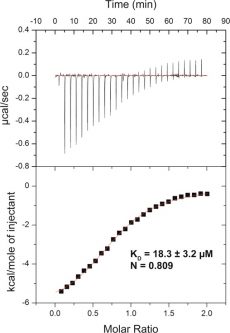
Compound 1 binds SETD8 with a KD of 18.3 ± 3.2 μM (n = 3) in ITC studies.J Med Chem.2014 Aug 14;57(15):6822-33. |
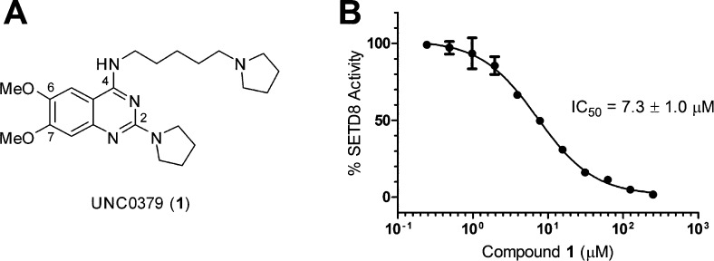
Compound1was identified as an inhibitor of SETD8 by cross-screening a quinazoline-based inhibitor set. |
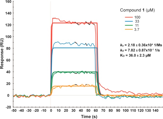
Compound1exhibits rapid on and off rates in SPR studies. |
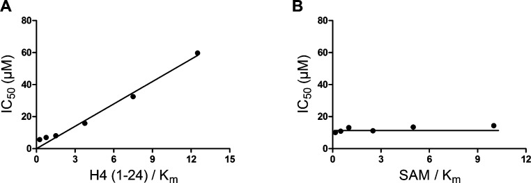
MOA studies of compound1.J Med Chem.2014 Aug 14;57(15):6822-33. |
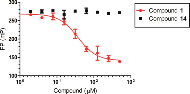
Peptide displacement assay.J Med Chem.2014 Aug 14;57(15):6822-33. |
Products are for research use only; We do not sell to patients
Copyright 2020 InvivoChem LLC | All Rights Reserved


 COA
COA