| Size | Price | Stock | Qty |
|---|---|---|---|
| 2mg |
|
||
| 5mg |
|
||
| 10mg |
|
||
| 25mg |
|
||
| 50mg |
|
||
| 100mg |
|
||
| 250mg |
|
||
| 500mg | |||
| Other Sizes |


Purity: ≥98%
| Targets |
MEK (IC50 = 3.2 nM)
Mitogen-activated protein kinase kinase 1 (MEK1) and MEK2, serine/threonine kinases in the MAPK pathway. For TAK-733, literature [1] reported: MEK1 (IC50 = 0.7 nM), MEK2 (IC50 = 0.9 nM) via HTRF kinase assay; Ki = 0.3 nM (MEK1), Ki = 0.5 nM (MEK2) via equilibrium binding assay. It showed no inhibition of 40 other kinases (e.g., ERK1, JNK, p38, PI3K) at 1 μM, confirming MEK1/2 selectivity [1] |
|---|---|
| ln Vitro |
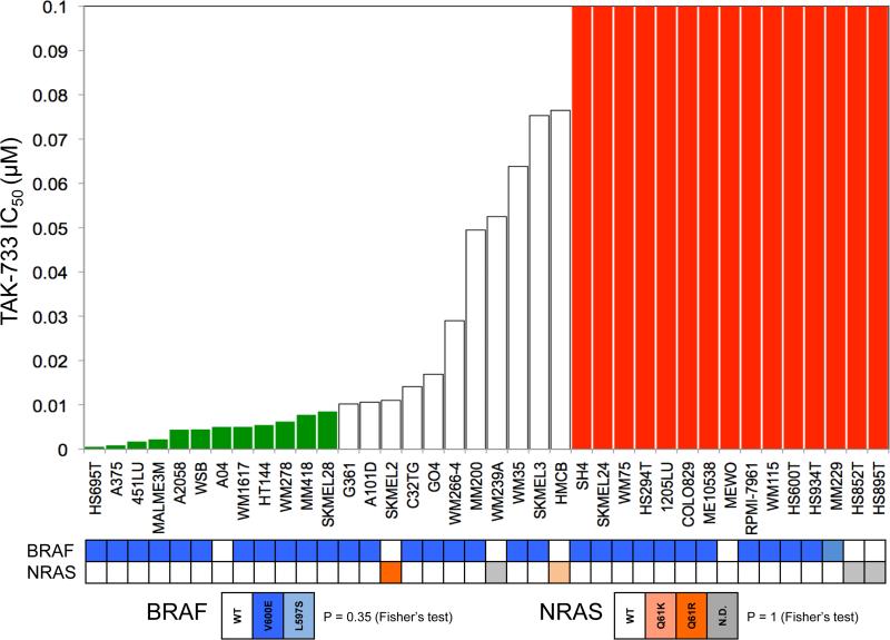
TAK-733 exhibits potent enzymatic and cellular activity, with an IC50 of 3.2 nM against constitutively active MEK enzyme and an EC50 of 1.9 nM against ERK phosphorylation in cells. Other kinases, receptors, and ion channels tested with inhibitor concentrations up to 10 μM are unaffected by TAK-733. TAK-733 exhibits high permeability and high microsomal stability across species, and it is found to moderately bind plasma protein (about 97% for humans and 96% for mice). Up to 30 μM, it doesn't inhibit P450s[1]. TAK-733 exhibits widespread activity in the majority of melanoma cell lines, with relative resistance noted at IC50 > 0.1 μM in vitro. For 72 hours, 34 different melanoma cell lines are incubated with progressively higher doses of TAK-733. Seven of the 34 cell lines are wild-type, while 27 have the BRAFV600E mutation. SRB proliferation assays were carried out, and the IC50 concentrations obtained allowed cell lines to be divided into three groups: relatively resistant, intermediate, and highly sensitive. Based on an IC50 that differs by at least 10 fold[2,] relatively resistant and highly sensitive lines are assigned.
Enzymatic & MAPK Pathway Inhibition: TAK-733 (0.001 nM–100 nM) dose-dependently inhibited recombinant MEK1/2 and blocked EGF-induced ERK phosphorylation in A375 (melanoma) cells: 50% p-ERK reduction at 0.1 nM (Western blot, 1 h) [1]. In COS-7 cells, 1 nM TAK-733 reduced serum-induced p-ERK by 95% (2 h) without affecting MEK protein expression [1] - Melanoma Cell Proliferation: In BRAF V600E-mutant (A375, SK-MEL-28) and NRAS-mutant (WM1366) melanoma cells, TAK-733 (0.001 μM–10 μM) inhibited proliferation with IC50 = 0.01 μM (A375), 0.02 μM (SK-MEL-28), 0.03 μM (WM1366) (MTT assay, 72 h) [2]. It induced apoptosis in A375 cells: Annexin V staining showed 40% apoptotic cells at 0.1 μM (48 h) and increased cleaved caspase-3 (3.0-fold) [2] - Patient-Derived Tumor Explants (PDX) In Vitro: In 8 melanoma PDX explant cultures (4 BRAF-mutant, 4 NRAS-mutant), TAK-733 (0.05 μM–0.5 μM) reduced cell viability by 55%–70% (CCK-8 assay, 72 h) and downregulated Ki-67 (45%–60% reduction, immunocytochemistry) [2] |
| ln Vivo |
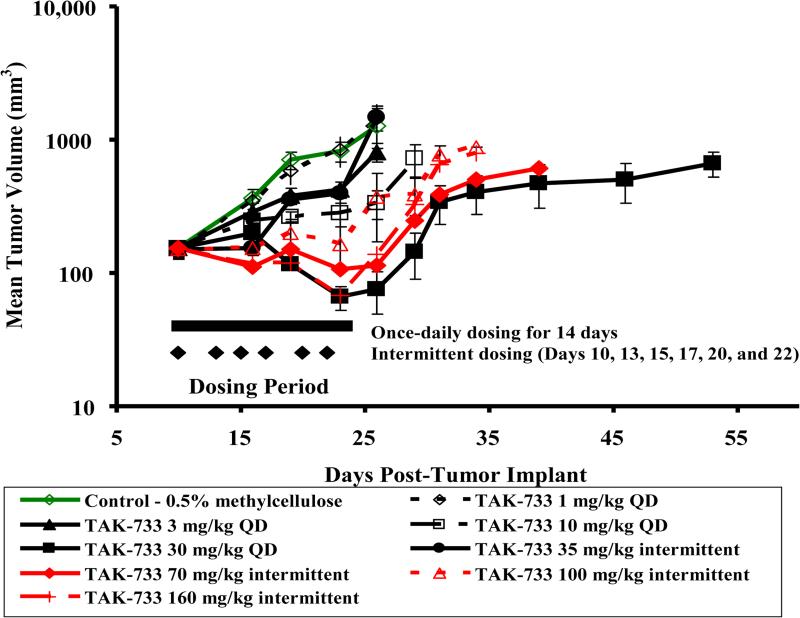
TAK-733's pharmacokinetics are examined in naked mice, rats, dogs, and monkeys. All species exhibit low clearance and high oral bioavailability. In mouse xenograft models of human cancer, including melanoma, colorectal, NSCLC, pancreatic, and breast cancer models, TAK-733 exhibits broad antitumor activity[1]. In the mice (5/group), implanted with A375 cells, daily oral administration of 1, 3, 10, and 30 mg/kg of TAK-733 for 14 days (Days 10 to 23) results in a delay in tumor growth. On an intermittent dosing schedule of 3 days per week for 2 weeks (Days 10, 13, 15, 17, 20, and 22), TAK-733 (35, 70, 100, and 160 mg/kg) also significantly inhibits tumor growth. In mice given 30 mg/kg of TAK-733 daily and mice given 160 mg/kg of TAK-733 intermittently, three partial regressions (PR) with a 60% response rate were seen. Mice that were given intermittent doses of 70, 100, and 160 mg/kg of TAK-733 also showed responses, complete regressions (CR), and partial regressions (PR). The tumor regression rate is more pronounced with the intermittent administration regimen; the largest reduction in tumor volume is seen at 160 mg/kg (57.29%), compared to a maximum reduction of 46.97% at 30 mg/kg once daily. In mice given 3, 10, and 30 mg/kg once daily or 35, 70, 100, and 160 mg/kg intermittently, tumor growth is significantly (p<0.05 for %T/C, Student's t-test) inhibited by the final day of administration[2].
Melanoma Xenograft Model: Female nude mice (6 weeks old) bearing A375 xenografts were randomized into 3 groups (n=8/group): vehicle (0.5% methylcellulose + 0.1% Tween 80), TAK-733 1 mg/kg, TAK-733 3 mg/kg. Drugs were administered orally once daily for 21 days. Tumor volume reduction: 60% (1 mg/kg), 85% (3 mg/kg) vs. vehicle; tumor weight decreased by 55% (1 mg/kg) vs. 75% (3 mg/kg). Immunohistochemistry showed p-ERK reduction by 80% (3 mg/kg) [1] - PDX Xenograft Model: Female NSG mice (7 weeks old) implanted with BRAF-mutant melanoma PDX tissue were treated with TAK-733 3 mg/kg (oral, once daily) for 28 days. Tumor volume reduced by 70%, and serum S100B (melanoma marker) decreased from 45 ng/mL to 15 ng/mL [2] |
| Enzyme Assay |
K-733 is highly potent and selective MEK allosteric site inhibitor with IC50 of 3.2 nM. TAK-733 shows potent enzymatic and cell activity with an EC50 of 1.9 nM against ERK phosphorylation in cells.
MEK1/2 HTRF Kinase Assay (Literature [1]): Recombinant human MEK1 (residues 44–313) or MEK2 (residues 38–326) was incubated with biotinylated peptide substrate (MEK1: RRRVSYRRR, MEK2: RRRLSYRRR, 20 μM), Eu-labeled anti-phospho-peptide antibody, and ATP (10 μM) in kinase buffer (25 mM Tris-HCl pH 7.5, 10 mM MgCl₂, 1 mM DTT). Serial dilutions of TAK-733 (0.001 nM–100 nM) were added, incubated at 30°C for 60 min. Time-resolved fluorescence (excitation 340 nm, emission 620 nm) was measured, and IC50 values were calculated via four-parameter logistic regression [1] - MEK Binding Assay (Literature [1]): Recombinant MEK1/2 was incubated with TAK-733 (0.001 nM–100 nM) in binding buffer (25 mM Tris-HCl pH 7.5, 150 mM NaCl) at 37°C for 24 h. Equilibrium dialysis was performed to separate free and bound drug; free drug concentration was quantified via HPLC, and Ki values were derived [1] |
| Cell Assay |
The cutaneous melanoma cell lines are transferred to 96 well flat bottom plates with lids while they are still in the logarithmic growth phase. Prior to being exposed to TAK-733 at increasing concentrations (10, 20, 30, 40, 50, 60, 70, 80, 90, and 100 nM) for 72 hours, 100 μL cell suspensions containing 2000–3000 viable cells are plated into each well. After the medication has been administered, the media is removed, and cells are fixed for 30 minutes at 4°C in cold 10% trichloroacetic acid. Following a water wash, the cells are stained for 30 min at room temperature with 0.4% SRB and then again washed with 1% acetic acid. Finally, the stain is solubilized for 30 min at room temperature with 10 mM tris. A plate reader is used to measure the absorbance at 565 nm. The raw absorbance (OD) data are used to create cell proliferation curves. Version 5.00 of GraphPad Prism is used for statistical analysis and data visualization[2].
Melanoma Cell Proliferation & Apoptosis Assay (Literature [2]): A375/SK-MEL-28/WM1366 cells were seeded in 96-well plates (5×10³ cells/well) and treated with TAK-733 (0.001 μM–10 μM) for 72 h. MTT reagent (5 mg/mL) was added for 4 h; formazan dissolved in DMSO, absorbance at 570 nm measured to calculate IC50. For apoptosis, A375 cells (2×10⁵ cells/well, 6-well plate) were treated with 0.1 μM drug for 48 h, stained with Annexin V-FITC/PI, analyzed via flow cytometry. Western blot probed anti-p-ERK, anti-cleaved caspase-3, anti-GAPDH [2] - PDX Explant Assay (Literature [2]): Melanoma PDX tissues were minced into 1 mm³ fragments, cultured in 24-well plates with TAK-733 (0.05 μM–0.5 μM) for 72 h. CCK-8 assay measured viability; explants were fixed, sectioned, and immunostained for Ki-67 to assess proliferation [2] |
| Animal Protocol |
Mice: Five to six-week-old female athymic nude mice are used. Using Trypsin-EDTA, cells from mid-log phase cultures are harvested to create A375 human melanoma xenograft tumors. The right flank of 6–8-week-old mice is injected subcutaneously with approximately 5×10⁶ cells suspended in Hanks' balanced salt solution (HBSS). When all of the mice in an experiment have tumors that are between 100 and 200 mm3 in size for antitumor efficacy studies and between 300 and 500 mm3 in size for pharmacodynamic (PD) studies, TAK-733 (1 mg/kg or 10 mg/kg) is started orally[2].
A375 Xenograft Protocol (Literature [1]): Female nude mice (6 weeks old) were subcutaneously implanted with 5×10⁶ A375 cells. When tumors reached ~100 mm³, TAK-733 was dissolved in 0.5% methylcellulose + 0.1% Tween 80, administered orally once daily (1 mg/kg or 3 mg/kg) for 21 days. Tumor volume (length×width²/2) measured every 3 days; mice euthanized on day 21, tumors processed for p-ERK immunohistochemistry [1] - PDX Xenograft Protocol (Literature [2]): Female NSG mice (7 weeks old) were subcutaneously implanted with 2 mm³ melanoma PDX fragments. When tumors reached ~150 mm³, TAK-733 (3 mg/kg, dissolved in 0.5% hydroxypropyl methylcellulose) was administered orally once daily for 28 days. Serum S100B was measured weekly via ELISA; tumor volume recorded every 3 days [2] |
| ADME/Pharmacokinetics |
Rat pharmacokinetics (Reference [1]): Male Sprague-Dawley rats (8 weeks old) were orally administered TAK-733 3 mg/kg: oral bioavailability = 65%, Cmax = 3.1 μM, Tmax = 1.0 h, terminal half-life t₁/₂ = 6.8 h. Intravenous injection of 1 mg/kg: CL = 7.2 mL/min/kg, Vss = 0.9 L/kg [1] - Human plasma protein binding: 99% (equilibrium dialysis, [1])
|
| Toxicity/Toxicokinetics |
In vitro cytotoxicity: In normal human peripheral blood mononuclear cells (PBMCs) and foreskin fibroblasts, the cell viability of TAK-733 (at a concentration up to 10 μM, treated for 72 hours) was >85%, indicating low non-specific toxicity [1][2]
- Acute in vivo toxicity: After treatment with 3 mg/kg TAK-733 (orally, for 28 days), rats did not show significant weight loss, lethargy, or abnormalities in serum ALT/AST/creatinine. Histological examination of the liver and kidneys showed no inflammation or necrosis [1] |
| References |
|
| Additional Infomation |
TAK-733 belongs to the pyridopyrimidine class of compounds, with the structure pyrido[2,3-d]pyrimidine-4,7(3H,8H)-dione, substituted at positions 3, 5, 6, and 8 with (2R)-2,3-dihydroxypropyl, (2-fluoro-4-iodophenyl)amino, fluorine, and methyl, respectively. It is a non-ATP-competitive MEK1/2 allosteric inhibitor. TAK-733 possesses various pharmacological activities, including as an EC 2.7.11.24 (mitogen-activated protein kinase) inhibitor, an apoptosis inducer, an antitumor agent, and an anti-inflammatory agent. It is an organic iodine compound, pyridopyrimidine, monofluorobenzene compound, substituted aniline, diol, secondary alcohol, primary alcohol, and secondary amino compound. TAK-733 has been used in clinical trials to investigate the treatment of advanced metastatic melanoma, advanced non-hematologic malignancies, and advanced non-hematologic malignancies.
MEK inhibitor REC-4881 is an orally bioavailable, non-ATP-competitive, allosteric small molecule mitogen-activated protein kinase kinase (MAP2K; MAPK/ERK kinase; MEK) 1 and MEK2 inhibitor with potential antitumor activity. After oral administration, REC-4881 selectively binds to and inhibits the activity of MEK1/2. This prevents the activation of MEK1/2-dependent effector proteins and transcription factors, thereby inhibiting growth factor-mediated cell signaling and tumor cell proliferation. MEK1/2 (MAP2K1/K2) are bispecific threonine/tyrosine kinases that play a crucial role in the activation of the RAS/RAF/MEK/ERK pathway and are frequently upregulated in various tumor cell types. TAK-733 is a potent, selective MEK1/2 allosteric inhibitor developed specifically for the treatment of MAPK-driven cancers, such as BRAF/NRAS mutant melanoma [1][2] - Its mechanism of action involves binding to the allosteric sites of MEK1/2 (non-ATP competitive), stabilizing its inactive conformation, and blocking ERK phosphorylation, which is essential for the proliferation and survival of melanoma cells [1][2] - It has shown potent activity in melanoma PDX models, including BRAF and NRAS mutants, supporting its potential for treating a variety of melanoma subtypes [2] |
| Molecular Formula |
C17H15F2IN4O4
|
|
|---|---|---|
| Molecular Weight |
504.23
|
|
| Exact Mass |
504.01
|
|
| Elemental Analysis |
C, 40.49; H, 3.00; F, 7.54; I, 25.17; N, 11.11; O, 12.69
|
|
| CAS # |
1035555-63-5
|
|
| Related CAS # |
|
|
| PubChem CID |
24963252
|
|
| Appearance |
White to off-white solid powder
|
|
| Density |
1.9±0.1 g/cm3
|
|
| Boiling Point |
530.5±60.0 °C at 760 mmHg
|
|
| Flash Point |
274.6±32.9 °C
|
|
| Vapour Pressure |
0.0±1.5 mmHg at 25°C
|
|
| Index of Refraction |
1.710
|
|
| LogP |
0.28
|
|
| Hydrogen Bond Donor Count |
3
|
|
| Hydrogen Bond Acceptor Count |
8
|
|
| Rotatable Bond Count |
5
|
|
| Heavy Atom Count |
28
|
|
| Complexity |
772
|
|
| Defined Atom Stereocenter Count |
1
|
|
| SMILES |
IC1C([H])=C([H])C(=C(C=1[H])F)N([H])C1=C(C(N(C([H])([H])[H])C2=C1C(N(C([H])=N2)C([H])([H])[C@]([H])(C([H])([H])O[H])O[H])=O)=O)F
|
|
| InChi Key |
RCLQNICOARASSR-SECBINFHSA-N
|
|
| InChi Code |
InChI=1S/C17H15F2IN4O4/c1-23-15-12(16(27)24(7-21-15)5-9(26)6-25)14(13(19)17(23)28)22-11-3-2-8(20)4-10(11)18/h2-4,7,9,22,25-26H,5-6H2,1H3/t9-/m1/s1
|
|
| Chemical Name |
3-[(2R)-2,3-dihydroxypropyl]-6-fluoro-5-(2-fluoro-4-iodoanilino)-8-methylpyrido[2,3-d]pyrimidine-4,7-dione
|
|
| Synonyms |
TAK 733; TAK733; TAK-733
|
|
| HS Tariff Code |
2934.99.9001
|
|
| Storage |
Powder -20°C 3 years 4°C 2 years In solvent -80°C 6 months -20°C 1 month |
|
| Shipping Condition |
Room temperature (This product is stable at ambient temperature for a few days during ordinary shipping and time spent in Customs)
|
| Solubility (In Vitro) |
DMSO: ~101 mg/mL (~200.3 mM)
Water: 1 mg/mL (Insoluble) Ethanol: 1 mg/mL |
|---|---|
| Solubility (In Vivo) |
Solubility in Formulation 1: ≥ 2.5 mg/mL (4.96 mM) (saturation unknown) in 10% DMSO + 90% Corn Oil (add these co-solvents sequentially from left to right, and one by one), clear solution.
For example, if 1 mL of working solution is to be prepared, you can add 100 μL of 25.0 mg/mL clear DMSO stock solution to 900 μL of corn oil and mix evenly. (Please use freshly prepared in vivo formulations for optimal results.) |
| Preparing Stock Solutions | 1 mg | 5 mg | 10 mg | |
| 1 mM | 1.9832 mL | 9.9161 mL | 19.8322 mL | |
| 5 mM | 0.3966 mL | 1.9832 mL | 3.9664 mL | |
| 10 mM | 0.1983 mL | 0.9916 mL | 1.9832 mL |
*Note: Please select an appropriate solvent for the preparation of stock solution based on your experiment needs. For most products, DMSO can be used for preparing stock solutions (e.g. 5 mM, 10 mM, or 20 mM concentration); some products with high aqueous solubility may be dissolved in water directly. Solubility information is available at the above Solubility Data section. Once the stock solution is prepared, aliquot it to routine usage volumes and store at -20°C or -80°C. Avoid repeated freeze and thaw cycles.
Calculation results
Working concentration: mg/mL;
Method for preparing DMSO stock solution: mg drug pre-dissolved in μL DMSO (stock solution concentration mg/mL). Please contact us first if the concentration exceeds the DMSO solubility of the batch of drug.
Method for preparing in vivo formulation::Take μL DMSO stock solution, next add μL PEG300, mix and clarify, next addμL Tween 80, mix and clarify, next add μL ddH2O,mix and clarify.
(1) Please be sure that the solution is clear before the addition of next solvent. Dissolution methods like vortex, ultrasound or warming and heat may be used to aid dissolving.
(2) Be sure to add the solvent(s) in order.
| NCT Number | Recruitment | interventions | Conditions | Sponsor/Collaborators | Start Date | Phases |
| NCT00948467 | Completed | Drug: TAK-733 | Advanced Metastatic Melanoma Advanced Non-hematologic Malignancies |
Millennium Pharmaceuticals, Inc. |
December 2009 | Phase 1 |
| NCT01613261 | Withdrawn | Drug: TAK-733 and alisertib | Advanced Nonhematologic Malignancies |
Millennium Pharmaceuticals, Inc. |
August 2013 | Phase 1 |
|
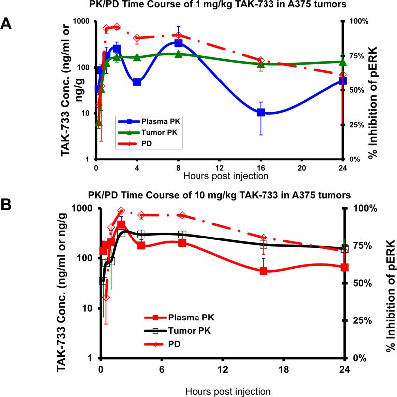
Pharmacokinetic and pharmacodynamic effects of TAK-733 in A375 tumor-bearing mice following oral administration of (A) 1 mg/kg or (B) 10 mg/kg dose. A375 tumor cells were implanted subcutaneously in the flank of nu/nu mice (3/time point). Administration was initiated when all mice had tumors ranging in size from 300 to 500 mm3(Day 10). TAK-733 was given as a single oral dose.Mol Cancer Ther.2015 Feb;14(2):317-25. |
Effects of TAK-733 on growth of A375 melanoma tumor xenografts in athymic nude mice. A375 melanoma cells were implanted subcutaneously in the flank of nu/nu mice (5/group). Administration was initiated 10 days after the tumor implant, when all mice had tumors ranging in size from 80 to 225 mm3(Day 10). TAK-733 was given orally daily for 14 days (QD 1-14) or 3 days per week for 2 cycles (M, TH, SAT ×2).Mol Cancer Ther.2015 Feb;14(2):317-25. |
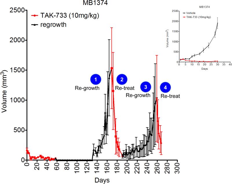
Tumor regrowth kinetics for PDTX model MB1374 administered with vehicle or TAK-733 (10 mg/kg). Inset, original tumor growth inhibition study for 30 days. Red lines indicate TAK-733 administration times, black lines indicate cessation of TAK-733 for tumor regrowth periods.Mol Cancer Ther.2015 Feb;14(2):317-25. |
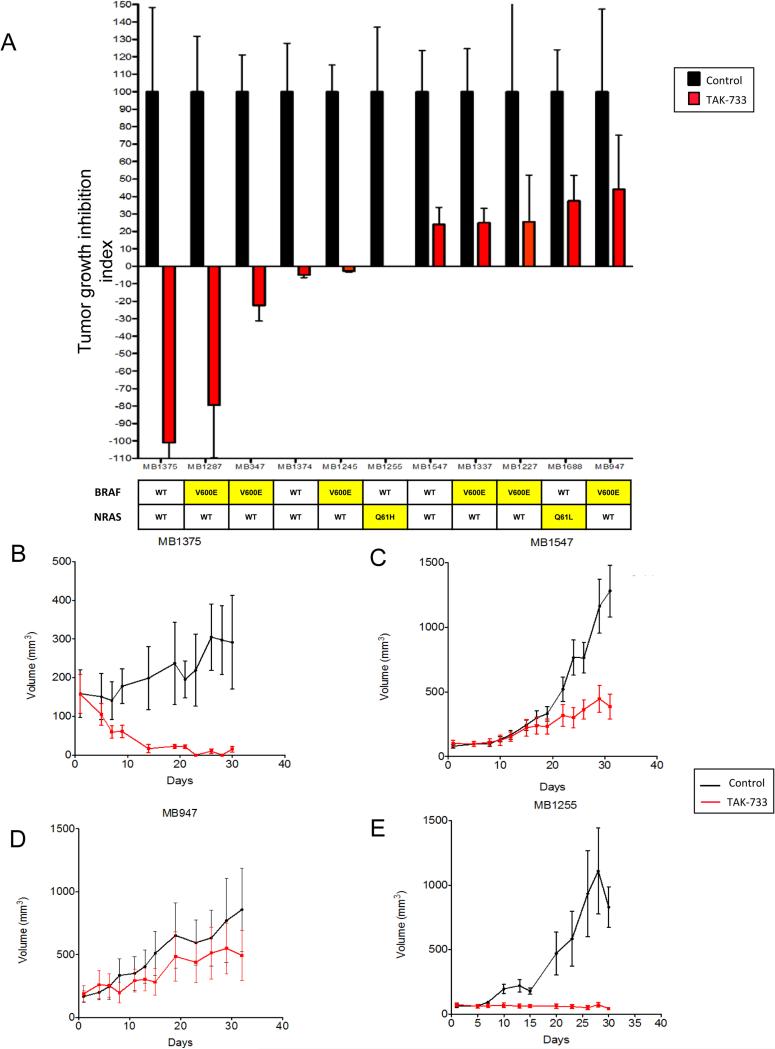
Antiproliferative effects of TAK-733 against a panel of human cutaneous melanoma cell lines. Cell lines were treated with increasing doses of TAK-733 for 72 hours and proliferation was assessed using the SRB assay. IC50values are depicted. BRAF and NRAS mutational status in depicted in the table below.Mol Cancer Ther.2015 Feb;14(2):317-25. |
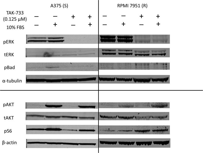
Immunoblot analysis of downstream effectors upon administration of TAK-733. A375 and RPMI 7951 cell lines were serum starved overnight and then administered TAK-733 for 1 hr. Cells were then challenged with 10% FBS for 30 mins, harvested and immunoblotted with the indicated antibodies.Mol Cancer Ther.2015 Feb;14(2):317-25. |
Products are for research use only; We do not sell to patients
Copyright 2020 InvivoChem LLC | All Rights Reserved


 COA
COA