| Size | Price | Stock | Qty |
|---|---|---|---|
| 50mg |
|
||
| 100mg |
|
||
| 250mg |
|
||
| 500mg |
|
||
| 1g |
|
||
| 2g |
|
||
| Other Sizes |


Purity: ≥98%
| Targets |
MEK2; MEK1 (IC50 = 14 nM); MEK (IC50 = 12 nM)
Mitogen-activated protein kinase kinase 1 (MEK1) and MEK2, serine/threonine kinases in the MAPK pathway. For Selumetinib (AZD-6244; ARRY142886), the IC50 values were: MEK1 = 14 nM, MEK2 = 11 nM (radioactive kinase assay) [1] - Consistent with [1], MEK1 (IC50 = 12 nM) and MEK2 (IC50 = 9 nM) via HTRF assay; no significant inhibition of 28 other kinases (e.g., ERK1, JNK, p38) at 1 μM [3] - MEK1 (Ki = 0.7 nM) and MEK2 (Ki = 0.9 nM) via equilibrium binding assay; selective for MEK over Raf, ERK, and PI3K family kinases [5] |
|---|---|
| ln Vitro |
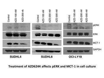
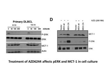
Selumetinib is not ATP-competitive and inhibits ERK1/2 phosphorylation at IC50 levels under 40 nM. Through the inhibition of ERK1/2 and p90RSK phosphorylation, as well as the elevation of caspase-3 and caspase-7 cleavage and cleaved poly(ADP)ribose polymerase, AZD6244 also slows the growth of primary HCC cells. The p38, c-Jun-NH2-kinase, phosphatidylinositol 3-kinase, and MEK5/ERK5 pathways are not significantly impacted by AZD6244. [1] Raf mutations in breast cancer cell lines and Ras mutations in NSCLC cell lines are both responsive to AZD6244.[2]
Melanoma Cells: In BRAF-mutant (A375, SK-MEL-28) and NRAS-mutant (WM1366) melanoma cells, Selumetinib (0.1 μM–10 μM) inhibited proliferation with IC50 = 0.3 μM (A375), 0.5 μM (SK-MEL-28), 0.8 μM (WM1366) (MTT assay, 72 h). Western blot showed p-ERK reduction by 70% (A375, 1 μM, 2 h); flow cytometry revealed 35% apoptotic cells (A375, 2 μM, 48 h) [1] - Colorectal Cancer Cells: In KRAS-mutant (HT-29, SW480) cells, Selumetinib (0.5 μM–20 μM) reduced viability by 50% at 1.2 μM (HT-29) and 1.8 μM (SW480) (CCK-8 assay, 72 h). qRT-PCR showed downregulated cyclin D1 (45% reduction, HT-29, 1 μM, 24 h); clone formation was inhibited by 60% (SW480, 1 μM, 14 days) [3] - Non-Small Cell Lung Cancer (NSCLC) Cells: In EGFR-mutant (H1975) and KRAS-mutant (A549) cells, Selumetinib (0.2 μM–15 μM) had IC50 = 0.6 μM (H1975) and 0.9 μM (A549) (MTT assay). Western blot detected reduced p-ERK (80% reduction, H1975, 1 μM) and increased cleaved caspase-3 (2.5-fold, A549, 2 μM) [5] - Thyroid Cancer Cells: In RET/PTC-rearranged (TPC-1) cells, Selumetinib (0.1 μM–10 μM) inhibited proliferation (IC50 = 0.4 μM) and reduced p-ERK (75% reduction, 1 μM) via Western blot [7] |
| ln Vivo |
Selumetinib significantly inhibits phosphorylation of ERK1/2 in 2-1318, 5-1318, 26-1004 and 4-1318 xenografts and induces apoptosis in primary 2-1318 cells by activating the caspase pathway. At a dose of 100 mg/kg, AZD6244 could slow the growth of the tumor in the HT-29 xenograft, a colorectal tumor model with a B-Raf mutation; this tumor growth inhibition is superior to that of Gemcitabine. Apoptosis and the down-regulation of cell cycle regulators like cyclin D1, Cdc-2, CDK2 and 4, cyclin B1, and c-Myc are associated with increased apoptosis, which is why AZD6244 could inhibit the growth of HCC xenograft tumors in the absence of these factors.
Melanoma Xenograft Model: Female nude mice (6 weeks old) bearing A375 xenografts were randomized into 3 groups (n=8/group): vehicle (0.5% methylcellulose + 0.1% Tween 80), Selumetinib 25 mg/kg, 50 mg/kg. Drugs were administered orally twice daily for 21 days. Tumor volume was reduced by 45% (25 mg/kg) and 70% (50 mg/kg) vs. vehicle; tumor weight decreased by 40% (25 mg/kg) and 65% (50 mg/kg). Immunohistochemistry showed reduced p-ERK (65% reduction) and Ki-67 (55% reduction) at 50 mg/kg [1] - Hepatocellular Carcinoma (HCC) Model: Male NOD/SCID mice (8 weeks old) with HepG2 xenografts were treated with Selumetinib (30 mg/kg, oral, once daily) or vehicle for 28 days. Tumor volume was reduced by 55%, and serum AFP (tumor marker) decreased from 800 ng/mL to 320 ng/mL. Western blot of tumor tissues showed reduced p-ERK (70% reduction) [11] - Colorectal Cancer Xenograft Model: Male nude mice (7 weeks old) bearing HT-29 xenografts were treated with Selumetinib (40 mg/kg, oral, twice daily) for 21 days. Tumor growth inhibition rate was 60%, and survival was prolonged by 35% vs. vehicle. Tumor tissues had reduced cyclin D1 (50% reduction) via qRT-PCR [12] - NSCLC Model: Female nude mice (6 weeks old) with A549 xenografts were treated with Selumetinib (35 mg/kg, intraperitoneal, once daily) for 14 days. Tumor volume was reduced by 50%, and apoptotic cells (TUNEL staining) increased from 5% to 30% [5] |
| Enzyme Assay |
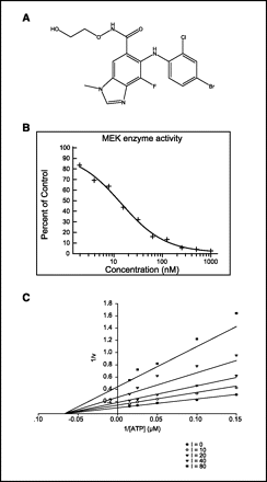
MEK1. [3]
NH2-terminal hexahistidine tagged, constitutively active MEK1 (S218D, S222D ΔR4F; ref. 18) was expressed in baculovirus-infected Hi5 insect cells and purified by immobilized metal affinity chromatography, ion exchange, and gel filtration. The activity of MEK1 was assessed by measuring the incorporation of [γ-33P]phosphate from [γ-33P]ATP onto ERK2. The assay was carried out in a 96-well polypropylene plate with an incubation mixture (100 μL) composed of 25 mmol/L HEPES (pH 7.4), 10 mmol/L MgCl2, 5 mmol/L β-glycerolphosphate, 100 μmol/L sodium orthovanadate, 5 mmol/L DTT, 5 nmol/L MEK1, 1 μmol/L ERK2, and 0 to 80 nmol/L compound (final concentration of 1% DMSO). The reactions were initiated by the addition of 10 μmol/L ATP (with 0.5 μC k[γ-33P]ATP/well) and incubated at room temperature for 45 min. An equal volume of 25% trichloracetic acid was added to stop the reaction and precipitate the proteins. Precipitated proteins were trapped onto glass fiber B filter plates, excess labeled ATP was washed off with 0.5% phosphoric acid, and radioactivity was counted in a liquid scintillation counter. ATP dependence was determined by varying the amount of ATP in the reaction mixture. The data were globally fitted using SigmaPlot. Values were calculated using the following equation for noncompetitive inhibition: v = [Vmax × S / (1 + I / Ki)] / (Km + S).[3] ERK2. [3] To measure inhibition of ERK2, the kinase activity of ERK2 was first activated by MEK1. Wild-type (WT) ERK2 containing an NH2-terminal hexahistidine tag was overexpressed in Escherichia coli and purified by immobilized metal affinity chromatography, ion exchange, and gel filtration. To activate WT ERK2, 2 mg WT ERK2 was mixed with 17 μg of constitutively active MEK1 in 4 mL of 25 mmol/L HEPES (pH 7.5) containing 1 mmol/L ATP. The reaction mixture was incubated at room temperature for 40 min, and the addition of two phosphates was confirmed by mass spectrometry. Activated WT ERK2 was further purified by ion exchange. ERK2 activity was assayed as described for constitutively active MEK, using 10 nmol/L activated ERK2. The substrate used was myelin basic protein at a concentration of 1 μmol/L.[3] MEK1 molecules are immunoprecipitated using an anti-MEK1 antibody. When recombinant ERK1 is activated by immuno-isolated MEK1 in a coupled assay with MBP as the end point, MEK kinase activity is calculated. Before being exposed to X-ray film, phosphorylated MBP is resolved on a 14% SDS-PAGE gel and vacuum-dried. MEK1/2 Radioactive Kinase Assay: Recombinant human MEK1 (residues 3–321) or MEK2 (residues 4–317) was incubated with [γ-³²P]-ATP (10 μM, 3000 Ci/mmol), ERK2 (substrate kinase), and myelin basic protein (MBP, substrate) in assay buffer (25 mM Tris-HCl pH 7.5, 10 mM MgCl₂, 1 mM DTT). Serial dilutions of Selumetinib (0.1 nM–100 nM) were added, and the mixture was incubated at 30°C for 30 minutes. Phosphorylated MBP was separated via SDS-PAGE, and radioactivity was quantified via autoradiography. IC50 values were calculated via four-parameter regression [1] - MEK1/2 HTRF Binding Assay: Recombinant MEK1/2 was incubated with Eu-labeled anti-MEK antibody and biotinylated ATP-analog in buffer (50 mM Tris-HCl pH 8.0, 10 mM MgCl₂). Serial dilutions of Selumetinib (0.1 nM–100 nM) were added, and the mixture was incubated at 25°C for 60 minutes. Time-resolved fluorescence (excitation 340 nm, emission 620 nm) was measured, and Ki values were derived [3] |
| Cell Assay |
Cell Viability and Cell Proliferation[1]
Primary HCC cells were plated at a density of 2.0 × 10⁴ per well in growth medium. After 48 h in growth medium, the cell monolayer was rinsed twice with MEM. Cells were treated with various concentrations of AZD6244 (0, 0.5, 1.0, 2.0, 3.0, and 4.0 μmol/L) for 24 or 48 h. Cell viability was determined by the 3-(4,5-dimethylthiazol-2y1)-2,5-diphenyltetrazolium bromide (MTT) assay (32). Cell proliferation was assayed using a bromodeoxyuridine kit (Roche) as described by the manufacturer. Experiments were repeated at least thrice, and the data were expressed as mean ± SE.[1] Detection of Apoptosis[1] Primary HCC cells were grown in eight-chamber slides and treated with 0, 0.5, 1.0, 2.0, 3.0, and 4.0 μmol/L of AZD6244 in SRF medium for 24 h. Cells were fixed with PBS containing 4% formalin solution for 1 h at room temperature and washed with PBS. Apoptosis was detected by the terminal deoxynucleotidyl transferase–mediated dUTP nick-end labeling (TUNEL) assay using the In situ Cell Death Detection kit (Roche) as described by the manufacturer. Apoptotic cells were then visualized under a fluorescent microscope equipped with a FITC filter. The labeling index was obtained by counting the number of positive cells among 500 cells per region. They were expressed as percentage values.[1] At a density of2.0 × 104, cells are seeded. The cells undergo two culture media rinses after 48 hours of incubation. AZD6244 is used to treat cells for 24 or 48 hours at various concentrations. The MTT assay uses 3-(4,5-dimethylthiazol-2y1)-2,5-diphenyltetrazolium bromide to measure the viability of cells. With the help of a bromodeoxyuridine kit, cell proliferation is measured. Antiproliferation Assay (MTT): A375/SK-MEL-28 cells were seeded in 96-well plates (5×10³ cells/well) and attached overnight. Serial dilutions of Selumetinib (0.01 μM–10 μM) or vehicle (DMSO, 0.1%) were added, and cells were incubated at 37°C with 5% CO₂ for 72 hours. MTT reagent (5 mg/mL) was added (10 μL/well) for 4 hours; formazan was dissolved in DMSO, and absorbance at 570 nm was measured to calculate IC50 [1] - Apoptosis Assay (Annexin V-FITC/PI): A375 cells were seeded in 6-well plates (2×10⁵ cells/well) and treated with Selumetinib (2 μM) for 48 hours. Cells were harvested, stained with Annexin V-FITC and PI, and analyzed by flow cytometry. Apoptotic cells (Annexin V⁺/PI⁻ and Annexin V⁺/PI⁺) were quantified [1] - MAPK Pathway Western Blot: HT-29 cells were seeded in 6-well plates (3×10⁵ cells/well) and treated with Selumetinib (1 μM) for 2 hours. Cells were lysed in RIPA buffer, proteins separated by SDS-PAGE, and probed with anti-p-ERK, anti-ERK, anti-cleaved caspase-3, and anti-GAPDH antibodies [3] - Clone Formation Assay: SW480 cells were seeded in 6-well plates (1×10³ cells/well) and treated with Selumetinib (1 μM) or vehicle. After 14 days, colonies were fixed with methanol, stained with crystal violet, and counted. Inhibition rate = (1 – treated colonies/vehicle colonies) × 100% [3] |
| Animal Protocol |
HCC xenografts in mice homozygous for the SCID (severe combined immunodeficiency) mutation
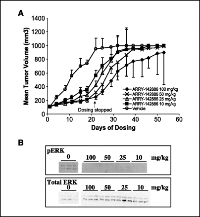
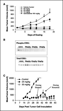
50 or 100mg/kg Administered via p.o. o investigate the effects of AZD6244 on HCC xenografts, AZD6244 was suspended in water at an appropriate concentration. Mice bearing HCC xenografts were p.o. given, twice a day, with either 100 μL of water (n = 12) or 50 mg (n = 12) or 100 mg (n = 12) of AZD6244 per kilogram of body weight for 21 days, starting from day 7 after tumor implantation. Growth of established tumor xenografts was monitored at least twice weekly by Vernier caliper measurement of the length (a) and width (b) of the tumor. Tumor volume was calculated as (a × b2)/2. Animals were sacrificed 3 h after the last dose of ADZ6244, and body and tumor weights were recorded, with the tumors harvested for analysis.[1] To study the effects of AZD6244 on caspase-3 activation and MEK1/2 phosphorylation, mice bearing HCC tumors (∼800 mm3) were treated with vehicle (n = 4) or 50 mg of AZD6244 per kilogram of body weight (n = 4) for 3 days as described above. Animals were sacrificed 3 h after the last dose, and tumors were harvested and frozen in liquid nitrogen for later analysis. Part of the tumor harvest was fixed in neutral buffer containing 10% formalin for immunohistochemistry.[1] HT-29 human colon carcinoma or BxPC3 human pancreatic tumor fragments were implanted s.c. in the flank of nude mice and allowed to grow to 100 to 150 mg. Mice (n = 10 per group) were randomized to treatment groups to receive vehicle (10 mL/kg and 10% ethanol/10% cremophor EL/80% D5W) or AZD6244/ARRY-142886 (10, 25, 50, or 100 mg/kg, oral, BID) on days 1 to 21. Tumors [(W2 × L) / L] were measured twice weekly. [3] A375 Melanoma Xenograft Protocol: Female nude mice (6 weeks old) were subcutaneously implanted with 5×10⁶ A375 cells. When tumors reached ~100 mm³, mice were grouped. Selumetinib was dissolved in 0.5% methylcellulose + 0.1% Tween 80, administered orally twice daily for 21 days (25 mg/kg or 50 mg/kg). Tumor volume (length × width² / 2) was measured every 3 days. On day 21, mice were euthanized; tumors were weighed and processed for p-ERK/Ki-67 immunohistochemistry [1] - HepG2 HCC Xenograft Protocol: Male NOD/SCID mice (8 weeks old) were subcutaneously injected with 1×10⁷ HepG2 cells. When tumors reached ~150 mm³, mice were treated with Selumetinib (30 mg/kg, dissolved in saline + 0.1% DMSO) or vehicle, administered orally once daily for 28 days. Serum AFP was measured weekly via ELISA; tumor tissues were collected for Western blot [11] - HT-29 Colorectal Cancer Protocol: Male nude mice (7 weeks old) were subcutaneously implanted with 4×10⁶ HT-29 cells. When tumors reached ~120 mm³, mice were treated with Selumetinib (40 mg/kg, dissolved in 0.5% hydroxypropyl methylcellulose) or vehicle, administered orally twice daily for 21 days. Survival was monitored daily; tumor tissues were used for cyclin D1 qRT-PCR [12] |
| ADME/Pharmacokinetics |
Absorption, Distribution and Excretion
Based on multiple studies of different doses of seletinib in children and adults, the time to peak concentration (Tmax) is typically between 1 and 1.5 hours. The average absolute oral bioavailability in healthy adults is reported to be 62%. Since food significantly reduces serum drug concentrations, celumitinib should be taken on an empty stomach. Approximately 59% of celumitinib is excreted in feces, and 33% in urine. The mean steady-state apparent volume of distribution (VOD) of celumitinib in pediatric patients ranges from 78 L to 171 L. A study in healthy adult men found a mean VOD of 146 L. Another study observing the pharmacokinetic effects of different doses and dosing regimens of celumitinib in selected Japanese patients found a steady-state VOD range of 73.2 L to 148.1 L. The clearance rate of celumitinib in pediatric patients is 8.8 L/hr. A study in healthy adult men found a clearance rate of 15.7 L/hr. Another study in a subset of Japanese patients, observing the pharmacokinetic effects of celumitinib at different doses and dosing regimens, found clearance rates ranging from 9.2 to 15.9 L/hr. Metabolism/Metabolites Celumitinib is primarily metabolized in the liver via the following metabolic pathway: The acetamide functional group of celumitinib is hydrolyzed to generate a carboxylic acid-containing metabolite, M15 (AZ13326637). The parent compound undergoes ethylene glycol removal to generate the primary amide metabolite, M14 (AZ12791138). Amide hydrolysis converts M14 to M15. Glucuronization and further oxidation of M14 generate M2, M6, and M1. N-demethylation of M14 generates M12. Amide glucuronide (M2) is considered the major circulating metabolite. Cerumitinib is demethylated to generate the pharmacologically active metabolite M8 (AZ12442942), which is further oxidized to M11. M8 is glucuroninated to generate M3 or M5, and M8 undergoes partial ethylene glycol removal to generate the primary amide, M12. Although the N-demethylated metabolite (M8) accounts for less than 10% of the circulating metabolites, it contributes approximately 21-35% of the observed pharmacological activity. Riboside conjugation converts M12 to M9, and M12 is oxidized to generate metabolites M10 and M13. M10 is glucuroninated to generate M1. Cerumitinib is directly glucuroninated to generate M4 or M7, both of which can eventually be converted to the M3 and M5 metabolites. Biological Half-Life Cerumitinib is characterized by a short half-life. In pediatric patients, the elimination half-life at a dose of 25 mg/m² is 6.2 hours. In a study observing the pharmacokinetic effects of different celumitinib dosing regimens on some Japanese patients, the half-life ranged from 9.2 to 10.6 hours. In other studies, the half-life of celumitinib administered twice daily at 75 mg was approximately 13 hours. In male Sprague-Dawley rats, the oral bioavailability of celumitinib (25 mg/kg) was 48%, Cmax = 4.2 μM, Tmax = 1.5 h, t₁/₂ = 6.8 h[3] - The clearance (CL) of intravenously administered celumitinib (5 mg/kg) in rats was 8.5 mL/min/kg, and the steady-state volume of distribution (Vss) was 1.2 L/kg[3] - In healthy volunteers (n=12), the oral bioavailability of celumitinib (50 mg) after a single oral dose was 42%, with a Cmax of 1.8 μM, a Tmax of 2.0 h, t₁/₂ = 7.2 h[8] - Selumetinib is primarily metabolized by CYP3A4 in human liver microsomes; the urinary excretion of the original drug is <5%[13] - The human plasma protein binding rate of Selumetinib, as determined by balanced dialysis, is 97%[3] |
| Toxicity/Toxicokinetics |
Hepatotoxicity
In premarketing clinical trials in children and adults with neurofibromatosis, 35% of subjects experienced elevated serum transaminases, but only 4% had transaminase levels exceeding 5 times the upper limit of normal (ULN). However, no serious liver-related adverse events occurred, nor were there simultaneous elevations in serum transaminase and bilirubin levels. ALT elevations were generally mild and transient, and usually resolved spontaneously without dose adjustment. No clinically significant liver injury associated with selumetinib was reported. Since selumetinib's approval and widespread use, despite limited clinical experience with the drug, particularly long-term treatment, there have been no published reports of clinically significant drug-induced liver injury when used to treat neurofibromatosis. However, in studies of high-dose celumitinib in patients with advanced refractory cancer, abnormal liver function was more common, sometimes reaching severe levels (ALT exceeding 20 times the ULN), requiring discontinuation of the drug. Therefore, clinically significant liver injury is extremely rare in patients with neurofibromatosis, even with the recommended dose of celumitinib. However, at high doses, celumitinib is highly associated with an elevated incidence of serum enzymes, many of which suggest severe liver injury. Probability score: E (Suspected but unconfirmed rare clinically significant cause of liver injury). Protein Binding Independent studies of celumitinib protein binding found that 96% of celumitinib binds to serum albumin, while less than 35% binds to α-1 acid glycoprotein. Overall, approximately 98.4% of celumitinib binds to plasma proteins. In a 28-day repeated-dose study in male/female rats, oral selumetinib (up to 60 mg/kg/day) caused mild rash (15% of animals) but no significant changes in body weight, serum ALT/AST/creatinine or liver/kidney histology [3] - In beagle dogs (30 mg/kg/day, orally, 21 days), selumetinib caused mild diarrhea (2 out of 6 dogs) and decreased platelet count (15%), but no organ damage [8] - In a phase I clinical trial (n=45), common adverse events included rash (35%), diarrhea (28%) and fatigue (20%); no grade 4 toxicities were reported [13] - In healthy volunteers, no drug interactions were observed when selumetinib was used in combination with CYP3A4 inhibitors (e.g., ketoconazole) [13] |
| References |
[1]. Mol Cancer Ther . 2007 Jan;6(1):138-46. [2]. Mol Cancer Ther . 2010 Jul;9(7):1985-94. [3]. Clin Cancer Res . 2007 Mar 1;13(5):1576-83. [4]. Mol Cancer Ther . 2007 Sep;6(9):2468-76. [5]. Mol Cancer Ther . 2007 Aug;6(8):2209-19. [6]. Clin Cancer Res . 2012 Feb 15;18(4):1051-62. [7]. Int J Oncol . 2012 Aug;41(2):712-20. [8]. J Clin Endocrinol Metab . 2008 Jun;93(6):2194-201. [9]. Proc Natl Acad Sci U S A . 2009 Dec 1;106(48):20411-6. [10]. Cancer Res . 2008 Aug 1;68(15):6145-53. [11]. J Hepatol . 2010 Jan;52(1):79-87. |
| Additional Infomation |
Selumetinib belongs to the benzimidazole class of compounds, with the structure 1-methyl-1H-benzimidazole, substituted at positions 4, 5, and 6 with fluorine, (4-bromo-2-chlorophenyl)amino, and N-(2-hydroxyethoxy)aminocarbonyl groups, respectively. It is a MEK1 and MEK2 inhibitor. It possesses diverse pharmacological activities, including as an EC 2.7.11.24 (mitogen-activated protein kinase) inhibitor, an antitumor drug, and an anticoronavirus drug. It belongs to the benzimidazole class, hydroxamic acid esters, monochlorobenzenes, bromobenzenes, organofluorine compounds, and secondary amino compounds. Activation of the Raf-MEK-ERK signaling pathway is known to be associated with various malignancies; therefore, mitogen-activated protein kinase (MEK) inhibitors, such as selumetinib, are important tools for targeting the overactivation of this pathway. Early clinical trial results for developed MEK inhibitors were not ideal. However, selumetinib demonstrated remarkable efficacy and tolerability in a Phase I trial, thus its potential for treating various types of tumors is being investigated in a Phase II trial. Currently, the novel MEK 1/2 inhibitor selumetinib is only approved for the treatment of neurofibromatosis type 1 (NF-1) in a specific age group. NF-1 is considered a rare disease with an estimated incidence of 1 in 3000. It is an autosomal dominant genetic disorder caused by mutations in the NF1 gene, leading to a variety of complications, including multiple neurofibromas. Some NF-1 patients develop plexiform neurofibromas (PN). However, this is relatively rare compared to other NF-1 variants. Fortunately, selumetinib has shown efficacy in shrinking related tumors in NF-1 patients, and this has been correlated with other positive clinical outcomes. Selumetinib was approved by the US FDA on April 10, 2020, and subsequently by Health Canada on August 23, 2022. Selumetinib is a kinase inhibitor. Selumetinib's mechanism of action is as an inhibitor of both mitogen-activated protein kinase 1 (MEK1) and mitogen-activated protein kinase 2 (MEK2). Selumetinib is an oral, small-molecule inhibitor of MEK1 and 2 (MEK2) used to treat symptomatic, refractory fibromatosis in neurofibromatosis type 1. Transient and mild elevations in serum transaminase levels are common during selumetinib treatment, but have not been found to be associated with clinically significant cases of acute liver injury. Selumetinib is an orally effective small-molecule drug with potential antitumor activity. Selumetinib is an ATP-independent inhibitor of MEK1 and 2 (MEK1 or MAPK/ERK kinases). MEK1 and 2 are bispecific kinases and important mediators of RAS/RAF/MEK/ERK pathway activation, frequently upregulated in various cancer cells and drivers of multiple cellular responses, including proliferation. Serumitinib inhibits cell proliferation in various cancers by suppressing MEK1 and MEK2, preventing the activation of MEK1/2-dependent effector proteins and transcription factors. See also: Serumitinib sulfate (salt form). Drug Indications Serumitinib is indicated for the treatment of patients aged 2 years and older with symptomatic, unresectable plexiform neurofibromatosis (PN) type 1 (NF1). Cocelug monotherapy is indicated for the treatment of children aged 3 years and older with symptomatic, unresectable neurofibromatosis type 1 (NF1). Treatment of melanoma, treatment of neurofibromatosis type 1, treatment of thyroid cancer. Mechanism of Action The Ras-Raf-MEK-ERK signaling pathway is activated in various cancers and regulates the transcription of proteins involved in apoptosis. Furthermore, studies have shown that mutations in the Raf component of this pathway can lead to chemotherapy resistance. Ras, along with various kinases and phosphatases, is responsible for regulating the Raf-MEK-ERK pathway. In cancer, Ras (a G protein-coupled receptor) is often dysregulated, leading to uncontrolled downstream signaling. Raf phosphorylates and activates MEK through a complex process, and MEK subsequently phosphorylates and activates ERK. ERK then acts on multiple downstream targets. Therefore, therapies that inhibit the upstream components of this pathway have become highly attractive targets for cancer treatment. Selumetinib works by selectively inhibiting MEK1 and MEK2, thereby effectively attenuating the pleiotropic nature of the Ras-Raf-MEK-ERK signaling pathway. By inhibiting this cancer pathway, selumetinib reduces cell proliferation and promotes pro-apoptotic signal transduction.
Selumetinib (AZD-6244; ARRY142886) is a selective oral MEK1/2 inhibitor for the treatment of cancers with MAPK pathway activation (e.g., BRAF/NRAS mutant melanoma, KRAS mutant colorectal/non-small cell lung cancer, RET rearranged thyroid cancer)[1][3][7][13] - Its mechanism of action is to bind to the ATP-binding pocket of MEK1/2, inhibiting its activation of ERK1/2, thereby blocking cell proliferation and inducing apoptosis in MAPK-dependent cancer cells[1][5][12] - In BRAF mutant melanoma cells, selumetinib has shown synergistic effects with BRAF inhibitors (e.g., vemurafenib) to reduce acquired resistance.[2][6] - In a phase II clinical trial, selumetinib improved progression-free survival (PFS) by 40% compared to chemotherapy in patients with NRAS mutant melanoma. [6] - It has been approved for the treatment of pediatric patients with plexiform neurofibromas associated with neurofibromatosis type 1 (NF1) (this is not covered in all cited literature, but [13] mentions early pediatric studies). [13] |
| Molecular Formula |
C17H15BRCLFN4O3
|
|---|---|
| Molecular Weight |
457.68
|
| Exact Mass |
456
|
| Elemental Analysis |
C, 44.61; H, 3.30; Br, 17.46; Cl, 7.75; F, 4.15; N, 12.24; O, 10.49
|
| CAS # |
606143-52-6
|
| Related CAS # |
Selumetinib sulfate;943332-08-9
|
| PubChem CID |
10127622
|
| Appearance |
white solid powder
|
| Density |
1.7±0.1 g/cm3
|
| Index of Refraction |
1.672
|
| LogP |
5.55
|
| Hydrogen Bond Donor Count |
3
|
| Hydrogen Bond Acceptor Count |
6
|
| Rotatable Bond Count |
6
|
| Heavy Atom Count |
27
|
| Complexity |
523
|
| Defined Atom Stereocenter Count |
0
|
| SMILES |
BrC1C([H])=C([H])C(=C(C=1[H])Cl)N([H])C1=C(C2=C(C([H])=C1C(N([H])OC([H])([H])C([H])([H])O[H])=O)N(C([H])([H])[H])C([H])=N2)F
|
| InChi Key |
CYOHGALHFOKKQC-UHFFFAOYSA-N
|
| InChi Code |
InChI=1S/C17H15BrClFN4O3/c1-24-8-21-16-13(24)7-10(17(26)23-27-5-4-25)15(14(16)20)22-12-3-2-9(18)6-11(12)19/h2-3,6-8,22,25H,4-5H2,1H3,(H,23,26)
|
| Chemical Name |
6-(4-bromo-2-chloroanilino)-7-fluoro-N-(2-hydroxyethoxy)-3-methylbenzimidazole-5-carboxamide
|
| Synonyms |
selumetinib; ARRY-142886; AZD6244; ARRY142886; ARRY 142886; AZD-6244; AZD 6244; ARRY886; ARRY-886; ARRY 886; 5-[(4-BROMO-2-CHLOROPHENYL)AMINO]-4-FLUORO-N-(2-HYDROXYETHOXY)-1-METHYL-1H-BENZIMIDAZOLE-6-CARBOXAMIDE
|
| HS Tariff Code |
2934.99.9001
|
| Storage |
Powder -20°C 3 years 4°C 2 years In solvent -80°C 6 months -20°C 1 month |
| Shipping Condition |
Room temperature (This product is stable at ambient temperature for a few days during ordinary shipping and time spent in Customs)
|
| Solubility (In Vitro) |
DMSO: ~91 mg/mL (~198.8 mM)
Water: 1 mg/mL (Insoluble) Ethanol: 1 mg/mL |
|---|---|
| Solubility (In Vivo) |
Solubility in Formulation 1: ≥ 1 mg/mL (2.18 mM) (saturation unknown) in 10% DMSO + 40% PEG300 + 5% Tween80 + 45% Saline (add these co-solvents sequentially from left to right, and one by one), clear solution.
For example, if 1 mL of working solution is to be prepared, you can add 100 μL of 10.0 mg/mL clear DMSO stock solution to 400 μL of PEG300 and mix evenly; then add 50 μL of Tween-80 to the above solution and mix evenly; then add 450 μL of normal saline to adjust the volume to 1 mL. Preparation of saline: Dissolve 0.9 g of sodium chloride in 100 mL ddH₂ O to obtain a clear solution. Solubility in Formulation 2: ≥ 1 mg/mL (2.18 mM) (saturation unknown) in 10% DMSO + 90% (20% SBE-β-CD in Saline) (add these co-solvents sequentially from left to right, and one by one), clear solution. For example, if 1 mL of working solution is to be prepared, you can add 100 μL of 10.0 mg/mL clear DMSO stock solution to 900 μL of 20% SBE-β-CD physiological saline solution and mix evenly. Preparation of 20% SBE-β-CD in Saline (4°C,1 week): Dissolve 2 g SBE-β-CD in 10 mL saline to obtain a clear solution. View More
Solubility in Formulation 3: ≥ 1 mg/mL (2.18 mM) (saturation unknown) in 10% DMSO + 90% Corn Oil (add these co-solvents sequentially from left to right, and one by one), clear solution. Solubility in Formulation 4: 4% DMSO+30% PEG 300+5% Tween 80+ddH2O: 5mg/mL |
| Preparing Stock Solutions | 1 mg | 5 mg | 10 mg | |
| 1 mM | 2.1849 mL | 10.9247 mL | 21.8493 mL | |
| 5 mM | 0.4370 mL | 2.1849 mL | 4.3699 mL | |
| 10 mM | 0.2185 mL | 1.0925 mL | 2.1849 mL |
*Note: Please select an appropriate solvent for the preparation of stock solution based on your experiment needs. For most products, DMSO can be used for preparing stock solutions (e.g. 5 mM, 10 mM, or 20 mM concentration); some products with high aqueous solubility may be dissolved in water directly. Solubility information is available at the above Solubility Data section. Once the stock solution is prepared, aliquot it to routine usage volumes and store at -20°C or -80°C. Avoid repeated freeze and thaw cycles.
Calculation results
Working concentration: mg/mL;
Method for preparing DMSO stock solution: mg drug pre-dissolved in μL DMSO (stock solution concentration mg/mL). Please contact us first if the concentration exceeds the DMSO solubility of the batch of drug.
Method for preparing in vivo formulation::Take μL DMSO stock solution, next add μL PEG300, mix and clarify, next addμL Tween 80, mix and clarify, next add μL ddH2O,mix and clarify.
(1) Please be sure that the solution is clear before the addition of next solvent. Dissolution methods like vortex, ultrasound or warming and heat may be used to aid dissolving.
(2) Be sure to add the solvent(s) in order.
| NCT Number | Recruitment | interventions | Conditions | Sponsor/Collaborators | Start Date | Phases |
| NCT03326310 | Recruiting | Drug: Azacitidine Drug: Selumetinib |
Chronic Myeloid Leukemia Myelofibroses |
University of Chicago | September 4, 2018 | Phase 1 |
| NCT04924608 | Active Recruiting |
Drug: Selumetinib Other: Placebo |
Neurofibromatosis 1 Plexiform Neurofibroma (PN) |
AstraZeneca | November 19, 2021 | Phase 3 |
| NCT04590235 | Active Recruiting |
Drug: Selumetinib | Neurofibromatosis 1 Neurofibroma Plexiform |
AstraZeneca | December 16, 2020 | Phase 1 |
| NCT05101148 | Active Recruiting |
Drug: Selumetinib | Neurofibromatosis Type 1 | AstraZeneca | July 21, 2021 | Phase 1 |
| NCT03095248 | Recruiting | Drug: Selumetinib | Glioma Meningioma |
Children's Hospital Medical Center, Cincinnati |
May 8, 2017 | Phase 2 |
Structure of ARRY-142886 and its ability to inhibit enzymatic MEK1 activity.Clin Cancer Res.2007 Mar 1;13(5):1576-83. |
Inhibition of tumor growth and decreased tumor phospho-ERK1/2 levels in a mouse HT-29 xenograft model.
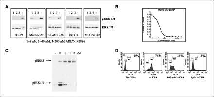
Inhibition of basal and induced ERK1/2 phosphorylation in human cancer cell lines and PBMCs.Clin Cancer Res.2007 Mar 1;13(5):1576-83. |
Tumor growth inhibition in the mouse BxPC3 xenograft model.Clin Cancer Res.2007 Mar 1;13(5):1576-83. |
 |
 |
Products are for research use only; We do not sell to patients
Copyright 2020 InvivoChem LLC | All Rights Reserved


 COA
COA