| Size | Price | Stock | Qty |
|---|---|---|---|
| 5mg |
|
||
| 10mg |
|
||
| 25mg |
|
||
| 50mg |
|
||
| 100mg | |||
| Other Sizes |


| Targets |
TRPC3/transient receptor potential canonical channel 3 (IC50 = 700 nM, for TRPC3-mediated Ca2+ influx)
Pyr3 specifically targets the transient receptor potential canonical channel 3 (TRPC3). It acts as a selective antagonist at this channel, inhibiting TRPC3-mediated calcium influx. The compound has an IC50 of 0.05 μM for suppressing the activation of the nuclear factor of activated T cells (NFAT) and an IC50 of 0.7 μM for inhibiting TRPC3-mediated Ca2+ influx. It is selective for TRPC3 over other TRPC family members. |
|---|---|
| ln Vitro |
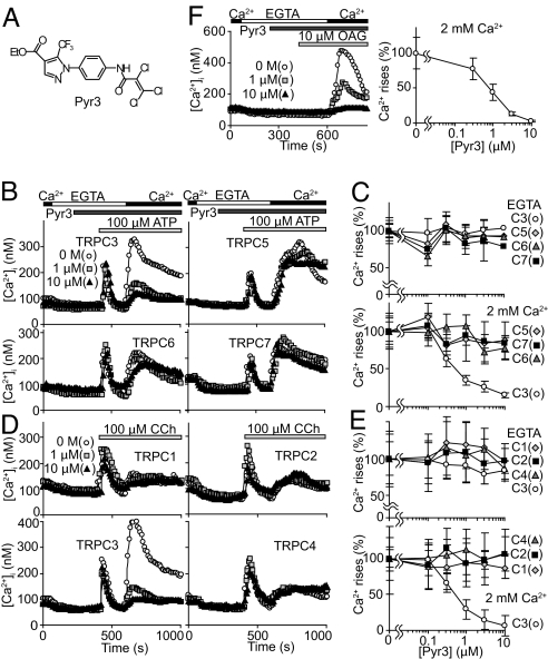
In TRPC family members, Pyr3 directly and selectively inhibits TRPC3 channels. With an IC50 value of 700 nM, Pyr3 application suppressed TRPC3-mediated Ca2+ influx in a dose-dependent manner. At 0.3 μM, Pyr3 becomes noticeable, and at 3 μM, it is nearly finished. It's interesting to note that cells coexpressing TRPC3 and TRPC6 were able to suppress Ca2+ influx using Pyr3, but not cells coexpressing TRPC1 and TRPC5. Pyr3 inhibits Ang II-induced NFAT translocation, but Pyr2's inhibition is mild and concentration-dependent (Pyr3's IC50 value is 0.05 μM, while Pyr2's IC50 value is 2 μM) [1].
In vitro, Pyr3 inhibits TRPC3-mediated Ca2+ influx in a dose-dependent manner. It has an IC50 of 700 nM for this effect. At concentrations around 3 µM, it almost completely blocks the channel activity. In cells co-expressing TRPC3 and TRPC6, Pyr3 inhibits Ca2+ influx, but it has no effect in cells co-expressing TRPC1 and TRPC5, demonstrating its selectivity. It also suppresses the activation of NFAT. |
| ln Vivo |
In this study, researchers examined Pyr3 in pressure overload-induced cardiac hypertrophy in vivo. Importantly, systolic and diastolic blood pressure, heart rate, mortality, body weight, and weight for liver, lung, and heart were unaffected by chronic treatment with Pyr3 (0.1 mg·kg−1·day−1) in sham operated mice (Fig. S16A and Tables S1 and S2 in SI Appendix). In addition, transverse aortic constriction (TAC) operation significantly increased left ventricular end-systolic pressure (ESP) in mice treated with vehicle or Pyr3 (Tables S1 and S2 in SI Appendix), suggesting that pressure overload was equally induced in these mice. Strikingly, increased size of the heart by 1-week TAC operation was significantly attenuated by Pyr3 (Fig. 6 A and B and Fig. S16B in SI Appendix). The Pyr3 effect refers to concentric hypertrophy, because the ratio of internal ventricular radius at end diastole (r) to ventricular wall thickness (h) was significantly decreased in echocardiography in mid transverse heart sections (Fig. 6A and Fig. S16C in SI Appendix), in contrast to fractional shortening (FS) and the right ventricle unaffected by TAC (Fig. S16 D and E in SI Appendix). The TAC-induced increase in expression of atrial natriuretic peptide (ANP) mRNA, a reliable marker for cardiac hypertrophy, was also suppressed by Pyr3 (Fig. 6C). Six-weeks TAC operation induced an r/h ratio increase characteristic of dilated hypertrophy (Fig. S16F in SI Appendix), and deterioration of FS and elevation of weight-to-tibia length ratio of the left ventricle (LVW/TL) in good correlation with systolic pressure gradient. These symptoms were suppressed by Pyr3 (Fig. 6 D and E). Thus, Pyr3 is potent against concentric and dilated cardiac hypertrophy[1].
In vivo, Pyr3 has been used to study the role of TRPC3 in various physiological and pathological processes. As a TRPC3 antagonist, it can be used to investigate the channel's function in animal models of disease, such as cardiovascular and neurological disorders. However, specific in vivo data for Pyr3, such as its efficacy in specific disease models, is not detailed in the available literature. Its use as a research tool suggests it has been applied in such studies. |
| Enzyme Assay |
Electrophysiology. [1]
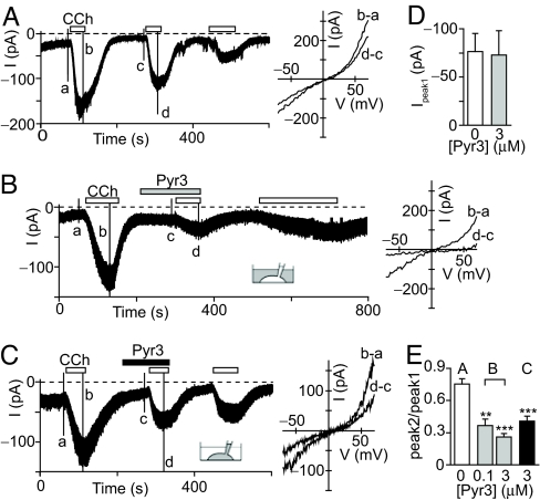
Whole-cell mode of the patch-clamp technique was performed on HEK293 or HEK293T cells at room temperature (22–25°C) with an EPC-9 or Axopatch 200B patch-clamp amplifier as previously described. Voltage-clamp experiments were performed at a holding potential of –50 mV or –60 mV, and recordings were sampled at 2.0 kHz and filtered at 2.9 kHz. The I-V relationships were determined using a 50-, 200-, or 400-ms voltage ramp from –100 mV to +100 mV or from –120 mV to +80 mV. 6 Pipette resistance ranged from 2 to 6 megohm when filled with the pipette solution described below. The series resistance was electronically compensated to > 50%. An external solution contained (in mM): For TRPC3 and C6, 140 NaCl, 5 KCl, 2 CaCl2, 1 MgCl2, 10 glucose, 10 HEPES (pH 7.4 adjusted with NaOH). For TRPM4b, 110 NaCl, 5 CaCl2, 10 glucose, 10 HEPES (pH 7.4 adjusted with NaOH). The pipette solution contained (in mM): For mAChR-activated TRPC3 current, 95 CsOH, 95 aspartate, 40 CsCl, 4 MgCl2, 5 EGTA, 2 ATPNa2, 5 HEPES, 8 creatine phosphate (pH 7.2 adjusted with CsOH). For OAG-induced TRPC3 current, 130 CsOH, 130 glutamate, 3.1 MgCl2, 2.8 CaCl2, 10 EGTA, 2 ATPNa2, 0.3 GTPNa2, 10 HEPES (pH 7.2 adjusted with CsOH). For TRPC6 current, 145 CsOH, 145 aspartate, 2 MgCl2, 0.3 CaCl2, 10 EGTA, 10 HEPES (pH 7.2 adjusted with CsOH). For TRPM4b current, 110 CsOH, 110 aspartate, 4 MgCl2, 8.4 CaCl2, 10 EGTA, 4 ATPNa2, 10 HEPES (pH 7.4 adjusted with CsOH). The osmolarity of the external solutions was adjusted to about 300 mOSM. No significant difference (P = 0.74) was observed in membrane capacitance between the control (22.7 ± 2.4 pF) and Pyr3-treated (21.2 ± 2.5 pF) HEK293 cells expressing TRPC3. Cell-free assays for Pyr3 are not the primary method for characterizing its activity, as it is an ion channel blocker whose function depends on the channel being in a membrane. However, its binding affinity to TRPC3 could be studied using radioligand binding assays if a suitable radiolabeled ligand is available. Alternatively, its effect could be measured in a cell-free system using isolated membrane patches in electrophysiological studies. |
| Cell Assay |
Measurement of NFAT activity and hypertrophic growth of cardiomyocytes was performed as described. Briefly, neonatal rat cardiomyocytes were isolated from 1−2 day-old Sprague-Dawley rats, and cultured on the gelatin-coated dishes or laminin-coated silicon rubber dishes. The cDNAs (pNFAT-Luc, pBNP-Luc and pRL-SV40) were transfected by Fugene 6 and a recombinant adenovirus encoding GFP-fused amino terminal region of NFAT4 isoform was infected at 100 MOI in serum-free medium. Forty-eight h after transfection, cells were stimulated with Ang II (100 nM) or mechanical stretch by 20%. Cells were treated with Pyr2 or Pyr3 for 20 min prior to the stimulation with Ang II or mechanical stretch. The cells were washed, fixed, and then stained with Alexa Fluor 594-phalloidin to visualize actin filaments. Protein synthesis was measured by [3 H]leucine incorporation. Cells were treated with Pyr2 or Pyr3 for 20 min prior to the stimulation with Ang II (100 nM), and [3 H]leucine (1 10 μCi/ml) was add to the culture medium and further incubated for 6 h. The incorporated [3 H]leucine was measured using liquid-scintillation counter. IC50 values were determined by using GraphPad Prism.
In vitro cell-based assays for Pyr3 are performed to measure its inhibitory effects on TRPC3-mediated calcium influx. A common method is to use cells expressing TRPC3 and a calcium-sensitive fluorescent dye, such as Fura-2 or Fluo-4. Cells are loaded with the dye, and the fluorescence is measured over time. The cells are then stimulated with a TRPC3 agonist (e.g., a diacylglycerol analog) to induce calcium influx, and the increase in fluorescence is recorded. The cells are then treated with varying concentrations of Pyr3, and the inhibition of the calcium signal is measured. The IC50 for inhibition is calculated from the dose-response curve. |
| Animal Protocol |
TAC surgery, hemodynamic measurements, and histological analyses were performed as described. Briefly, pressure overload was evoked by the surgical TAC. TAC was performed on 6-week-old male C57BL6j mice. A mini-osmotic pump filled with polyethylene glycol or Pyr3 (0.1 mg/kg/day) was implanted intraperitoneally in mice 3 d before 1 week of TAC. In the case of 6 weeks of TAC, Pyr3 was treated 3 d after TAC. Six weeks after TAC operation, transthoracic echocardiography was performed using ALOKA ultrasonic image analyzing system (SSD-5500) equipped with 7.5 MHz imaging transducer. Hemodynamic measurements were taken by inserting a micronanometer catheter from the right common carotid artery into the aorta and then the left ventricle. To calculate pressure gradient, distal arterial pressure was measured using tail-cuff detection system. After heart weight was measured, total RNA was isolated. The quantitative measurement of ANP mRNA expression was performed by real time RT-PCR. Primers and probe for mouse ANP mRNA are as follow: forward primer, 5’-CATCACCCTGGGCTTCTTCCT-3’; reverse primer, 5’-TGGGCTCCAATCCTGTCAATC-3’; TaqMan® probe, 5’-ATTTCAAGAACCTGCTAGACCACCTGGA-3’.
In vivo animal experiments for Pyr3 would be designed to study the role of TRPC3 in disease. For example, in a model of cardiac hypertrophy, an animal model of heart disease, the compound could be administered, and its effect on the progression of hypertrophy and heart function would be assessed. In a model of neurological disease, its effect on neuronal function or behavior could be evaluated. Specific protocols for Pyr3 are not detailed in the available literature. Its use is limited to research. |
| ADME/Pharmacokinetics |
Pharmacokinetic (PK) data for Pyr3 is not provided in the available literature. Its properties, such as bioavailability, half-life, and tissue distribution, would be important for its use as an in vivo research tool. For storage, the compound is typically kept as a powder.
|
| Toxicity/Toxicokinetics |
Toxicological data for Pyr3 is not available in the public literature. As a research compound that blocks a calcium channel, its safety profile would be a critical consideration. However, no specific LD50, organ toxicity, or genotoxicity data are reported. Its use is strictly for research purposes, and it is not intended for human therapeutic use.
|
| References | |
| Additional Infomation |
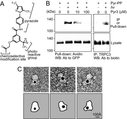
Classical transient receptor potential (TRPC) channels control the influx of Ca²⁺ and other cations, which, upon coupling with phospholipase C (PLC) to plasma membrane receptors, can induce a variety of cellular processes. Developing TRPC subtype-specific inhibitors is crucial for distinguishing different TRPC channels that play specific physiological roles in native systems. This paper identifies a pyrazole compound (Pyr3) that selectively inhibits TRPC3 channels. Structure-function studies of the pyrazole compound show that the trichloroacrylamide group is essential for the TRPC3 selectivity of Pyr3. Electrophysiological and photoaffinity labeling experiments revealed the direct action of Pyr3 on the TRPC3 protein. In DT40 B lymphocytes, Pyr3 effectively inhibited Ca²⁺ influx-dependent PLC translocation to the plasma membrane and the late oscillatory phase of the B cell receptor-induced Ca²⁺ response. In addition, Pyr3 can attenuate the activation of activated T cell nuclear factor (a Ca²⁺-dependent transcription factor), inhibit the hypertrophic growth of neonatal rat cardiomyocytes, and inhibit stress overload-induced cardiac hypertrophy in mice. These findings on the important role of the natural TRPC3 channel are highly consistent with previous genetic studies. Therefore, Pyr3, a selective inhibitor of TRPC3, is a powerful tool for studying the in vivo function of TRPC3, suggesting that Pyr3 has potential medicinal value in the treatment of TRPC3-related diseases such as cardiac hypertrophy. [1]
Background and Objectives: Pyrazole derivatives have recently been identified as selective blockers of transient receptor potential cation (TRPC) channels, but their ability to distinguish TRPC and Orai pore complexes remains unclear. This study aimed to characterize the TRPC/Orai selectivity of a series of pyrazole derivatives and to elucidate the effects of selective inhibition of these pathways on mast cell activation. Experimental Methods: Pyrazole compounds were prepared by microwave-assisted synthesis and their effects on Ca²⁺ influx were detected by Fura-2 imaging and their effects on membrane current were detected by patch-clamp recording. Experiments were conducted in HEK293 cells overexpressing TRPC3 and RBL-2H3 mast cells expressing canonical Orai channel-mediated storage-operating Ca²⁺ influx. The inhibitory effects of pyrazole compounds on Ca²⁺ signaling in RBL-2H3 cells were investigated, including degranulation and activation of nuclear factor T (NFAT) activation. Main results: Pyr3, a previously reported selective inhibitor of TRPC3, inhibited Orai1 and TRPC3-mediated Ca²⁺ influx and current, as well as mast cell activation, with similar potency. In contrast, Pyr6 was 37-fold more potent in inhibiting Orai1-mediated Ca²⁺ influx than in inhibiting TRPC3-mediated Ca²⁺ influx and effectively inhibited mast cell activation. The novel pyrazole compound Pyr10 exhibited significant selectivity (18-fold) for the TRPC3-mediated response, and the selective blocking of TRPC3 channels by Pyr10 had almost no effect on mast cell activation. Conclusions and Significance: Pyrazole derivatives Pyr6 and Pyr10 can distinguish between TRPC- and Orai-mediated Ca²⁺ influxes, serving as effective tools for analyzing the cellular function of related Ca²⁺ channels. https://pubmed.ncbi.nlm.nih.gov/22862290/ Pyr3 is a research-grade compound used as a selective tool to study the function of the TRPC3 channel. Its high selectivity for TRPC3 over other TRPC family members makes it invaluable for dissecting the specific roles of TRPC3 in calcium signaling and cellular physiology. It is particularly useful for studying the role of TRPC3 in the cardiovascular and nervous systems. It has not been approved for clinical use. All information is for research reference and not for diagnostic or clinical use. |
| Molecular Formula |
C16H11CL3F3N3O3
|
|---|---|
| Molecular Weight |
456.62
|
| Exact Mass |
454.982
|
| Elemental Analysis |
C, 42.09; H, 2.43; Cl, 23.29; F, 12.48; N, 9.20; O, 10.51
|
| CAS # |
1160514-60-2
|
| PubChem CID |
56964346
|
| Appearance |
White to off-white solid powder
|
| LogP |
4.964
|
| Hydrogen Bond Donor Count |
1
|
| Hydrogen Bond Acceptor Count |
7
|
| Rotatable Bond Count |
6
|
| Heavy Atom Count |
28
|
| Complexity |
621
|
| Defined Atom Stereocenter Count |
0
|
| InChi Key |
RZHGONNSASQOAY-UHFFFAOYSA-N
|
| InChi Code |
InChI=1S/C16H11Cl3F3N3O3/c1-2-28-15(27)10-7-23-25(12(10)16(20,21)22)9-5-3-8(4-6-9)24-14(26)11(17)13(18)19/h3-7H,2H2,1H3,(H,24,26)
|
| Chemical Name |
ethyl 1-[4-(2,3,3-trichloroprop-2-enoylamino)phenyl]-5-(trifluoromethyl)pyrazole-4-carboxylate
|
| Synonyms |
Pyr 3; Pyr-3; 1160514-60-2; 1H-Pyrazole-4-carboxylic acid, 1-[4-[(2,3,3-trichloro-1-oxo-2-propen-1-yl)amino]phenyl]-5-(trifluoromethyl)-, ethyl ester; CHEMBL4177187; ethyl 1-(4-(2,3,3-trichloroacrylamido)phenyl)-5-(trifluoromethyl)-1h-pyrazole-4-carboxylate; RZHGONNSASQOAY-UHFFFAOYSA-N; GTPL4293; SCHEMBL12274132; Pyr3
|
| HS Tariff Code |
2934.99.9001
|
| Storage |
Powder -20°C 3 years 4°C 2 years In solvent -80°C 6 months -20°C 1 month |
| Shipping Condition |
Room temperature (This product is stable at ambient temperature for a few days during ordinary shipping and time spent in Customs)
|
| Solubility (In Vitro) |
DMSO : ≥ 125 mg/mL (~273.74 mM)
|
|---|---|
| Solubility (In Vivo) |
Solubility in Formulation 1: ≥ 2.08 mg/mL (4.56 mM) (saturation unknown) in 10% DMSO + 40% PEG300 + 5% Tween80 + 45% Saline (add these co-solvents sequentially from left to right, and one by one), clear solution.
For example, if 1 mL of working solution is to be prepared, you can add 100 μL of 20.8 mg/mL clear DMSO stock solution to 400 μL PEG300 and mix evenly; then add 50 μL Tween-80 to the above solution and mix evenly; then add 450 μL normal saline to adjust the volume to 1 mL. Preparation of saline: Dissolve 0.9 g of sodium chloride in 100 mL ddH₂ O to obtain a clear solution. Solubility in Formulation 2: ≥ 2.08 mg/mL (4.56 mM) (saturation unknown) in 10% DMSO + 90% (20% SBE-β-CD in Saline) (add these co-solvents sequentially from left to right, and one by one), clear solution. For example, if 1 mL of working solution is to be prepared, you can add 100 μL of 20.8 mg/mL clear DMSO stock solution to 900 μL of 20% SBE-β-CD physiological saline solution and mix evenly. Preparation of 20% SBE-β-CD in Saline (4°C,1 week): Dissolve 2 g SBE-β-CD in 10 mL saline to obtain a clear solution. View More
Solubility in Formulation 3: ≥ 2.08 mg/mL (4.56 mM) (saturation unknown) in 10% DMSO + 90% Corn Oil (add these co-solvents sequentially from left to right, and one by one), clear solution. |
| Preparing Stock Solutions | 1 mg | 5 mg | 10 mg | |
| 1 mM | 2.1900 mL | 10.9500 mL | 21.9000 mL | |
| 5 mM | 0.4380 mL | 2.1900 mL | 4.3800 mL | |
| 10 mM | 0.2190 mL | 1.0950 mL | 2.1900 mL |
*Note: Please select an appropriate solvent for the preparation of stock solution based on your experiment needs. For most products, DMSO can be used for preparing stock solutions (e.g. 5 mM, 10 mM, or 20 mM concentration); some products with high aqueous solubility may be dissolved in water directly. Solubility information is available at the above Solubility Data section. Once the stock solution is prepared, aliquot it to routine usage volumes and store at -20°C or -80°C. Avoid repeated freeze and thaw cycles.
Calculation results
Working concentration: mg/mL;
Method for preparing DMSO stock solution: mg drug pre-dissolved in μL DMSO (stock solution concentration mg/mL). Please contact us first if the concentration exceeds the DMSO solubility of the batch of drug.
Method for preparing in vivo formulation::Take μL DMSO stock solution, next add μL PEG300, mix and clarify, next addμL Tween 80, mix and clarify, next add μL ddH2O,mix and clarify.
(1) Please be sure that the solution is clear before the addition of next solvent. Dissolution methods like vortex, ultrasound or warming and heat may be used to aid dissolving.
(2) Be sure to add the solvent(s) in order.
|
|
|
Products are for research use only; We do not sell to patients
Copyright 2020 InvivoChem LLC | All Rights Reserved