| Size | Price | Stock | Qty |
|---|---|---|---|
| 1mg |
|
||
| 5mg |
|
||
| 10mg | |||
| Other Sizes |


| Targets |
ProTAME targets the Anaphase-Promoting Complex/Cyclosome (APC/C), specifically inhibiting its two co-activators, APC/CCdc20 and APC/CFzr. The APC/C is a multi-subunit E3 ubiquitin ligase that orchestrates the progression through mitosis by tagging specific cell cycle regulators for degradation. By inhibiting the APC/C, ProTAME prevents the ubiquitination and subsequent destruction of its substrates, thereby causing a mitotic arrest.
|
|---|---|
| ln Vitro |
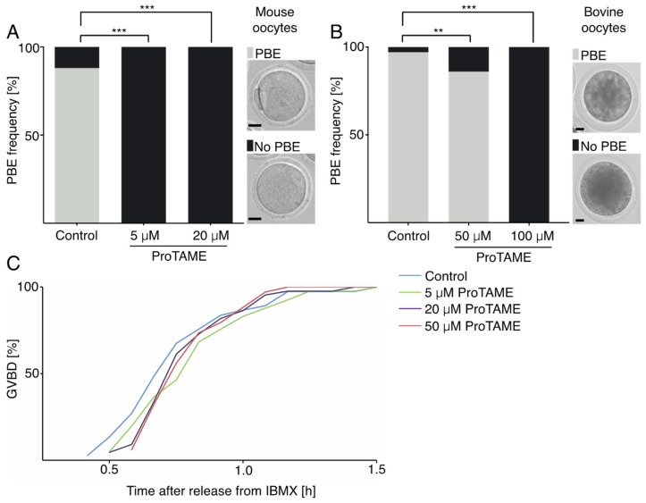
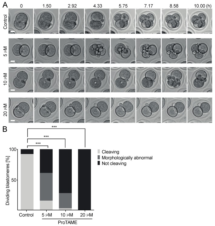
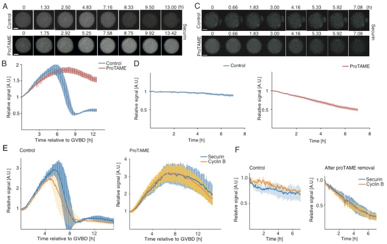
ProTAME stops mouse and bovine oocytes, as well as mouse 2-cell embryos, from entering anaphase. Mammalian oocytes treated with proTAME (0-100 μM) exhibit dose-dependent metaphase arrest and early cleavage embryos. Furthermore, spindle assembly checkpoint (SAC) activity is not necessary for the drug-induced metaphase arrest [1]. Because ProTAME inhibits APC/C, it prevents mouse oocyte meiosis I. The arrest of oocytes and embryos is irreversible, in contrast to somatic cells [1]. In oocytes and embryos, proTAME (0–20 μM) dose-dependently alters spindle morphological characteristics [1]. ProTAME efficiently overcomes resistance resulting from Polo-like kinase 1 (PLK1)-based resistance in ovarian cancer cells, CDH1 hyperphosphorylation-based resistance in glioblastoma cells, and CDC20-based resistance in diffuse large B-cell lymphoma cells. therapeutic qualities [1]. OVCAR-3 cell growth is inhibited by proTAME, with an IC50 of 12.5 μM[2].
ProTAME has demonstrated in vitro efficacy in enhancing the effects of other anticancer agents. Specifically, combinations of ProTAME with topoisomerase inhibitors, such as doxorubicin and etoposide, significantly increase cell death in primary cells and Multiple Myeloma (MM) cell lines. This synergistic effect is particularly pronounced when topoisomerase II alpha (TOPIIα) levels are first increased through pre-treatment with ProTAME. This suggests a potential strategy to sensitize cancer cells to conventional chemotherapeutics. |
| ln Vivo |
In vivo, ProTAME's activity is inferred from its mechanism as an APC/C inhibitor. By inducing a metaphase arrest, it is expected to have antiproliferative effects in tumor models. However, specific in vivo data on ProTAME, such as its effects in xenograft models, are limited in the available literature. Its primary use remains as a research tool to study the cell cycle and to validate the APC/C as a therapeutic target. The compound's cell-permeable nature makes it suitable for in vivo administration in preclinical studies.
|
| Enzyme Assay |
The typical assay for measuring the activity of ProTAME and its active metabolite, TAME, involves a cell-free reconstituted system. The APC/C is immunoprecipitated from cell lysates, and its ubiquitination activity is measured in the presence of recombinant E1 and E2 enzymes, ubiquitin, and a specific substrate (e.g., cyclin B1 or securin). The reaction is carried out in a buffer containing ATP. The inhibition of this ubiquitination reaction by ProTAME or TAME is then quantified, often by monitoring the formation of polyubiquitinated substrates via Western blotting or by using a fluorescently labeled ubiquitin. ProTAME is typically used at concentrations ranging from 1 to 100 µM in such assays.
|
| Cell Assay |
In vitro cell-based assays for ProTAME are performed to evaluate its effects on cell cycle progression and viability. Cells, such as HeLa or multiple myeloma cell lines, are treated with varying concentrations of ProTAME (typically 1-50 µM) for different time points (e.g., 24-72 hours). The effects are then assessed using several methods. Cell cycle analysis is performed via flow cytometry after propidium iodide staining to determine the accumulation of cells in metaphase. Cell viability is measured using assays like MTT or CellTiter-Glo. Furthermore, the synergistic effect of ProTAME with other drugs, such as doxorubicin, can be evaluated by co-treatment and calculating the combination index.
|
| Animal Protocol |
In vivo animal experiments for ProTAME are conducted to study its antitumor activity and its potential to enhance the efficacy of other chemotherapies. The compound is typically administered via intraperitoneal (IP) or intravenous (IV) injection in mouse xenograft models of cancer, such as multiple myeloma. Tumor-bearing mice are treated with ProTAME, either alone or in combination with drugs like doxorubicin or etoposide. Tumor growth is monitored by caliper measurements, and endpoints include tumor volume, tumor weight, and survival. The studies also assess the induction of apoptosis and mitotic arrest in tumor tissues via immunohistochemistry and Western blotting. Pharmacokinetic (PK) studies of ProTAME are limited. As a prodrug, it is converted intracellularly to the active inhibitor TAME by esterases. Its bioavailability and half-life are influenced by this conversion process. Data on its tissue distribution, protein binding, and metabolic stability are not extensively published in the public domain. As a cell-permeable small molecule, it is presumed to have reasonable membrane permeability. However, for detailed PK parameters like Cmax, Tmax, and AUC, one would need to consult proprietary data from the compound's developer or conduct specialized studies. The compound is supplied as a powder and is typically stored at -20°C.
|
| ADME/Pharmacokinetics |
Toxicological data for ProTAME is primarily derived from its mechanism of action. By inhibiting the APC/C, it arrests cells in mitosis, which can lead to cell death. This on-target effect is the basis for its potential anticancer activity but also indicates that it would be toxic to rapidly dividing normal cells, such as those in the bone marrow and gastrointestinal tract. There are no specific toxicity studies (e.g., LD50, organ toxicity) for ProTAME available in the public literature. Its use is restricted to laboratory research, and it is not approved for human therapeutic use.
|
| Toxicity/Toxicokinetics |
ProTAME is a research-grade compound not approved for clinical use. It is an important tool for studying the role of the APC/C in cell cycle regulation and for validating the APC/C as a target for cancer therapy. Its prodrug nature, requiring intracellular conversion to its active form, TAME, is a key aspect of its design. The compound has been used in studies to understand the mechanisms of chemoresistance and to explore synergistic drug combinations, particularly with topoisomerase inhibitors. It is also available in a fluorescently labeled form (proTAME-Fluorescein) for studying its cellular uptake and localization. ProTAME is for research use only and not for human or veterinary use. All information is for research reference and not for diagnostic or clinical use.
|
| References |
|
| Additional Infomation |
ProTAME is a research-grade compound not approved for clinical use. It is an important tool for studying the role of the APC/C in cell cycle regulation and for validating the APC/C as a target for cancer therapy. Its prodrug nature, requiring intracellular conversion to its active form, TAME, is a key aspect of its design. The compound has been used in studies to understand the mechanisms of chemoresistance and to explore synergistic drug combinations, particularly with topoisomerase inhibitors. It is also available in a fluorescently labeled form (proTAME-Fluorescein) for studying its cellular uptake and localization. ProTAME is for research use only and not for human or veterinary use. All information is for research reference and not for diagnostic or clinical use.
|
| Molecular Formula |
C34H38N4O12S
|
|---|---|
| Molecular Weight |
726.75
|
| Exact Mass |
726.22
|
| Elemental Analysis |
C, 56.19; H, 5.27; N, 7.71; O, 26.42; S, 4.41
|
| CAS # |
1362911-19-0
|
| PubChem CID |
56924780
|
| Appearance |
White to off-white solid powder
|
| Density |
1.3±0.1 g/cm3
|
| Index of Refraction |
1.587
|
| LogP |
6.05
|
| Hydrogen Bond Donor Count |
3
|
| Hydrogen Bond Acceptor Count |
14
|
| Rotatable Bond Count |
23
|
| Heavy Atom Count |
51
|
| Complexity |
1210
|
| Defined Atom Stereocenter Count |
1
|
| SMILES |
C(OC)(=O)[C@@H](NS(C1=CC=C(C)C=C1)(=O)=O)CCC/N=C(\NC(OCOC(CC1=CC=CC=C1)=O)=O)/NC(=O)OCOC(=O)CC1=CC=CC=C1
|
| InChi Key |
MHYOVHULCQSDRZ-NDEPHWFRSA-N
|
| InChi Code |
InChI=1S/C34H38N4O12S/c1-24-15-17-27(18-16-24)51(44,45)38-28(31(41)46-2)14-9-19-35-32(36-33(42)49-22-47-29(39)20-25-10-5-3-6-11-25)37-34(43)50-23-48-30(40)21-26-12-7-4-8-13-26/h3-8,10-13,15-18,28,38H,9,14,19-23H2,1-2H3,(H2,35,36,37,42,43)/t28-/m0/s1
|
| Chemical Name |
methyl (2S)-5-[bis[(2-phenylacetyl)oxymethoxycarbonylamino]methylideneamino]-2-[(4-methylphenyl)sulfonylamino]pentanoate
|
| Synonyms |
pro-Tosyl-L-Arginine Methyl Ester ProTAME
|
| HS Tariff Code |
2934.99.9001
|
| Storage |
Powder -20°C 3 years 4°C 2 years In solvent -80°C 6 months -20°C 1 month |
| Shipping Condition |
Room temperature (This product is stable at ambient temperature for a few days during ordinary shipping and time spent in Customs)
|
| Solubility (In Vitro) |
DMSO : ~100 mg/mL (~137.60 mM)
|
|---|---|
| Solubility (In Vivo) |
Solubility in Formulation 1: ≥ 3.75 mg/mL (5.16 mM) (saturation unknown) in 10% DMSO + 90% Corn Oil (add these co-solvents sequentially from left to right, and one by one), clear solution.
For example, if 1 mL of working solution is to be prepared, you can add 100 μL of 37.5 mg/mL clear DMSO stock solution to 900 μL of corn oil and mix evenly. (Please use freshly prepared in vivo formulations for optimal results.) |
| Preparing Stock Solutions | 1 mg | 5 mg | 10 mg | |
| 1 mM | 1.3760 mL | 6.8799 mL | 13.7599 mL | |
| 5 mM | 0.2752 mL | 1.3760 mL | 2.7520 mL | |
| 10 mM | 0.1376 mL | 0.6880 mL | 1.3760 mL |
*Note: Please select an appropriate solvent for the preparation of stock solution based on your experiment needs. For most products, DMSO can be used for preparing stock solutions (e.g. 5 mM, 10 mM, or 20 mM concentration); some products with high aqueous solubility may be dissolved in water directly. Solubility information is available at the above Solubility Data section. Once the stock solution is prepared, aliquot it to routine usage volumes and store at -20°C or -80°C. Avoid repeated freeze and thaw cycles.
Calculation results
Working concentration: mg/mL;
Method for preparing DMSO stock solution: mg drug pre-dissolved in μL DMSO (stock solution concentration mg/mL). Please contact us first if the concentration exceeds the DMSO solubility of the batch of drug.
Method for preparing in vivo formulation::Take μL DMSO stock solution, next add μL PEG300, mix and clarify, next addμL Tween 80, mix and clarify, next add μL ddH2O,mix and clarify.
(1) Please be sure that the solution is clear before the addition of next solvent. Dissolution methods like vortex, ultrasound or warming and heat may be used to aid dissolving.
(2) Be sure to add the solvent(s) in order.
|
|
|
Products are for research use only; We do not sell to patients
Copyright 2020 InvivoChem LLC | All Rights Reserved