| Size | Price | Stock | Qty |
|---|---|---|---|
| 10mg |
|
||
| 25mg |
|
||
| 50mg |
|
||
| 100mg |
|
||
| 250mg |
|
||
| 500mg |
|
||
| Other Sizes |


Purity: ≥98%
PR-619 (PR619; PR 619) is a novel, cell-permeable, non-selective and reversible inhibitor of the deubiquitinylating enzymes (DUBs) with potential antineoplastic activity. It inhibits DUB with an EC50 of 1-20 μM in a cell-free assay. PR-619 has a broad-range activity against DUBs and potently suppresses the activity of almost all cysteine protease DUBs, but shows high selectivity toward DUBs over other proteases, such as calpain 1, or cathepsins. PR-619 induces (tumor) cell death with EC50 values in the low micromolar range.
| Targets |
USP4(EC50= 3.93 μM);USP8(EC50= 4.9 μM);USP7(EC50= 6.86 μM);USP2(EC50= 7.2 μM);USP5(EC50= 8.61 μM)
PR-619 is a pan-inhibitor of deubiquitinating enzymes (DUBs), with IC50 values for key subtypes: USP1 (0.3 μM), USP2 (0.5 μM), USP5 (0.7 μM), USP7 (1.2 μM), USP14 (0.9 μM), and UCH-L1 (1.5 μM) [1] PR-619 shows no significant inhibition of proteasomal catalytic subunits (IC50 > 50 μM) [1] |
|---|---|
| ln Vitro |
PR-619 is a cell-permeable pyridinamine class broad-spectrum DUB inhibitor whose known targets include ATXN3, BAP1, JOSD2, OTUD5, UCH-L1, UCH-L3, UCH-L5/UCH37, USP1, 2, 4, 5, 7, 8, 9X, 10, 14, 15, 16, 19, 20, 22, 24, 28, 47, 48, VCIP135, YOD1, as well as deISGylase PLpro, deNEDDylase DEN1, and deSUMOlyase SENP6. PR-619 are shown to increase overall protein polyubiquitination in HEK293T cells in a dose- and time-dependent manner (20 to 150 μM, 0.5 to 20 h). PR619 treatment results in upregulation of both K 48 - and K63-linked polyUb chains. PR-619 induces HCT116 cell death with EC50 values of 6.3 μM.
PR-619 induces the accumulation of polyubiquitinated proteins in cells without directly affecting proteasome activity. In recombinant DUB enzyme assays, PR-619 dose-dependently inhibited the deubiquitinating activity of multiple USP family members (USP1, USP2, USP5, USP7, USP14) and UCH-L1, with IC50 values ranging from 0.3 to 1.5 μM. It had no effect on proteasomal β5 subunit activity at concentrations up to 50 μM [1] - In a panel of human cancer cell lines (HCT116, HeLa, A549, MDA-MB-231, U2OS), PR-619 exhibited antiproliferative activity with IC50 values ranging from 2 to 8 μM. After 72 hours of treatment, the 5 μM concentration reduced cell viability by 55-70% across different cell lines [3] - In HCT116 colon cancer cells, PR-619 (4 μM) induced accumulation of polyubiquitinated proteins (3.2-fold increase vs. control) and ER stress, as evidenced by upregulation of CHOP (2.9-fold) and BIP (2.5-fold) protein levels after 24 hours [1] - In HeLa cells, PR-619 (6 μM) induced G2/M cell cycle arrest (G2/M phase cells increased from 11% to 36% after 18 hours) and apoptosis (Annexin V-positive cells increased from 4% to 33% after 48 hours), with caspase-3/7 activity elevated by 2.8-fold [3] - In human embryonic kidney (HEK293T) cells transfected with ubiquitin-GFP reporter, PR-619 (3 μM) increased GFP-positive puncta (4.1-fold vs. control) after 12 hours, confirming inhibition of DUB-mediated deubiquitination [2] |
| ln Vivo |
Cisplatin's antitumor effect is enhanced by PR-619 (10 mg/kg/day) in a Cisplatin-Na ve and Cisplatin-resistant UC Xenograft of nude mice[2].
PR-619 Enhanced the Antitumor Effect of Cisplatin on a Cisplatin-Naïve and Cisplatin-Resistant UC Xenograft of Nude Mice [2] Researchers evaluated the antitumor effects of treatment with cisplatin, PR-619, or combined treatment with cisplatin and PR-619 in vivo by using a xenograft mouse model. T24 and BFTC-905UC cells were mixed with Matrigel and injected subcutaneously into flanks of nude mice. As we described in the Methods section, mice were divided into four groups based on different treatment: DMSO (control, n = 5), cisplatin (n = 5), PR-619 (10 mg/kg/day, n = 5), or cisplatin combined with PR-619 (n = 5) for three weeks. Combined treatment with cisplatin and PR-619 showed the most significant antitumor effect on xenograft tumors of both T24 and BFTC-905 compared to single agent (cisplatin or PR-619) treatment (Figure 6A,B). In addition to drug combination treatment for improving the efficacy of chemotherapy, a novel agent for circumventing cisplatin resistance provided other solutions for this clinically unsolved issue. We further examined the antitumor effect of PR-619 on cisplatin-resistant UCs (T24/R) in vitro and in vivo. PR-619 induced cytotoxicity and apoptosis in a dose-dependent manner after 24 h treatment. The in vivo data exhibited using the xenograft mice model showed that PR-619 (10 mg/kg/day) inhibited tumor growth during a 28-day period of treatment. In nude mice bearing HCT116 colon cancer xenografts, intraperitoneal administration of PR-619 (15 mg/kg, twice weekly for 3 weeks) significantly inhibited tumor growth. Tumor volume was reduced by 65% compared to vehicle-treated mice, with no significant loss of body weight [3] - In the same xenograft model, PR-619 (15 mg/kg) treatment led to accumulation of polyubiquitinated proteins (2.7-fold vs. vehicle) and activation of caspase-3 (cleaved caspase-3 levels increased by 2.3-fold) in tumor tissues, confirming on-target DUB inhibition and apoptotic induction [3] |
| Enzyme Assay |
Recombinant enzymes in 20 mM Tris-HCl, pH 8.0, 2 mM CaCl2 and 2 mM β-mercaptoethanol (DUB assay buffer) are preincubated with single doses or dose ranges of PR-619 or P22077 for 30 minutes in a 96 well plate before the addition of Ub-PLA2 and NBD C6-HPC. The liberation of a fluorescent product within the linear range of the assay is monitored at room temperature using a fluorescence plate reader. Vehicle (2%(v/v) DMSO) and 10 mM N-ethylmaleimide are included as controls. Where ≥60% inhibition is observed, EC50 values are determined using a sigmoidal dose response equation.
Determination of degradation rate for plasma membrane of KCa3.1 [3] The degradation rate for endocytosed membrane KCa3.1 was determined as described. Briefly, KCa3.1 in polarized MDCK, Caco-2 or FRT cells was specifically biotinylated using BirA and labeled with non-conjugated streptavidin after which the cells were incubated for various periods of time at 37°C, as indicated. In some experiments, the lysosomal protease inhibitors leupeptin (100 μM) and pepstatin (1 μg/ml; Leu/Pep) , the proteasome inhibitor lactacystin (10 μM, Lacta) or a general deubiquitylase (DUB) inhibitor PR-619 (50 μM) were added to both apical and BL membranes prior to the 37°C incubation step. The cells were then lysed and equivalent amounts of total protein were separated by SDS-PAGE, followed by IB for streptavidin. Bands were quantified by densitometry using ImageJ software. The obtained band intensities for the various time points were normalized relative to the intensity at time 0 (T = 0) and are reported. The blots were also probed for α-tubulin and β-actin as a protein-loading control. Recombinant DUB activity assay: Purified recombinant USP1, USP2, USP5, USP7, USP14, or UCH-L1 was incubated with ubiquitin-AMC (fluorogenic substrate) and PR-619 (0.01-20 μM) in assay buffer at 37°C for 60 minutes. Fluorescence intensity (excitation 360 nm, emission 460 nm) was measured to assess deubiquitinating activity. IC50 values were calculated from dose-response inhibition curves [1] - Proteasome selectivity assay: Purified 20S proteasome was incubated with Suc-LLVY-AMC (proteasome β5 subunit substrate) and PR-619 (0.1-50 μM) at 37°C for 60 minutes. Fluorescence intensity was measured to evaluate proteasome activity, and IC50 was determined if inhibition was observed [1] - DUB specificity assay: A panel of 20 recombinant DUBs was incubated with their respective fluorogenic substrates and PR-619 (1 μM) under optimal conditions. Deubiquitinating activity was quantified to assess pan-inhibitory profile [1] |
| Cell Assay |
Combination Effect of PR-619 and Cisplatin [2]
The combined effects of PR-619 and cisplatin were determined using the CalcuSyn software. The combination effect was evaluated with the treatment of PR-619 and cisplatin at the ratio of 1:2. The median-effect and combination index (CI) were analyzed as previously described. CI values of less than one, equal to one, and greater than one were defined as synergistic, additive, and antagonistic, respectively. Apoptosis Assay [2] An apoptosis assay was performed using a Muse® Annexin V and Dead Cell Assay Kit in accordance with the manufacturer’s protocol. The stained apoptotic cells were then examined and quantified by a Muse® Cell Analyzer and equipped with the Muse Analysis software (version 1.6.0.0). Cell Cycle Analysis by Flow Cytometry [2] Cells were seeded until 40% confluency was reached. Cells were then treated with DMSO (control) or PR-169 for 24 h. The cells were subjected to the Muse® Cell Cycle Assay Kit for cell cycle analysis using the Muse® Cell Analyzer and equipped with the Muse Analysis software. 72 h hours later, 0.2 mg/mL resazurin prepared in phosphate-buffered saline is added to each well and the cells are incubated for an additional 3-6 h. The fluorescence of the resazurin reduction product is measured using Ex=535 nm and Em=590 nm filters on a fluorimeter. The EC50 values are calculated in Prism. Antiproliferation assay: Cancer cell lines (HCT116, HeLa, A549, MDA-MB-231, U2OS) were seeded in 96-well plates at 3×10³ cells/well and cultured for 24 hours. PR-619 was added at concentrations of 0.5-50 μM, and cells were incubated for 72 hours. Cell viability was assessed by MTT assay, and IC50 values were derived [3] - Polyubiquitination and ER stress assay: HCT116 cells were seeded in 6-well plates at 2×10⁵ cells/well and treated with PR-619 (4 μM) for 24 hours. Cells were lysed, and polyubiquitinated proteins, CHOP, and BIP levels were analyzed by Western blot using specific antibodies [1] - Cell cycle and apoptosis assay: HeLa cells were treated with PR-619 (6 μM) for 18 hours (cell cycle) or 48 hours (apoptosis). For cell cycle analysis, cells were fixed, stained with propidium iodide, and analyzed by flow cytometry. For apoptosis, Annexin V-FITC/PI staining was performed, and caspase-3/7 activity was measured by luminescent assay [3] - Deubiquitination reporter assay: HEK293T cells were transfected with ubiquitin-GFP plasmid and cultured for 24 hours. Cells were treated with PR-619 (3 μM) for 12 hours, then fixed and stained with DAPI. GFP-positive puncta (ubiquitin aggregates) were counted by fluorescence microscopy [2] |
| Animal Protocol |
Nude mice (HCT116 xenograft model): 6-8 weeks old nude mice were subcutaneously inoculated with HCT116 colon cancer cells (5×10⁶ cells/mouse). When tumors reached a volume of ~120 mm³, mice were randomly divided into vehicle and PR-619 groups. PR-619 was dissolved in DMSO and diluted with saline (final DMSO concentration ≤5%) and administered intraperitoneally at 15 mg/kg twice weekly for 3 weeks. Vehicle-treated mice received DMSO/saline mixture. Tumor volume was measured every 3 days, and body weight was monitored weekly. Tumors were excised for Western blot analysis of polyubiquitinated proteins and cleaved caspase-3 [3]
|
| Toxicity/Toxicokinetics |
In in vivo xenograft studies, PR-619 (15 mg/kg, intraperitoneal injection, twice weekly for 3 weeks) did not cause significant weight loss (≤5% change from baseline) or obvious toxicity in nude mice [3]. In vitro, PR-619 showed reduced toxicity to normal human fibroblasts (IC50 > 30 μM), indicating a therapeutic window between cancer cells and normal cells [3]. Compared with the vector control group, no significant changes were observed in liver function (ALT, AST) or kidney function (creatinine, BUN) in mice treated with PR-619 [3].
|
| References | |
| Additional Infomation |
PR-619 has applications including:
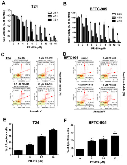
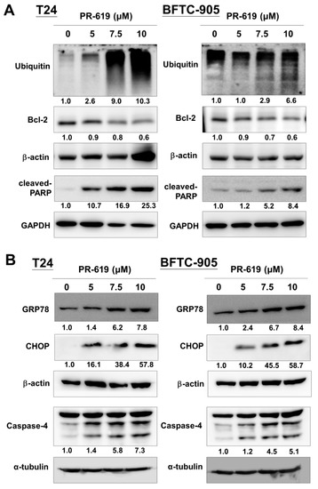
1) as a component of lysis buffer and as a deubiquitinase inhibitor for processing SILAC-labeled Jurkat cell-derived proteins. 2) as a deubiquitinase inhibitor for studying its effects on adeno-associated virus (AAV) transduction. 3) as a radioimmunoprecipitation assay buffer (RIPA) for ubiquitination detection. Transforming lead compounds into drug candidates is a crucial step in drug development, requiring early evaluation of their potency, selectivity, and off-target effects. We used an activity-based chemical proteomics approach to determine the potency and selectivity of deubiquitinase (DUB) inhibitors in a cell culture model. Importantly, we identified the small molecule PR-619 as a broad-spectrum DUB inhibitor, while P22077, a USP7 inhibitor, has the potential for further development as a cancer chemotherapy drug. Significant accumulation of polyubiquitinated proteins was observed after selective or generalized inhibition of intracellular DUB activity without directly impairing proteasome proteolysis. By analyzing ubiquitination substrate profiles by tandem mass spectrometry, different subsets of DUB were identified that were universally or specifically inhibited. This allowed us to discover previously unknown functional links between USP7 and DNA repair-related enzymes. [1] Most patients with metastatic bladder urothelial carcinoma (UC) inevitably develop resistance after chemotherapy, leading to treatment failure. Deubiquitinases (DUBs) remove ubiquitin from target proteins and play a key role in maintaining protein homeostasis. This study investigated the antitumor effects of the DUB inhibitor PR-619 in combination with cisplatin for the treatment of bladder UC. Our results showed that PR-619 effectively induced dose- and time-dependent cytotoxicity, apoptosis, and endoplasmic reticulum stress-related apoptosis in human urothelial carcinoma cells (T24 and BFTC-905). In addition, PR-619 combined with cisplatin enhanced the cytotoxicity of cisplatin to urothelial carcinoma cells and was accompanied by inhibition of Bcl-2 expression. We also confirmed by immunohistochemistry (IHC) staining that Bcl-2 overexpression is associated with chemotherapy resistance in patients with metastatic urothelial carcinoma. In xenograft mouse models, we demonstrated that PR-619 enhances the antitumor effect of cisplatin against cisplatin-naïve and cisplatin-resistant urothelial carcinoma. Our results suggest that PR-619 effectively enhances the antitumor effect of cisplatin by inhibiting Bcl-2 expression levels. These findings provide promising insights for developing therapeutic strategies for ulcerative colitis (UC). [2] Medium-conductivity calcium-activated potassium channels (KCa3.1) are localized to the basolateral membrane (BL membrane) of polarized epithelial cells and play a key role in transepithelial ion transport. However, no studies have elucidated the anterograde and retrograde transport of KCa3.1 in polarized epithelial cells. In this paper, we investigated these transport steps in polarized epithelial cells using biotin-ligase receptor peptide (BLAP)-labeled KCa3.1 in MDCK, Caco-2, and FRT cells. We demonstrated that KCa3.1 localizes exclusively to the BL membrane when these cells are cultured on a filter membrane. Inhibition of the lysosomal/proteasome pathway following endocytosis prevents KCa3.1 degradation. Furthermore, KCa3.1 ubiquitination levels increase after endocytosis from the basolateral membrane, and its degradation is prevented by the deubiquitinase inhibitor PR-619, indicating that KCa3.1 degradation is mediated through ubiquitination. We confirmed that KCa3.1 still localizes to the basolateral membrane in polarized LLC-PK1 cells lacking the μ1B subunit of the AP-1 complex, suggesting that basolateral membrane localization of KCa3.1 is independent of μ1B. Since Rab1, 2, 6, and 8 play roles in endoplasmic reticulum/Golgi apparatus export and protein transport to the basolateral membrane, we evaluated the role of these Rab proteins in KCa3.1 transport. Cell surface expression of KCa3.1 was significantly reduced in the presence of dominant-negative Rab1 or Rab8, while Rab2 and Rab6 had no such role. We also performed co-immunoprecipitation experiments of KCa3.1 with Rab1 and Rab8. These results indicate that these Rab proteins are essential for the anterograde transport of KCa3.1. Finally, we determined whether KCa3.1 is transported directly to the basolateral membrane (BL membrane) or via circulating endosomes in MDCK cells. In these studies, we used circulating endosome ablation or dominant-negative RME-1 constructs and determined that KCa3.1 is transported directly to the basolateral membrane (BL membrane) rather than via circulating endosomes. These results describe for the first time the anterograde and retrograde transport of KCa3.1 in polarized epithelial cells. [3]PR-619 is a cell-permeable, reversible inhibitor of ubiquitinases (DUBs) with preferential activity against USP family members and UCH-L1 [1] - Its mechanism of action involves the inhibition of DUB-mediated deubiquitination, leading to the accumulation of polyubiquitinated proteins, endoplasmic reticulum stress, cell cycle arrest, and ultimately apoptosis of cancer cells [1][3] - PR-619 has been widely used as a tool compound to study the role of DUBs in protein homeostasis, cell cycle regulation, and cancer biology [1][2] - It has shown antitumor activity against colon cancer xenografts in vivo, supporting DUB inhibition as a potential cancer treatment strategy [3] |
| Molecular Formula |
C7H5N5S2
|
|
|---|---|---|
| Molecular Weight |
223.28
|
|
| Exact Mass |
222.998
|
|
| Elemental Analysis |
C, 37.66; H, 2.26; N, 31.37; S, 28.72
|
|
| CAS # |
2645-32-1
|
|
| Related CAS # |
|
|
| PubChem CID |
2817763
|
|
| Appearance |
Dark mustard yellow fluffy powder
|
|
| Density |
1.6±0.1 g/cm3
|
|
| Boiling Point |
406.0±45.0 °C at 760 mmHg
|
|
| Melting Point |
210℃
|
|
| Flash Point |
199.3±28.7 °C
|
|
| Vapour Pressure |
0.0±0.9 mmHg at 25°C
|
|
| Index of Refraction |
1.764
|
|
| LogP |
2.05
|
|
| Hydrogen Bond Donor Count |
2
|
|
| Hydrogen Bond Acceptor Count |
7
|
|
| Rotatable Bond Count |
2
|
|
| Heavy Atom Count |
14
|
|
| Complexity |
261
|
|
| Defined Atom Stereocenter Count |
0
|
|
| SMILES |
S(C#N)C1C(N([H])[H])=NC(=C(C=1[H])SC#N)N([H])[H]
|
|
| InChi Key |
ZXOBLNBVNROVLC-UHFFFAOYSA-N
|
|
| InChi Code |
InChI=1S/C7H5N5S2/c8-2-13-4-1-5(14-3-9)7(11)12-6(4)10/h1H,(H4,10,11,12)
|
|
| Chemical Name |
(2,6-diamino-5-thiocyanatopyridin-3-yl) thiocyanate
|
|
| Synonyms |
|
|
| HS Tariff Code |
2934.99.9001
|
|
| Storage |
Powder -20°C 3 years 4°C 2 years In solvent -80°C 6 months -20°C 1 month |
|
| Shipping Condition |
Room temperature (This product is stable at ambient temperature for a few days during ordinary shipping and time spent in Customs)
|
| Solubility (In Vitro) |
DMSO : 3~21 mg/mL ( 13.43~94.05 mM)
|
|---|---|
| Solubility (In Vivo) |
Solubility in Formulation 1: 2.5 mg/mL (11.20 mM) in 10% DMSO + 40% PEG300 +5% Tween-80 + 45% Saline (add these co-solvents sequentially from left to right, and one by one), suspension solution; with sonication.
For example, if 1 mL of working solution is to be prepared, you can add 100 μL of 25.0 mg/mL clear DMSO stock solution to 400 μL PEG300 and mix evenly; then add 50 μL Tween-80 + to the above solution and mix evenly; then add 450 μL normal saline to adjust the volume to 1 mL. Preparation of saline: Dissolve 0.9 g of sodium chloride in 100 mL ddH₂ O to obtain a clear solution. (Please use freshly prepared in vivo formulations for optimal results.) |
| Preparing Stock Solutions | 1 mg | 5 mg | 10 mg | |
| 1 mM | 4.4787 mL | 22.3934 mL | 44.7868 mL | |
| 5 mM | 0.8957 mL | 4.4787 mL | 8.9574 mL | |
| 10 mM | 0.4479 mL | 2.2393 mL | 4.4787 mL |
*Note: Please select an appropriate solvent for the preparation of stock solution based on your experiment needs. For most products, DMSO can be used for preparing stock solutions (e.g. 5 mM, 10 mM, or 20 mM concentration); some products with high aqueous solubility may be dissolved in water directly. Solubility information is available at the above Solubility Data section. Once the stock solution is prepared, aliquot it to routine usage volumes and store at -20°C or -80°C. Avoid repeated freeze and thaw cycles.
Calculation results
Working concentration: mg/mL;
Method for preparing DMSO stock solution: mg drug pre-dissolved in μL DMSO (stock solution concentration mg/mL). Please contact us first if the concentration exceeds the DMSO solubility of the batch of drug.
Method for preparing in vivo formulation::Take μL DMSO stock solution, next add μL PEG300, mix and clarify, next addμL Tween 80, mix and clarify, next add μL ddH2O,mix and clarify.
(1) Please be sure that the solution is clear before the addition of next solvent. Dissolution methods like vortex, ultrasound or warming and heat may be used to aid dissolving.
(2) Be sure to add the solvent(s) in order.
|
|
|
|
Products are for research use only; We do not sell to patients
Copyright 2020 InvivoChem LLC | All Rights Reserved


 COA
COA