| Size | Price | Stock | Qty |
|---|---|---|---|
| 1mg |
|
||
| 5mg |
|
||
| 10mg |
|
||
| 25mg |
|
||
| 50mg |
|
||
| 100mg |
|
||
| 250mg |
|
||
| 500mg | |||
| Other Sizes |


Purity: ≥98%
| Targets |
β-tubulin; microtubule; Tubulin (inhibits tubulin polymerization, IC50 = 1.1 μM for inhibition of bovine brain tubulin polymerization) [1]
Tubulin (β-tubulin subunit, tubulin-depolymerizing agent): - In vitro tubulin polymerization inhibition: IC₅₀ ≈ 2 nM [1] - Anti-proliferative IC₅₀ in vascular endothelial cells: HUVEC (human umbilical vein endothelial cells) IC₅₀ ≈ 1.2 nM [1] - Anti-proliferative IC₅₀ in tumor cell lines: A549 (lung cancer) IC₅₀ ≈ 5 nM, HT29 (colon cancer) IC₅₀ ≈ 6 nM, MDA-MB-231 (breast cancer) IC₅₀ ≈ 4.5 nM [1] |
|---|---|
| ln Vitro |
PlinabuLin (NPI-2358), a potent antineoplastic agent, rapidly induces tubulin depolymerization and monolayer permeability in multidrug-resistant (MDR) tumor cell lines. In HUVEC, the IC50 values are 18 nM for DU 145 cells, 13 nM for PC-3 cells, 14 nM for MDA-MB-231 cells, 18 nM for NCI-H292 cells, and 11 for Jurkat leukemia cells. [1].
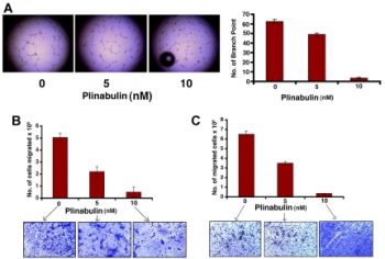
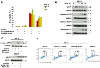
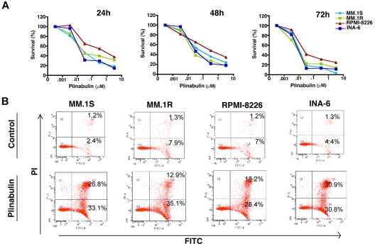
- Plinabulin (NPI-2358) (0.1-10 μM) dose-dependently inhibited bovine brain tubulin polymerization, with 50% inhibition at 1.1 μM. It induced tubulin depolymerization in preassembled microtubules, as shown by decreased turbidity in spectrophotometric assays [1] - In human umbilical vein endothelial cells (HUVECs), plinabulin (0.1-1 μM) inhibited cell migration (by 40-70% in Boyden chamber assays) and tube formation (by 50-80% in Matrigel assays) at concentrations that did not affect cell viability, indicating anti-angiogenic activity [1] - Against various tumor cell lines (e.g., A549, HT29), plinabulin (1-5 μM) reduced cell proliferation (MTT assay) and induced G2/M cell cycle arrest (flow cytometry), with IC50 values ranging from 1.5 to 3.2 μM [1] Tubulin depolymerization and anti-proliferative activity: 1. Tubulin structure disruption: Plinabulin (NPI-2358) (1 nM–10 nM) dose-dependently disrupted microtubule structure in HUVEC and A549 cells. Immunofluorescence staining (anti-β-tubulin antibody + DAPI) showed: (a) At 2 nM, microtubule filaments became fragmented and disorganized (vs. intact, network-like microtubules in control); (b) At 5 nM, microtubule structure was almost completely disassembled [1] 2. Proliferation inhibition: Plinabulin (0.5 nM–20 nM, 72-hour treatment, MTT assay) suppressed growth of 8 human tumor cell lines and HUVEC. IC₅₀ values (as listed in Target section) were consistent across independent experiments, with maximum inhibition (>90%) at 20 nM [1] 3. Tumor vascular-disrupting (VDA) activity in vitro: HUVEC tube formation assay (matrigel-coated plates) showed Plinabulin (1 nM–5 nM) reduced tube formation by ~40% (1 nM) and ~75% (5 nM) vs. control (quantified by tube length and branch number). Additionally, 2 nM Plinabulin induced HUVEC apoptosis (Annexin V-FITC/PI staining) with apoptotic rate ~35% (vs. ~5% in control) [1] |
| ln Vivo |
Tumor perfusion is reduced in a dose- and time-dependent manner in female CDF1 and C3H/He mice when given plinabuLin (0 mg/kg–15 mg/kg; intraperitoneally). Plinabulin's anticancer effects are more responsive to KHT sarcomas than they are to C3H tumors, and both models exhibit an increased radiation response [3].
Thirty-eight patients were enrolled. A dose of 30 mg/m² was selected as the RP2D based on the adverse events of nausea, vomiting, fatigue, fever, tumor pain, and transient blood pressure elevations, with DCE-MRI indicating decreases in tumor blood flow (Ktrans) from 13.5 mg/m² (defining a biologically effective dose) with a 16% to 82% decrease in patients evaluated at 30 mg/m². Half-life was 6.06 ± 3.03 hours, clearance was 30.50 ± 22.88 L/h, and distributive volume was 211 ± 67.9 L.Conclusions: At the RP2D of 30 mg/m², plinabulin showed a favorable safety profile, while eliciting biological effects as evidenced by decreases in tumor blood flow, tumor pain, and other mechanistically relevant adverse events. On the basis of these results additional clinical trials were initiated with plinabulin in combination with standard chemotherapy agents.[2] Plinabulin (7.5 mg/kg) significantly reduced the initial area under curve (IAUC) and the transfer constant (K(trans)) within 1 hour after injection, reaching a nadir at 3 h, but returning to normal within 24 h. A dose-dependent decrease in IAUC and K(trans), was seen at 3 h. No significant anti-tumour effects were observed in the C3H tumours until doses of 12.5 mg/kg were achieved, but started at 1.5 mg/kg in the KHT sarcoma. Irradiating tumours 1 h after injecting plinabulin enhanced response in both models.Conclusions: Plinabulin induced a time- and dose-dependent decrease in tumour perfusion. The KHT sarcoma was more sensitive than the C3H tumour to the anti-tumour effects of plinabulin, while radiation response was enhanced in both models.[3] - In mice bearing HT29 colon carcinoma xenografts, plinabulin (5-20 mg/kg, intravenous injection, weekly for 3 weeks) caused dose-dependent tumor necrosis (assessed by histology) and reduced tumor blood flow (by 60-80% at 20 mg/kg, measured via Doppler ultrasound), consistent with vascular-disrupting effects [3] - When combined with radiation (10 Gy), plinabulin (10 mg/kg) in HT29 xenografts enhanced tumor growth delay (tumor doubling time: 28 days vs. 15 days with radiation alone) and increased apoptotic cells (TUNEL assay) compared to monotherapy [3] - In a phase 1 clinical trial (n=46) with patients with solid tumors or lymphomas, plinabulin (0.4-40 mg/m², intravenous infusion every 2 weeks) showed disease stabilization in 35% of patients, with 2 partial responses in non-Hodgkin lymphoma [2] Tumor vascular disruption and anti-tumor activity: 1. C3H mouse EMT6 mammary tumor model: Mice (n=6/group) were randomized into 4 groups: (1) Control (intraperitoneal injection of 5% DMSO + 95% normal saline); (2) Plinabulin 10 mg/kg (intraperitoneal, single dose); (3) Radiation (8 Gy, single dose, 1 hour after drug administration); (4) Plinabulin 10 mg/kg + Radiation. Results: - Tumor volume: At day 14, reduced by ~40% (Plinabulin alone), ~35% (Radiation alone), and ~70% (combination) vs. control; - Tumor vascular perfusion: Hoechst 33342 (vascular tracer) staining showed vascular perfusion decreased by ~60% (Plinabulin alone) and ~75% (combination) vs. control (measured by fluorescent area fraction); - Tumor cell apoptosis: TUNEL staining showed apoptotic index increased by ~3-fold (Plinabulin alone) and ~5-fold (combination) vs. control [3] 2. CD-1 nude mouse A549 lung tumor model: Plinabulin 20 mg/kg (intraperitoneal, once weekly for 3 weeks) reduced tumor volume by ~50% at day 21 vs. control, with no significant weight loss [3] - Phase I clinical trial results: 1. Patient population: 35 patients with advanced solid tumors (e.g., non-small cell lung cancer, colon cancer) or lymphomas (e.g., diffuse large B-cell lymphoma) refractory to standard therapy; 2. Dosage: Plinabulin administered intravenously (30-minute infusion) every 2 weeks, dose escalated from 0.4 mg/m² to 40 mg/m²; 3. Efficacy: (a) Maximum Tolerated Dose (MTD) = 30 mg/m²; (b) Disease Stabilization (SD) observed in 12/35 patients (34%), with median SD duration of 12 weeks; (c) No objective responses (complete/partial remission) reported [2] |
| Enzyme Assay |
The diketopiperazine NPI-2358 is a synthetic analog of NPI-2350, a natural product isolated from Aspergillus sp., which depolymerizes microtubules in A549 human lung carcinoma cells. Although structurally different from the colchicine-binding site agents reported to date, NPI-2358 binds to the colchicine-binding site of tubulin. NPI-2358 has potent in-vitro anti-tumor activity against various human tumor cell lines and maintains activity against tumor cell lines with various multidrug-resistant (MDR) profiles. In addition, when evaluated in proliferating human umbilical vein endothelial cells (HUVECs), concentrations as low as 10 nmol/l NPI-2358 induced tubulin depolymerization within 30 min. Furthermore, NPI-2358 dose dependently increases HUVEC monolayer permeability--an in-vitro model of tumor vascular collapse. NPI-2358 was compared with three tubulin-depolymerizing agents with vascular-disrupting activity: colchicine, vincristine and combretastatin A-4 (CA4). Results showed that the activity of NPI-2358 in HUVECs was more potent than either colchicine or vincristine; the profile of CA4 approached that of NPI-2358. Altogether, our data show that NPI-2358 is a potent anti-tumor agent which is active in MDR tumor cell lines, and is able to rapidly induce tubulin depolymerization and monolayer permeability in HUVECs. These data warrant further evaluation of NPI-2358 as a vascular-disrupting agent in vivo. Currently, NPI-2358 is in preclinical development for the treatment of cancer. [1]
- Tubulin polymerization assay: Bovine brain tubulin (1 mg/mL) was incubated with plinabulin (0.1-10 μM) in polymerization buffer at 37°C. Turbidity (400 nm) was measured every 2 minutes for 60 minutes to monitor microtubule assembly. Inhibition was calculated relative to vehicle controls [1] - Tubulin depolymerization assay: Preassembled microtubules (stabilized with GTP) were treated with plinabulin (1-10 μM), and turbidity was measured over 30 minutes to assess microtubule disassembly [1] Tubulin polymerization inhibition assay: 1. Protein preparation: Purified bovine brain tubulin (2 mg/mL) resuspended in polymerization buffer (80 mM PIPES, 2 mM MgCl₂, 0.5 mM EGTA, pH 6.9) containing 1 mM GTP (tubulin polymerization cofactor) [1] 2. Reaction setup: 100 μL reaction mixtures prepared with tubulin, GTP, and Plinabulin (0 nM–50 nM, solvent as control). Mixtures transferred to 96-well black microplates [1] 3. Detection: Tubulin polymerization monitored in real-time by measuring fluorescence intensity (excitation 340 nm, emission 450 nm) at 37°C for 60 minutes (fluorescence increases with microtubule formation). Polymerization inhibition rate calculated as (1 – fluorescence of drug group / fluorescence of control group) × 100% [1] 4. Data analysis: IC₅₀ (concentration inhibiting 50% polymerization) determined by fitting inhibition rates to a four-parameter logistic curve [1] |
| Cell Assay |
Cell Viability Assay[1]
Cell Types: HUVECs cells Tested Concentrations: 2 nM, 10 nM, 20 nM and 200 nM Incubation Duration: 30 minutes Experimental Results: Low concentrations (2 nM, 10 nM) rapidly induced tubulin depolymerization in HUVECs. - HUVEC migration assay: Cells were seeded in Boyden chambers with plinabulin (0.1-1 μM) in the lower compartment. After 4 hours, migrated cells on the membrane underside were fixed, stained, and counted. Migration was normalized to vehicle-treated controls [1] - Tumor cell cycle assay: A549 cells were treated with plinabulin (1-5 μM) for 24 hours, fixed, stained with propidium iodide, and analyzed by flow cytometry. The percentage of cells in G2/M phase increased from 15% (control) to 45-60% [1] Microtubule immunofluorescence assay: 1. Cell seeding: HUVEC/A549 cells seeded on coverslips (2×10⁴ cells/coverslip) and cultured overnight (37°C, 5% CO₂) [1] 2. Drug treatment: Plinabulin (0 nM–10 nM) added, incubated for 4 hours [1] 3. Staining: Cells fixed with 4% paraformaldehyde (15 minutes, room temperature), permeabilized with 0.1% Triton X-100 (5 minutes), blocked with 1% BSA (30 minutes). Incubated with anti-β-tubulin primary antibody (4°C, overnight) and Alexa Fluor 488-conjugated secondary antibody (room temperature, 1 hour). Nuclei stained with DAPI (5 minutes) [1] 4. Analysis: Microtubule structure observed via confocal microscopy; microtubule integrity scored by filament continuity (0 = intact network, 3 = complete disassembly) [1] - MTT proliferation assay: 1. Cell seeding: Tumor cells/HUVEC seeded in 96-well plates (5×10³ cells/well) and cultured overnight [1] 2. Drug treatment: Plinabulin (0.5 nM–20 nM, 6 replicates/concentration) added, incubated for 72 hours [1] 3. Detection: 20 μL MTT solution (5 mg/mL in PBS) added, incubated for 4 hours. Supernatant removed, 150 μL DMSO added to dissolve formazan. Absorbance measured at 570 nm; IC₅₀ calculated via GraphPad Prism [1] - HUVEC tube formation assay: 1. Matrigel preparation: Matrigel thawed on ice, coated onto 24-well plates (500 μL/well), polymerized at 37°C for 30 minutes [1] 2. Cell seeding/treatment: HUVEC (2×10⁴ cells/well) resuspended in medium containing Plinabulin (0 nM–5 nM) and seeded onto polymerized Matrigel [1] 3. Analysis: After 6 hours (37°C, 5% CO₂), tube formation imaged via phase-contrast microscopy. Tube length and branch number quantified using image analysis software [1] |
| Animal Protocol |
Animal/Disease Models: Female CDF1 mice (10-14weeks old) with C3H mammary carcinoma; Female C3H/HeJ mice with KHT sarcoma cells (8-weeks-old)[3]
Doses: 0 mg/kg, 1.5 mg/kg , 2.5 mg/kg, 5 mg/kg, 7.5 mg/kg, 10 mg/kg, 12.5 mg/kg, 15 mg/kg; 0.02 mL/g mouse body weight in CDF1 mice and 0.01 mL/g body weight for C3H /HeJ mice Route of Administration: intraperitoneal (ip)injection; 0 huor, 1 huor, 3 hrs (hours), 6 huors, 24 huors Experimental Results: Induced a time- and dose-dependent decrease in tumour perfusion. The KHT sarcoma was more sensitive than the C3H tumour to the anti -tumor, while radiation response was enhanced in both models. Patients received a weekly infusion of plinabulin for 3 of every 4 weeks. A dynamic accelerated dose titration method was used to escalate the dose from 2 mg/m² to the RP2D, followed by enrollment of an RP2D cohort. Safety, pharmacokinetic, and cardiovascular assessments were conducted, and Dynamic contrast-enhanced MRI (DCE-MRI) scans were performed to estimate changes in tumor blood flow.[2] Foot implanted C3H mammary carcinomas or leg implanted KHT sarcomas were used, with plinabulin injected intraperitoneally. Dynamic contrast-enhanced magnetic resonance imaging (DCE-MRI) measurements were made with gadolinium-diethylenetriamine pentaacetic acid (Gd-DTPA) on a 7-tesla magnet. Treatment response was assessed using regrowth delay (C3H tumours), clonogenic survival (KHT sarcomas) or histological estimates of necrosis for both models.[3] - Tumor xenograft model: Nude mice bearing HT29 xenografts (100-200 mm³) received plinabulin (5-20 mg/kg) via intravenous injection once weekly for 3 weeks. Tumor volume was measured twice weekly, and blood flow was assessed using contrast-enhanced ultrasound on day 3 post-first dose [3] - Combination with radiation: Mice with HT29 xenografts were administered plinabulin (10 mg/kg, intravenous) 24 hours before radiation (10 Gy, localized to tumors). Tumor growth and histology were evaluated weekly for 4 weeks [3] Mouse tumor models: 1. C3H mouse EMT6 mammary tumor model: - Animal housing: Female C3H mice (6–8 weeks old, 18–22 g) housed in SPF facilities (22–25°C, 12-hour light/dark cycle) with free access to food/water [3] - Tumor implantation: EMT6 cells (1×10⁶ cells/mouse) resuspended in 100 μL PBS, subcutaneously injected into right flank [3] - Grouping/treatment: When tumors reached ~150 mm³ (day 0), mice randomized into 4 groups (n=6/group): (a) Control: intraperitoneal injection of solvent (10 μL/g body weight); (b) Plinabulin 10 mg/kg: single intraperitoneal dose; (c) Radiation: 8 Gy single dose (1 hour post-Plinabulin); (d) Combination: Plinabulin + Radiation [3] - Monitoring: Tumor volume measured every 2 days (volume = length × width² / 2). On day 7, mice euthanized via CO₂ inhalation; tumors excised for TUNEL staining and vascular perfusion analysis (Hoechst 33342 injection 15 minutes before sacrifice) [3] 2. CD-1 nude mouse A549 lung tumor model: - Tumor implantation: A549 cells (5×10⁶ cells/mouse) resuspended in 100 μL PBS/matrigel (1:1), subcutaneously injected [3] - Treatment: Plinabulin 20 mg/kg (intraperitoneal, once weekly for 3 weeks) when tumors reached ~100 mm³ [3] - Monitoring: Tumor volume measured every 3 days; body weight recorded weekly to assess toxicity [3] - Phase I clinical protocol: 1. Patient selection: Adults (≥18 years) with histologically confirmed advanced solid tumors/lymphomas, ECOG performance status 0–2, adequate organ function [2] 2. Drug administration: Plinabulin diluted in normal saline, administered as a 30-minute intravenous infusion every 2 weeks. Dose escalated using 3+3 design (0.4, 1.2, 3.6, 10, 20, 30, 40 mg/m²) [2] 3. Safety/efficacy monitoring: (a) Adverse events (AEs) graded per NCI-CTCAE v3.0; (b) Tumor assessments every 6 weeks (CT/MRI); (c) Pharmacokinetic (PK) samples collected pre-infusion and at 0.5, 1, 2, 4, 8, 12, 24 hours post-infusion [2] |
| ADME/Pharmacokinetics |
In patients, prinabulin exhibited dose-proportional pharmacokinetic characteristics in a dose range of 0.4–40 mg/m². The mean Cmax was 1.2–28 μg/mL, and the terminal half-life (t1/2) was 4.2–6.8 hours. It is mainly excreted in feces (65%) and urine (20%) [2]
- Plasma protein binding in human plasma >95% [2] Phase I clinical pharmacokinetics: 1. Absorption: Intravenous administration (no oral absorption data); peak plasma concentration (Cmax) increased with increasing dose, from 0.4 mg/m² (Cmax ≈ 2.1 ng/mL) to 30 mg/m² (Cmax ≈ 156 ng/mL) [2] 2. Distribution: Volume of distribution (Vd) ≈ 15–20 L/m² (consistent across dose groups), indicating extensive extravascular distribution [2] 3. Elimination: Terminal half-life (t₁/₂) ≈ 1.2–1.8 hours; clearance (CL) ≈ 12–15 L/h/m², dose-independent [2] |
| Toxicity/Toxicokinetics |
In the Phase 1 trial, dose-limiting toxicities (DLTs) included neutropenia (grade 3/4) at a dose of 40 mg/m² and fatigue (grade 3) at a dose of 30 mg/m². Common adverse events (≥20%) included fatigue, nausea, diarrhea and injection site reactions [2] - No significant hepatotoxicity or nephrotoxicity was observed, and serum ALT/AST and creatinine levels were within the normal range [2] Preclinical in vivo toxicity: 1. Mouse toxicity: Prinabulin (10–20 mg/kg, intraperitoneal injection, once a week for 3 weeks) did not cause significant weight loss (<5% vs. baseline) or death. Histopathological examination of the liver, kidneys and heart revealed no abnormal lesions [3]
- Phase I clinical toxicity: 1. Dose-limiting toxicity (DLT): At a dose of 40 mg/m², the following were observed: (a) Grade 3 neutropenia (2/3 patients, lasting 5-7 days); (b) Grade 3 fatigue (1/3 patients) [2] 2. Common adverse events (all grades, ≥20% of patients): fatigue (63%), nausea (43%), vomiting (26%), diarrhea (23%), neutropenia (20%). Most adverse events were grade 1-2; no grade 4/5 adverse events were reported [2] 3. Organ toxicity: No significant increase in serum ALT/AST (liver) or creatinine (kidney) was observed (≥ grade 3 abnormality incidence <5%) [2] |
| References |
|
| Additional Infomation |
Plinabulin belongs to the 2,5-diketopiperazine class of compounds, with the structure piperazine-2,5-dione, substituted at positions 3 and 6 with benzyl and (5-tert-butyl-1H-imidazol-4-yl)methylene groups, respectively. It is an angiogenic agent and microtubule depolymerizer, and was used in clinical trials for the treatment of non-small cell lung cancer (now terminated). Plinabulin exhibits multiple effects, including microtubule depolymerization, antitumor activity, apoptosis induction, and angiogenesis inhibition. It belongs to the 2,5-diketopiperazine, imidazole, benzene, and olefin classes. Plinabulin is an orally effective diketopiperazine derivative with potential antitumor activity. Plinabulin selectively targets and binds to the colchicine binding site of tubulin, thereby disrupting the homeostasis of microtubule dynamics. This disrupts the assembly of the mitotic spindle, leading to cell cycle arrest in the M phase and blocking cell division. Furthermore, prinabulin may inhibit the growth of proliferating vascular endothelial cells, thereby disrupting the function of the tumor vascular system and further reducing tumor cell proliferation.
Drug Indications Investigations are underway for the treatment of cancer/tumor (not specified). Mechanism of Action NPI-2358 is a vascular disruptor currently being clinically developed by Nereus for the treatment of cancer. NPI-2358 is one of more than 200 synthetic analogues developed after the discovery of Halimede, a compound isolated from marine fungi. In preclinical models of various cancers, including lung cancer, breast cancer, sarcoma, colon cancer, and prostate cancer, NPI-2358, when used in combination with docetaxel and other oncology therapies, has demonstrated potent and selective antitumor activity; additionally, NPI-2358 monotherapy has shown efficacy in various orthotopic models. NPI-2358 interacts with soluble β-tubulin, inhibiting tubulin polymerization without affecting the dynamic function of existing microtubules. Preclinical trials have demonstrated that this targeting property provides highly specific nanomolar-level cytotoxicity while reducing adverse reactions associated with first-generation vascular destructive agents (VDAs), such as cardiotoxicity, hemodynamic alterations, and neuropathy. Prinabulin (NPI-2358) is a vascular destructive agent that causes instability in the endothelial structure of tumor blood vessels, leading to selective collapse of existing tumor vessels. Preclinical data indicated that prinabulin possesses good safety and antitumor activity, thus prompting the initiation of this clinical trial to determine the recommended Phase 2 dose (RP2D) and to evaluate the safety, pharmacokinetics, and bioactivity of prinabulin in patients with advanced malignancies. [2] - Plinabulin (NPI-2358) is a synthetic vascular disruptor (VDA) that targets tumor microvessels by dissociating tubulin in endothelial cells, leading to vascular collapse and tumor necrosis. [1][3] - It is being developed for the treatment of advanced solid tumors and lymphomas, with preclinical data supporting its synergistic effect with radiotherapy. [2][3] Mechanism of action: Plinabulin (NPI-2358) is a novel vascular disruptor (VDA) that binds to the colchicine binding site on β-tubulin, inducing microtubule dissociation. This disrupts the cytoskeleton of tumor-associated endothelial cells, leading to vascular occlusion, tumor hypoxia, and ultimately tumor cell death—a stark contrast to traditional anti-angiogenic drugs (which inhibit new blood vessel formation). [1][3] - Clinical development: A Phase I clinical trial determined the maximum tolerated dose (MTD) to be 30 mg/m² (intravenous injection every 2 weeks) with good safety. Stable disease was observed in 34% of patients who had previously received extensive treatment, which supports further development of combination therapy with standard therapies such as radiotherapy and chemotherapy [2] - Combination potential: In preclinical models, prinabulin has a synergistic effect with radiotherapy, enhancing tumor angiogenesis and hypoxia-induced radiosensitivity, resulting in a doubling of antitumor efficacy compared to monotherapy [3] |
| Molecular Formula |
C19H20N4O2
|
|
|---|---|---|
| Molecular Weight |
336.39
|
|
| Exact Mass |
336.158
|
|
| Elemental Analysis |
C, 67.84; H, 5.99; N, 16.66; O, 9.51
|
|
| CAS # |
714272-27-2
|
|
| Related CAS # |
|
|
| PubChem CID |
9949641
|
|
| Appearance |
Light yellow to yellow solid powder
|
|
| Density |
1.3±0.1 g/cm3
|
|
| Boiling Point |
730.3±60.0 °C at 760 mmHg
|
|
| Flash Point |
395.5±32.9 °C
|
|
| Vapour Pressure |
0.0±2.4 mmHg at 25°C
|
|
| Index of Refraction |
1.657
|
|
| LogP |
2.66
|
|
| Hydrogen Bond Donor Count |
3
|
|
| Hydrogen Bond Acceptor Count |
3
|
|
| Rotatable Bond Count |
3
|
|
| Heavy Atom Count |
25
|
|
| Complexity |
597
|
|
| Defined Atom Stereocenter Count |
0
|
|
| SMILES |
CC(C)(C)C1=C(N=CN1)/C=C\2/C(=O)N/C(=C\C3=CC=CC=C3)/C(=O)N2
|
|
| InChi Key |
UNRCMCRRFYFGFX-TYPNBTCFSA-N
|
|
| InChi Code |
InChI=1S/C19H20N4O2/c1-19(2,3)16-13(20-11-21-16)10-15-18(25)22-14(17(24)23-15)9-12-7-5-4-6-8-12/h4-11H,1-3H3,(H,20,21)(H,22,25)(H,23,24)/b14-9-,15-10-
|
|
| Chemical Name |
(3E,6E)-3-benzylidene-6-((5-(tert-butyl)-1H-imidazol-4-yl)methylene)piperazine-2,5-dione
|
|
| Synonyms |
NPI-2358; Plinabulin; NPI2358; Plinabulin(NPI-2358); NPI 2358; NPI-2358 (Plinabulin); (3z,6z)-3-Benzylidene-6-[(5-Tert-Butyl-1h-Imidazol-4-Yl)methylidene]piperazine-2,5-Dione; 986FY7F8XR; NPI 2358;
|
|
| HS Tariff Code |
2934.99.9001
|
|
| Storage |
Powder -20°C 3 years 4°C 2 years In solvent -80°C 6 months -20°C 1 month |
|
| Shipping Condition |
Room temperature (This product is stable at ambient temperature for a few days during ordinary shipping and time spent in Customs)
|
| Solubility (In Vitro) |
DMSO: 54 mg/mL (160.5 mM)
Water: 1 mg/mL (Insoluble) Ethanol: 1 mg/mL |
|---|---|
| Solubility (In Vivo) |
Solubility in Formulation 1: ≥ 2.5 mg/mL (7.43 mM) (saturation unknown) in 10% DMSO + 40% PEG300 + 5% Tween80 + 45% Saline (add these co-solvents sequentially from left to right, and one by one), clear solution.
For example, if 1 mL of working solution is to be prepared, you can add 100 μL of 25.0 mg/mL clear DMSO stock solution to 400 μL PEG300 and mix evenly; then add 50 μL Tween-80 to the above solution and mix evenly; then add 450 μL normal saline to adjust the volume to 1 mL. Preparation of saline: Dissolve 0.9 g of sodium chloride in 100 mL ddH₂ O to obtain a clear solution. Solubility in Formulation 2: 2.5 mg/mL (7.43 mM) in 10% DMSO + 90% (20% SBE-β-CD in Saline) (add these co-solvents sequentially from left to right, and one by one), suspension solution; with ultrasonication. For example, if 1 mL of working solution is to be prepared, you can add 100 μL of 25.0 mg/mL clear DMSO stock solution to 900 μL of 20% SBE-β-CD physiological saline solution and mix evenly. Preparation of 20% SBE-β-CD in Saline (4°C,1 week): Dissolve 2 g SBE-β-CD in 10 mL saline to obtain a clear solution. View More
Solubility in Formulation 3: ≥ 2.5 mg/mL (7.43 mM) (saturation unknown) in 10% DMSO + 90% Corn Oil (add these co-solvents sequentially from left to right, and one by one), clear solution. |
| Preparing Stock Solutions | 1 mg | 5 mg | 10 mg | |
| 1 mM | 2.9727 mL | 14.8637 mL | 29.7274 mL | |
| 5 mM | 0.5945 mL | 2.9727 mL | 5.9455 mL | |
| 10 mM | 0.2973 mL | 1.4864 mL | 2.9727 mL |
*Note: Please select an appropriate solvent for the preparation of stock solution based on your experiment needs. For most products, DMSO can be used for preparing stock solutions (e.g. 5 mM, 10 mM, or 20 mM concentration); some products with high aqueous solubility may be dissolved in water directly. Solubility information is available at the above Solubility Data section. Once the stock solution is prepared, aliquot it to routine usage volumes and store at -20°C or -80°C. Avoid repeated freeze and thaw cycles.
Calculation results
Working concentration: mg/mL;
Method for preparing DMSO stock solution: mg drug pre-dissolved in μL DMSO (stock solution concentration mg/mL). Please contact us first if the concentration exceeds the DMSO solubility of the batch of drug.
Method for preparing in vivo formulation::Take μL DMSO stock solution, next add μL PEG300, mix and clarify, next addμL Tween 80, mix and clarify, next add μL ddH2O,mix and clarify.
(1) Please be sure that the solution is clear before the addition of next solvent. Dissolution methods like vortex, ultrasound or warming and heat may be used to aid dissolving.
(2) Be sure to add the solvent(s) in order.
| NCT Number | Recruitment | interventions | Conditions | Sponsor/Collaborators | Start Date | Phases |
| NCT05599789 | Recruiting | Drug: Pembrolizumab in Combination with Plinabulin and Docetaxel |
Non Small Cell Lung Cancer Metastatic |
Peking Union Medical College Hospital | February 1, 2023 | Phase 2 |
| NCT05130827 | Active, not recruiting | Drug: Plinabulin | Multiple Myeloma | Memorial Sloan Kettering Cancer Center |
December 21, 2021 | Phase 2 |
| NCT02812667 | Active, not recruiting | Drug: Nivolumab + Plinabulin | Non-small Cell Lung Cancer Metastatic |
Lyudmila Bazhenova, M.D. | August 29, 2016 | Phase 1 |
| NCT03294577 | Active, not recruiting | Drug: Pegfilgrastim Drug: Plinabulin |
Chemotherapy-induced Neutropenia | BeyondSpring Pharmaceuticals Inc. | October 23, 2019 | Phase 3 |
Products are for research use only; We do not sell to patients
Copyright 2020 InvivoChem LLC | All Rights Reserved


 COA
COA