| Size | Price | Stock | Qty |
|---|---|---|---|
| 5mg |
|
||
| 10mg |
|
||
| 25mg |
|
||
| 50mg |
|
||
| 100mg |
|
||
| 250mg |
|
||
| 500mg | |||
| Other Sizes |


Purity: ≥98%
| Targets |
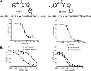
Chymotrypsin-like proteasome (IC50 = 27 nM)
Immunoproteasome subunits: - LMP7 (β5i, chymotrypsin-like activity): IC₅₀ ≈ 12 nM (recombinant human immunoproteasome) [1]; IC₅₀ ≈ 10 nM (purified mouse splenic immunoproteasome) [2] - MECL-1 (β2i, trypsin-like activity): IC₅₀ ≈ 35 nM (recombinant human immunoproteasome) [1] - Constitutive proteasome subunit β5 (β5c): IC₅₀ ≈ 250 nM (recombinant human constitutive proteasome), showing ~20-fold selectivity for LMP7 over β5c [1] |
|---|---|
| ln Vitro |
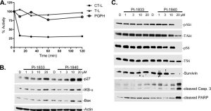
PI-1840 potently suppresses the proliferation and survival of human MDA-MB-468 cells, as well as the proteasomal CT-L activity, with an IC50 of 0.37 μM in intact cancer cells.[1] PI-1840 suppresses CT-L activity in intact cancer cells, causes apoptosis, blocks survival pathways and viability, and causes the proteasome substrates p27, Bax, and IκB-α to accumulate.[2]
Antiproliferative activity in hematologic malignancies (literature [1], [2]): 1. Multiple Myeloma (MM) cell lines (RPMI 8226, U266): PI-1840 (1 nM–100 nM, 72-hour MTT assay) concentration-dependently inhibited proliferation. IC₅₀ values: ~20 nM (RPMI 8226), ~25 nM (U266) [1]. At 50 nM, RPMI 8226 cell viability reduced by ~75% vs. solvent control [1] 2. Mantle Cell Lymphoma (MCL) cell line (Granta-519): IC₅₀ ≈ 22 nM (72-hour MTT assay); 40 nM PI-1840 reduced colony formation by ~80% (soft agar assay) [2] 3. Bortezomib-resistant MM cells (RPMI 8226/BtzR): IC₅₀ ≈ 35 nM (72-hour MTT), maintaining activity against refractory cells [1] - Apoptosis induction (literature [1], [2]): 1. RPMI 8226 cells: 25 nM PI-1840 treatment for 48 hours increased apoptotic rate from ~4% (control) to ~48% (Annexin V-FITC/PI staining, flow cytometry). Western blot showed cleaved caspase-3 (↑3.8-fold) and cleaved PARP (↑3.2-fold) vs. control [1] 2. Granta-519 cells: 30 nM PI-1840 for 48 hours induced ~42% apoptosis; TUNEL staining confirmed DNA fragmentation (↑4.5-fold vs. control) [2] - NF-κB pathway inhibition (literature [1], [2]): 1. RPMI 8226 cells: 20 nM PI-1840 (24-hour treatment) blocked TNF-α-induced NF-κB activation. Western blot revealed IκBα protein accumulation (↑4.0-fold) due to reduced proteasomal degradation; nuclear p-p65 (Ser536) levels decreased by ~65% (immunofluorescence staining) [1] 2. Granta-519 cells: 25 nM PI-1840 reduced LPS-induced TNF-α secretion by ~60% and IL-6 secretion by ~55% (ELISA) [2] - Immunoproteasome client protein downregulation: 1. RPMI 8226 cells: 25 nM PI-1840 (24-hour treatment) reduced immunoproteasome client proteins (c-Myc, Bcl-2) by ~70% and ~65%, respectively (Western blot); no significant effect on constitutive proteasome client protein Hsp70 [1] |
| ln Vivo |
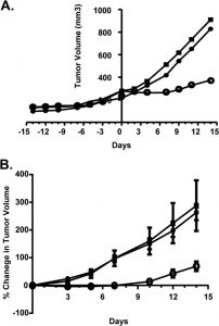
PI-1840 (150 mg/kg i.p.) inhibits the growth of MDA-MB-231 breast tumors in mice.[2]
Nude mouse MM xenograft models (literature [1], [2]): 1. RPMI 8226 xenograft: - Grouping: Mice (n=6/group) randomized into 4 groups: (1) Control (oral solvent: 5% DMSO + 10% Cremophor EL + 85% normal saline); (2) PI-1840 10 mg/kg; (3) PI-1840 30 mg/kg; (4) Bortezomib 0.5 mg/kg (intravenous injection, twice weekly) [1] - Treatment: Drugs administered via oral gavage once daily, starting when tumors reached ~100 mm³,持续21天 [1] - Efficacy: - Tumor volume: Reduced by ~42% (10 mg/kg), ~78% (30 mg/kg), and ~72% (bortezomib) vs. control; - Tumor weight: Decreased by ~38% (10 mg/kg), ~75% (30 mg/kg), and ~70% (bortezomib) at sacrifice; - Tumor immunoproteasome activity: LMP7 activity reduced by ~55% (10 mg/kg) and ~80% (30 mg/kg) (fluorescent substrate assay) [1] 2. U266 xenograft: - Treatment: PI-1840 25 mg/kg (oral gavage, once daily, 28 days) [2] - Efficacy: Tumor growth delay of ~20 days vs. control; serum M-protein (MM marker) decreased by ~65% (ELISA) [2] - Nude mouse MCL xenograft model: 1. Treatment: PI-1840 30 mg/kg (oral gavage, once daily, 21 days) [2] 2. Efficacy: Tumor volume reduced by ~70% vs. control; tumor apoptotic index (TUNEL) increased by ~4.0-fold [2] |
| Enzyme Assay |
In the high-throughput screen, the 50,000 compound library from ChemBridge is assayed (at 10 μM) for inhibitory activity against the CT-L proteolytic activity of the purified rabbit 20S proteasome using fluorogenic peptides as substrates. Using the matching fluorogenic peptides, selectivity for CT-L over T-L and PGPH-L is tested. In summary, 20 μM Suc-Leu-Leu-Val-Tyr-AMC for the CT-L activity, Bz-Val-Gly-Arg-AMC for the T-L activity, and benzyloxycarbonyl Z-Leu-Leu-Glu-AMC for the PGPH-L activity are incubated with 1 nM of purified 20S rabbit proteasome for 1 hour at 37 °C in 100 μL of assay buffer (50 mM Tris-HCl, pH 7.6) with or without compound. After incubation, a WALLAC Victor2 1420 Multilabel Counter equipped with an excitation filter of 355 nm and an emission filter of 460 nm is used to measure the production of hydrolyzed 7-amido-4-methyl-coumarin (AMC) groups. The released amount of AMC falls inside the linear range. As a positive control for IC50 calculations, bonetezomib is utilized. In order to assess proteasome activity in whole cells, the same assay described above is performed using extracts (5 μg) from cultured cell lysate rather than 20S rabbit proteasome.
Human immunoproteasome/constitutive proteasome activity inhibition assay (literature [1], [2]): 1. Protein preparation: - Recombinant human immunoproteasome (LMP7/β2i/β1i) and constitutive proteasome (β5/β2/β1) expressed in HEK293 cells, purified via affinity chromatography (using proteasome-specific antibody beads), resuspended in assay buffer (25 mM Tris-HCl, pH 7.5, 5 mM MgCl₂, 1 mM DTT) [1] - Mouse splenic immunoproteasome: Isolated from C57BL/6 mouse spleen via differential centrifugation and ion-exchange chromatography [2] 2. Reaction setup: 100 μL reaction mixture contained proteasome (0.2 μg), fluorescent substrate (Z-LLVY-AMC for β5i/β5, Z-ARR-AMC for β2i/β2, Z-nLPnLD-AMC for β1i/β1), and PI-1840 (0.1 nM–1000 nM, solvent as control) [1][2] 3. Incubation and detection: Incubated at 37°C for 90 minutes; fluorescence intensity measured at 15-minute intervals (excitation 380 nm, emission 460 nm). Inhibition rate = (1 – fluorescence of drug group / fluorescence of control group) × 100% [1][2] 4. Data analysis: IC₅₀ values calculated by fitting inhibition rates to a four-parameter logistic curve using GraphPad Prism [1][2] |
| Cell Assay |
In 96-well plates, cells are plated in 100 μL medium and left to adhere for the entire night. Next, different inhibitor concentrations are added to the cells and incubated for 120 hours. Aspirate the media, replace it with 100 μL of complete media containing 1 mg/ml of MTT, and incubate for three hours at 37 °C in an incubator with 5% CO2 humidification. After aspirating the media, DMSO is added. A μQuant spectrophotometric plate reader is used to measure the absorbance at 540 nm after the cells are shaken and incubated for 10 minutes at room temperature. Under the CT-L, T-L, and PGPH-L proteolytic activity tests, the IC50 values are calculated using the equation.
MTT antiproliferation assay (literature [1], [2]): 1. Cell seeding: MM/MCL cells (RPMI 8226/U266/Granta-519) seeded in 96-well plates (5×10³ cells/well) in RPMI 1640 medium (10% FBS, 1% penicillin-streptomycin) [1][2] 2. Drug treatment: PI-1840 (1 nM–100 nM, 6 replicates/concentration) added; incubated for 72 hours (37°C, 5% CO₂) [1][2] 3. Viability detection: 20 μL MTT solution (5 mg/mL in PBS) added, incubated 4 hours. Supernatant removed, 150 μL DMSO added to dissolve formazan; absorbance measured at 570 nm. IC₅₀ values calculated [1][2] - Apoptosis assay (Annexin V-FITC/PI, literature [1], [2]): 1. Cell treatment: RPMI 8226/Granta-519 cells (2×10⁵ cells/well, 6-well plates) treated with PI-1840 (10 nM–50 nM) for 48 hours [1][2] 2. Staining: Cells harvested, washed twice with cold PBS, resuspended in 100 μL binding buffer, stained with 5 μL Annexin V-FITC and 5 μL PI for 15 minutes in the dark [1][2] 3. Analysis: Apoptotic cells quantified via flow cytometry; early (Annexin V+/PI-) and late (Annexin V+/PI+) apoptosis percentages recorded [1][2] - Western blot (literature [1], [2]): 1. Cell treatment: Cells treated with PI-1840 (10 nM–30 nM) for 24–48 hours [1][2] 2. Lysate preparation: Cells lysed with RIPA buffer (含 protease/phosphatase inhibitors); protein concentration measured via BCA assay [1][2] 3. Blotting: 30 μg protein separated by 10% SDS-PAGE, transferred to PVDF membrane, blocked with 5% non-fat milk (1 hour, room temperature), probed with target antibodies (IκBα, p-p65, cleaved caspase-3, c-Myc, Bcl-2, β-actin). ECL chemiluminescence detected signals [1][2] |
| Animal Protocol |
Nude mice bearing human breast cancer MDA-MB-231 xenografts
~150 mg/kg daily i.p. Nude mouse RPMI 8226 xenograft protocol: 1. Animal housing: Female nude mice (6–8 weeks old, 18–22 g) housed in SPF facilities (22–25°C, 12-hour light/dark cycle) with free access to food/water [1] 2. Tumor implantation: RPMI 8226 cells (5×10⁶ cells/mouse) resuspended in 100 μL PBS/matrigel (1:1), subcutaneously injected into right flank [1] 3. Grouping and treatment: Tumors reaching ~100 mm³ (day 0) randomized into 4 groups. PI-1840 dissolved in solvent (5% DMSO + 10% Cremophor EL + 85% normal saline) and administered via oral gavage (10 μL/g body weight) at 10 mg/kg or 30 mg/kg, once daily for 21 days. Bortezomib (0.5 mg/kg) administered via intravenous injection twice weekly. Control received solvent alone [1] 4. Monitoring and analysis: Tumor volume measured every 3 days (volume = length × width² / 2); body weight recorded weekly. Mice euthanized via CO₂ inhalation; tumors excised, weighed, and lysed for proteasome activity assay (fluorescent substrate method) and Western blot [1] - Nude mouse Granta-519 xenograft protocol: 1. Tumor implantation: Granta-519 cells (2×10⁶ cells/mouse) resuspended in 100 μL PBS/matrigel (1:1), subcutaneously injected [2] 2. Treatment: PI-1840 30 mg/kg (oral gavage, once daily, 21 days) when tumors reached ~100 mm³ [2] 3. Analysis: Tumor volume measured every 4 days; tumors excised for TUNEL staining (apoptosis) and immunohistochemistry (c-Myc expression) [2] |
| ADME/Pharmacokinetics |
Pharmacokinetics of mice after oral administration (references [1], [2]):
1. Oral bioavailability: ~40% (mice, comparison of oral and intravenous doses of 30 mg/kg)[1] 2. Pharmacokinetic parameters (30 mg/kg orally, mice): - Cmax (peak plasma concentration): ~95 ng/mL (Tmax = 1.2 h); - AUC₀-24h (area under the curve): ~620 ng·h/mL; - Terminal half-life (t₁/₂): ~5.8 h; - Clearance (CL): ~16 mL/min/kg [1] 3. Tissue distribution (30 mg/kg orally, 2 h after administration): - Tumor (RPMI 8226): ~290 ng/g (tumor/plasma ratio ~3.1); - Liver: Approximately 180 ng/g; Spleen: Approximately 150 ng/g; Brain: <10 ng/g (low central nervous system permeability) [1][2] Metabolism: Primarily metabolized in mouse liver via CYP2D6 and CYP3A4; no major active metabolites were detected (LC-MS/MS analysis) [2] Excretion: Approximately 60% of the administered dose was excreted in feces within 72 hours (current drug: approximately 25%); approximately 18% was excreted in urine (primarily metabolites) [2] |
| Toxicity/Toxicokinetics |
In vitro toxicity (References [1], [2]):
1. Normal human cells: - Peripheral blood mononuclear cells (PBMCs): 50 nM PI-1840 (treatment for 72 hours) reduced cell viability by <15% (MTT method) [1]; - Bone marrow stromal cells (BMSCs): 50 nM PI-1840 showed no significant cytotoxicity (viability >85%, trypan blue exclusion method) [2] - In vivo toxicity (References [1], [2]): 1. Subacute toxicity (mice, 30 mg/kg orally, once daily for 21 days): - No death or clinical toxicity (e.g., lethargy, diarrhea); weight change <5% (compared to baseline) [1]; - Serum biochemical indicators (ALT, AST, creatinine, BUN) were all within the normal range; - No histopathological lesions were observed in the liver, kidneys, spleen and heart (HE staining) [1][2] 2. Acute toxicity (mice): A single oral dose of 100 mg/kg did not result in death; transient mild gastrointestinal symptoms (1/6 mice) subsided within 24 hours [2] - Plasma protein binding: approximately 94% (human plasma, balanced dialysis at 37°C) [1] - Drug interactions: no direct data, but metabolism by CYP2D6/CYP3A4 suggests possible interactions with inhibitors/inducers of these enzymes (not tested in this study) [2] |
| References | |
| Additional Infomation |
Background: PI-1840 is an orally effective selective immunoproteasome inhibitor designed to target hematologic malignancies with high expression of immunoproteasomes (e.g., multiple myeloma, mantle cell lymphoma) [1][2] - Mechanism of action: selectively inhibits the LMP7 (β5i) and MECL-1 (β2i) subunits of the immunoproteasome, blocking the degradation of ubiquitinated proteins (e.g., IκBα, c-Myc, Bcl-2). This leads to NF-κB pathway inhibition, downregulation of oncogenes and induction of cancer cell apoptosis[1][2]
- Therapeutic potential: 1. Preclinical efficacy has been demonstrated in bortezomib-sensitive and resistant multiple myeloma models and mantle cell lymphoma models, supporting its application in relapsed/refractory hematologic malignancies[1][2]; 2. Advantages compared to bortezomib: higher oral bioavailability (approximately 40%), higher selectivity to immunoproteasomes (reduced off-target toxicity to normal cells), and effectiveness against bortezomib-resistant cells[1][2] |
| Molecular Formula |
C22H26N4O3
|
|
|---|---|---|
| Molecular Weight |
394.47
|
|
| Exact Mass |
394.2
|
|
| Elemental Analysis |
C, 66.99; H, 6.64; N, 14.20; O, 12.17
|
|
| CAS # |
1401223-22-0
|
|
| Related CAS # |
|
|
| PubChem CID |
56945245
|
|
| Appearance |
White to off-white solid powder
|
|
| LogP |
3.9
|
|
| Hydrogen Bond Donor Count |
0
|
|
| Hydrogen Bond Acceptor Count |
6
|
|
| Rotatable Bond Count |
9
|
|
| Heavy Atom Count |
29
|
|
| Complexity |
496
|
|
| Defined Atom Stereocenter Count |
0
|
|
| SMILES |
O=C(N(C(C)C)CC1=NC(C2=CC=CN=C2)=NO1)COC3=CC=C(CCC)C=C3
|
|
| InChi Key |
ZVVXAODXPVWGMF-UHFFFAOYSA-N
|
|
| InChi Code |
InChI=1S/C22H26N4O3/c1-4-6-17-8-10-19(11-9-17)28-15-21(27)26(16(2)3)14-20-24-22(25-29-20)18-7-5-12-23-13-18/h5,7-13,16H,4,6,14-15H2,1-3H3
|
|
| Chemical Name |
N-propan-2-yl-2-(4-propylphenoxy)-N-[(3-pyridin-3-yl-1,2,4-oxadiazol-5-yl)methyl]acetamide
|
|
| Synonyms |
|
|
| HS Tariff Code |
2934.99.9001
|
|
| Storage |
Powder -20°C 3 years 4°C 2 years In solvent -80°C 6 months -20°C 1 month |
|
| Shipping Condition |
Room temperature (This product is stable at ambient temperature for a few days during ordinary shipping and time spent in Customs)
|
| Solubility (In Vitro) |
DMSO: ~78 mg/mL (~197.7 mM)
Water: 1 mg/mL (Insoluble) Ethanol: ~33 mg/mL (~83.7 mM) |
|---|---|
| Solubility (In Vivo) |
Solubility in Formulation 1: ≥ 2.5 mg/mL (6.34 mM) (saturation unknown) in 10% DMSO + 40% PEG300 + 5% Tween80 + 45% Saline (add these co-solvents sequentially from left to right, and one by one), clear solution.
For example, if 1 mL of working solution is to be prepared, you can add 100 μL of 25.0 mg/mL clear DMSO stock solution to 400 μL PEG300 and mix evenly; then add 50 μL Tween-80 to the above solution and mix evenly; then add 450 μL normal saline to adjust the volume to 1 mL. Preparation of saline: Dissolve 0.9 g of sodium chloride in 100 mL ddH₂ O to obtain a clear solution. Solubility in Formulation 2: ≥ 2.5 mg/mL (6.34 mM) (saturation unknown) in 10% DMSO + 90% Corn Oil (add these co-solvents sequentially from left to right, and one by one), clear solution. For example, if 1 mL of working solution is to be prepared, you can add 100 μL of 25.0 mg/mL clear DMSO stock solution to 900 μL of corn oil and mix evenly. (Please use freshly prepared in vivo formulations for optimal results.) |
| Preparing Stock Solutions | 1 mg | 5 mg | 10 mg | |
| 1 mM | 2.5350 mL | 12.6752 mL | 25.3505 mL | |
| 5 mM | 0.5070 mL | 2.5350 mL | 5.0701 mL | |
| 10 mM | 0.2535 mL | 1.2675 mL | 2.5350 mL |
*Note: Please select an appropriate solvent for the preparation of stock solution based on your experiment needs. For most products, DMSO can be used for preparing stock solutions (e.g. 5 mM, 10 mM, or 20 mM concentration); some products with high aqueous solubility may be dissolved in water directly. Solubility information is available at the above Solubility Data section. Once the stock solution is prepared, aliquot it to routine usage volumes and store at -20°C or -80°C. Avoid repeated freeze and thaw cycles.
Calculation results
Working concentration: mg/mL;
Method for preparing DMSO stock solution: mg drug pre-dissolved in μL DMSO (stock solution concentration mg/mL). Please contact us first if the concentration exceeds the DMSO solubility of the batch of drug.
Method for preparing in vivo formulation::Take μL DMSO stock solution, next add μL PEG300, mix and clarify, next addμL Tween 80, mix and clarify, next add μL ddH2O,mix and clarify.
(1) Please be sure that the solution is clear before the addition of next solvent. Dissolution methods like vortex, ultrasound or warming and heat may be used to aid dissolving.
(2) Be sure to add the solvent(s) in order.
|
|---|
|
|
Products are for research use only; We do not sell to patients
Copyright 2020 InvivoChem LLC | All Rights Reserved


 COA
COA