| Size | Price | Stock | Qty |
|---|---|---|---|
| 10mg |
|
||
| 50mg |
|
||
| 100mg |
|
||
| Other Sizes |
| Targets |
COX-2
Paradol targets cyclooxygenase-2 (COX-2), binding to its active site and inhibiting its activity. By inhibiting COX-2, it reduces the production of prostaglandins, which are mediators of inflammation and pain. It is an effective inhibitor of tumor promotion in mouse skin carcinogenesis. |
|---|---|
| ln Vitro |
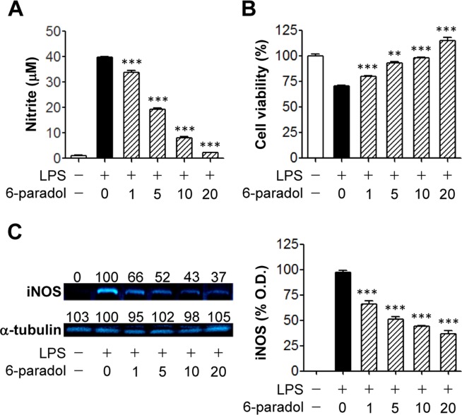
In the oral squamous carcinoma cell line KB, paradol ([6]-paradol) promotes apoptosis in a dose-dependent manner. Through a caspase-3-dependent mechanism, paradol promotes apoptosis [2].
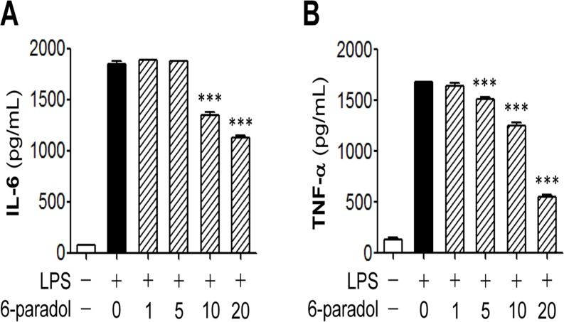
In vitro, paradol has been shown to bind to the COX-2 active site and inhibit its activity. It has antioxidant, anti-inflammatory, and anticancer activities in various cell-based assays. It inhibits the growth of cancer cells and induces apoptosis. |
| ln Vivo |
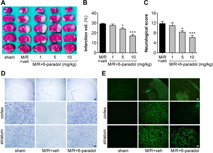
Giving 10 mg/kg of paradol (6-paradol) dramatically decreased the amount of Iba1-positive cells one and three days after the challenge. Moreover, even three days after the M/R challenge, paradol dramatically decreased the amount of Iba1-positive cells in the peri-ischemic region [3]. Of the many paradol compounds—2-, 4-, 6-, 8-, and 10-paradol—paradol (6-paradol) has the strongest anti-inflammatory activity. In addition, out of all the known analogues of Paradol, Paradol has the highest irritating qualities. Paradoll also demonstrated the longest exposure duration at the site of anti-obesity effect, according to the study's metabolite absorption results [4].
In vivo, paradol has been studied for its anticancer, anti-inflammatory, and neuroprotective effects in animal models. It effectively inhibits tumor promotion in mouse skin carcinogenesis. It is a natural product with potential therapeutic applications. |
| Enzyme Assay |
Ginger roots have been used to treat inflammation and have been reported to inhibit cyclooxygenase (COX). Ultrafiltration liquid chromatography mass spectrometry was used to screen a chloroform partition of a methanol extract of ginger roots for COX-2 ligands, and 10-gingerol, 12-gingerol, 8-shogaol, 10-shogaol, 6-gingerdione, 8-gingerdione, 10-gingerdione, 6-dehydro-10-gingerol, 6-paradol, and 8-paradol bound to the enzyme active site. Purified 10-gingerol, 8-shogaol and 10-shogaol inhibited COX-2 with IC(50) values of 32 μM, 17.5 μM and 7.5 μM, respectively. No inhibition of COX-1 was detected. Therefore, 10-gingerol, 8-shogaol and 10-shogaol inhibit COX-2 but not COX-1, which can explain, in part, the anti-inflammatory properties of ginger [1].
The in vitro COX-2 inhibition assay for paradol involves measuring its ability to inhibit COX-2 enzyme activity. This is typically done using a colorimetric or fluorometric assay that measures the conversion of arachidonic acid to prostaglandins. The IC50 is determined. |
| Cell Assay |
KB, human oral epidermoid carcinoma cell lines (ATCC CCL-17) are plated at a density of 5×10³ cells/200 μL/well into 96-well plate. After an overnight growth, the cells are treated with a series of paradol derivatives. All of the derivatives of paradol tested are dissolved in DMSO. The final concentration of DMSO in the culture medium is kept below 0.1% and the controls are treated with DMSO alone. Cell viability is assessed using MTT assay. In brief, after the cells are grown in the media in the absence or presence of the test compounds (e.g., Paradol, 10, 50, 100, 150, and 200 μM) for 48 h, they are then replaced to a 200 μL culture medium containing 0.5 mg/mL MTT for 3 h. The resulting MTT-formazan product is dissolved by an addition of the same volume of DMSO. The amount of formazan is determined by measuring the absorbance at 570 nm[2].
In vitro cellular assays for paradol are performed on various cancer cell lines to study its anticancer effects. Its effects on cell proliferation, apoptosis, and inflammation are measured. Its antioxidant activity is assessed by measuring ROS levels. |
| Animal Protocol |
Mice[3]
Male ICR mice (7 weeks old, 36±2 g) challenged with middle cerebral artery occlusion (MCAO)/reperfusion (M/R) are randomly divided into vehicle (10% Tween80)- or Paradol-administered groups (n=6~7 per group). Paradol dissolved in 10% Tween80 is orally administered (10 mg/kg) into mice at 1, 5, or 10 mg/kg immediately after reperfusion. Rats[4] Five-week-old Sprague-Dawley rats (male) are used. At 8 weeks of age, the rats are fasted for 14 h prior to the oral administration of olive oil (1 mL) containing zingerone or 6-, 8-, or 12-paradol (10 mg/kg). Three rats in each group are anesthetized with isoflurane, and samples (0.3 mL) of their blood are collected from their jugular vein using a heparinized needle and syringe at 0 (i.e., prior to the oral administration), 0.25, 0.5, 1, 3, 6, and 24 h after the oral administration of the olive oil containing test compounds. The AUC0-24h values determined using this time schedule are very similar compared with AUC0-24h that sampled the time points more minutely with other materials in our laboratory. In vivo animal experiments for paradol are conducted in models of cancer, inflammation, and neurodegeneration. The compound is administered orally or topically, and its effects on tumor growth, inflammation, and behavior are assessed. |
| ADME/Pharmacokinetics |
Paradol is a natural product and its pharmacokinetic properties are not extensively documented. It is a metabolite of shogaol found in ginger. Its bioavailability and metabolism are areas of ongoing study.
|
| Toxicity/Toxicokinetics |
Paradol is generally considered to be of low toxicity, as it is a natural compound found in food. Its safety profile has not been extensively studied in clinical trials. As with all research chemicals, standard laboratory safety practices should be followed.
|
| References | |
| Additional Infomation |
[6]- Paradoxol belongs to the phenol, ketone and monomethoxybenzene class of compounds.
It has been reported that paradoxol exists in African ginger (Aframomum melegueta), narrow-leaved African ginger (Aframomum angustifolium) and ginger (Zingiber officinale), and relevant data are available for reference. Paradol is a natural product found in ginger. It is also known as [6]-paradol, [6]-gingerone, and 1-(4-hydroxy-3-methoxyphenyl)decan-3-one. It is not an FDA-approved drug and is primarily used in research. |
| Molecular Formula |
C17H26O3
|
|---|---|
| Molecular Weight |
278.3865
|
| Exact Mass |
278.188
|
| Elemental Analysis |
C, 73.35; H, 9.41; O, 17.24
|
| CAS # |
27113-22-0
|
| Related CAS # |
(E)-[6]-Dehydroparadol;878006-06-5
|
| PubChem CID |
94378
|
| Appearance |
Light yellow to yellow liquid
|
| Density |
1.0±0.1 g/cm3
|
| Boiling Point |
406.5±30.0 °C at 760 mmHg
|
| Melting Point |
31 - 32 °C
|
| Flash Point |
140.0±18.1 °C
|
| Vapour Pressure |
0.0±1.0 mmHg at 25°C
|
| Index of Refraction |
1.507
|
| Source |
ginger and other Zingiberaceae plants
|
| LogP |
3.83
|
| Hydrogen Bond Donor Count |
1
|
| Hydrogen Bond Acceptor Count |
3
|
| Rotatable Bond Count |
10
|
| Heavy Atom Count |
20
|
| Complexity |
265
|
| Defined Atom Stereocenter Count |
0
|
| SMILES |
O=C(C([H])([H])C([H])([H])C1C([H])=C([H])C(=C(C=1[H])OC([H])([H])[H])O[H])C([H])([H])C([H])([H])C([H])([H])C([H])([H])C([H])([H])C([H])([H])C([H])([H])[H]
|
| InChi Key |
IKCUWBHTOKPRNS-UHFFFAOYSA-N
|
| InChi Code |
InChI=1S/C17H26O3/c1-3-4-5-9-15(18)10-7-6-8-14-11-12-16(19)17(13-14)20-2/h11-13,19H,3-10H2,1-2H3
|
| Chemical Name |
1-(4-Hydroxy-3-methoxyphenyl)decan-5-one
|
| Synonyms |
6-Paradol; [6]-Gingerone; [6]-Paradol
|
| HS Tariff Code |
2934.99.9001
|
| Storage |
Powder -20°C 3 years 4°C 2 years In solvent -80°C 6 months -20°C 1 month |
| Shipping Condition |
Room temperature (This product is stable at ambient temperature for a few days during ordinary shipping and time spent in Customs)
|
| Solubility (In Vitro) |
DMSO : ≥ 140 mg/mL (~502.89 mM)
|
|---|---|
| Solubility (In Vivo) |
Solubility in Formulation 1: ≥ 2.5 mg/mL (8.98 mM) (saturation unknown) in 10% DMSO + 40% PEG300 + 5% Tween80 + 45% Saline (add these co-solvents sequentially from left to right, and one by one), clear solution.
For example, if 1 mL of working solution is to be prepared, you can add 100 μL of 25.0 mg/mL clear DMSO stock solution to 400 μL PEG300 and mix evenly; then add 50 μL Tween-80 to the above solution and mix evenly; then add 450 μL normal saline to adjust the volume to 1 mL. Preparation of saline: Dissolve 0.9 g of sodium chloride in 100 mL ddH₂ O to obtain a clear solution. Solubility in Formulation 2: ≥ 2.5 mg/mL (8.98 mM) (saturation unknown) in 10% DMSO + 90% (20% SBE-β-CD in Saline) (add these co-solvents sequentially from left to right, and one by one), clear solution. For example, if 1 mL of working solution is to be prepared, you can add 100 μL of 25.0 mg/mL clear DMSO stock solution to 900 μL of 20% SBE-β-CD physiological saline solution and mix evenly. Preparation of 20% SBE-β-CD in Saline (4°C,1 week): Dissolve 2 g SBE-β-CD in 10 mL saline to obtain a clear solution. View More
Solubility in Formulation 3: ≥ 2.5 mg/mL (8.98 mM) (saturation unknown) in 10% DMSO + 90% Corn Oil (add these co-solvents sequentially from left to right, and one by one), clear solution. |
| Preparing Stock Solutions | 1 mg | 5 mg | 10 mg | |
| 1 mM | 3.5921 mL | 17.9604 mL | 35.9208 mL | |
| 5 mM | 0.7184 mL | 3.5921 mL | 7.1842 mL | |
| 10 mM | 0.3592 mL | 1.7960 mL | 3.5921 mL |
*Note: Please select an appropriate solvent for the preparation of stock solution based on your experiment needs. For most products, DMSO can be used for preparing stock solutions (e.g. 5 mM, 10 mM, or 20 mM concentration); some products with high aqueous solubility may be dissolved in water directly. Solubility information is available at the above Solubility Data section. Once the stock solution is prepared, aliquot it to routine usage volumes and store at -20°C or -80°C. Avoid repeated freeze and thaw cycles.
Calculation results
Working concentration: mg/mL;
Method for preparing DMSO stock solution: mg drug pre-dissolved in μL DMSO (stock solution concentration mg/mL). Please contact us first if the concentration exceeds the DMSO solubility of the batch of drug.
Method for preparing in vivo formulation::Take μL DMSO stock solution, next add μL PEG300, mix and clarify, next addμL Tween 80, mix and clarify, next add μL ddH2O,mix and clarify.
(1) Please be sure that the solution is clear before the addition of next solvent. Dissolution methods like vortex, ultrasound or warming and heat may be used to aid dissolving.
(2) Be sure to add the solvent(s) in order.
|
|
|
Products are for research use only; We do not sell to patients
Copyright 2020 InvivoChem LLC | All Rights Reserved