| Size | Price | Stock | Qty |
|---|---|---|---|
| 1mg |
|
||
| 5mg |
|
||
| 10mg |
|
||
| 100mg | |||
| Other Sizes |


| Targets |
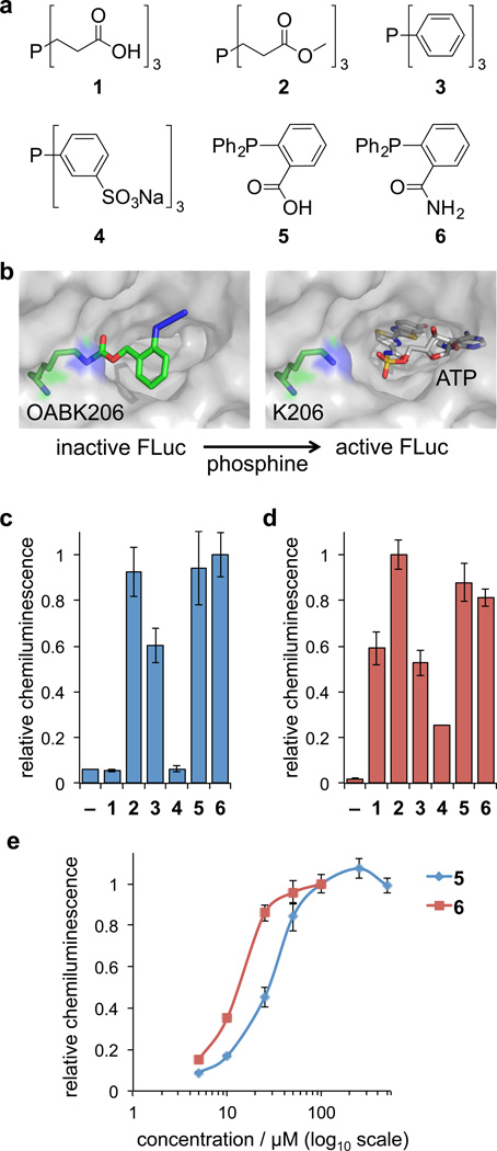
OABK does not have a traditional biological target like an enzyme or receptor. Instead, it is a chemical tool designed for site-specific incorporation into proteins. When introduced at a specific position, such as K206 in firefly luciferase (FLuc), it inhibits enzymatic activity by restricting the access of ATP to the active site. The enzyme can then be deprotected and activated through treatment with a phosphine. This allows for the precise control of protein function.
|
|---|---|
| ln Vitro |
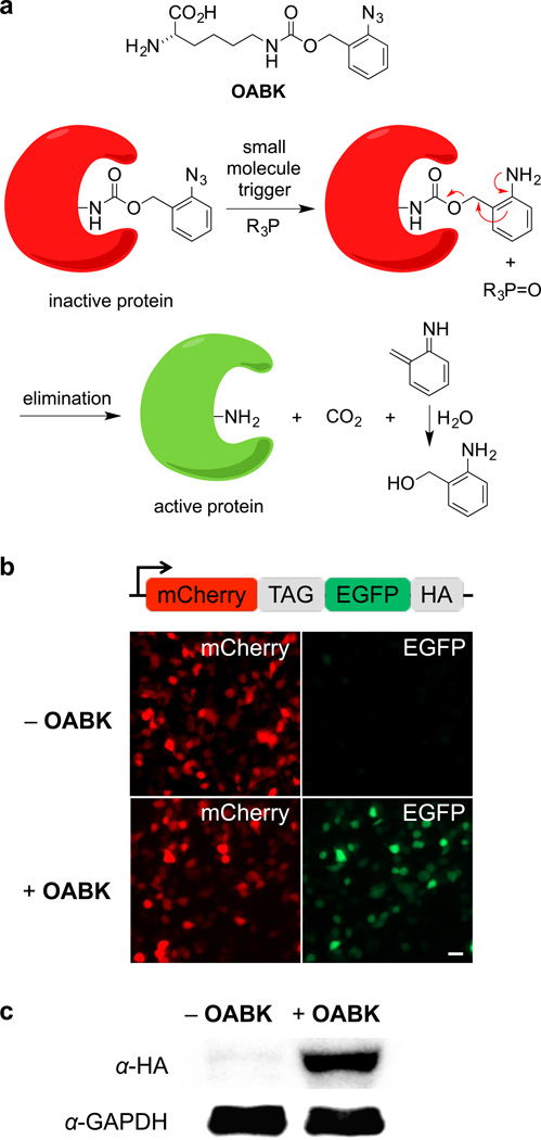
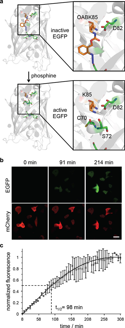
tiny molecular switch that causes the site-specific incorporation of o-azidobenzyloxycarbonyllysine (OABK) to activate protein activity. In three simple stages, 2-azidobenzyl alcohol can be converted to the amino acid OABK using succinimide carbonate ester. Lysine is created during deprotection, and upon incorporation of OABK, an active wild-type protein is produced. Conditional regulation of intracellular protein maturation can be achieved through the combination of small molecule activation and genetically encoded OABK. When OABK (0.5 mM) is added to EGFP at K85, it prevents the production of fluorophores until small molecule activation produces native lysine (PDB-based model). By reducing FLuc enzyme activity, OABK introduction at K206 decreases FLuc enzyme activity. Up until the enzyme is deprotected and activated by phosphine treatment, adenosine triphosphate (ATP) enters the active site. As in the Bright-Glo test's luciferase assay, OABK is added to FLuc in order to inhibit luciferase activity without small molecule activation [1].
In vitro, OABK is used as a small-molecule switch to control protein activity. Its primary application is the site-specific incorporation into proteins to create a "caged" or inhibited form. For example, introducing OABK at position K206 inhibits FLuc enzymatic activity by blocking ATP access to the active site. The protein's activity can then be restored by treating with a phosphine, which removes the OABK group. This provides a powerful tool for studying protein function with temporal control. |
| ln Vivo |
In vivo applications of OABK are not detailed in the provided literature. As a chemical biology tool, its use is primarily in vitro for studying protein function. Its potential for in vivo use would depend on efficient delivery and incorporation methods. The compound is intended for research use only.
|
| Enzyme Assay |
In vitro enzyme/receptor binding studies are not applicable to OABK in the traditional sense, as it is not a drug that binds to a target. Instead, its function is studied by assessing its incorporation into proteins and its subsequent effect on protein activity. For example, the activity of the modified enzyme (e.g., FLuc) can be measured using a luminescence assay. The ability of phosphine to deprotect and reactivate the enzyme is also assessed. These protocols are for research purposes only.
|
| Cell Assay |
In vitro cell-based assays for OABK involve its incorporation into proteins of interest within cells. This is typically achieved through genetic code expansion, where a specific codon is reassigned to encode OABK. The effect of OABK incorporation on the protein's function is then assessed. For example, the activity of a modified enzyme can be measured in cell lysates or in live cells. The reactivation of the protein upon treatment with phosphine can also be monitored. Standard cell culture and molecular biology techniques are used.
|
| Animal Protocol |
In vivo animal studies for OABK are not described in the provided literature. The compound's use is primarily as a research tool in cell biology and biochemistry. If used in vivo, it would likely involve the expression of a protein containing OABK in an animal model, followed by the controlled activation of the protein. This would be a complex and advanced application.
|
| ADME/Pharmacokinetics |
The pharmacokinetic properties of OABK are not characterized. It is a small molecule with a molecular weight of approximately 357.8 g/mol (free base). As a research chemical, it is not intended for systemic administration. Its stability and handling are typical for laboratory reagents.
|
| Toxicity/Toxicokinetics |
The toxicity profile of OABK is not detailed in the available literature. It is classified for research use only and not for human consumption. Standard safety precautions for handling chemical compounds should be followed.
|
| References |
[1]. Luo J, et al. Small-molecule control of protein function through Staudinger reduction. Nat Chem. 2016 Nov;8(11):1027-1034
|
| Additional Infomation |
Additional information: OABK has the CAS number 1984862-48-7. Its molecular formula is C₁₄H₂₀ClN₅O₄. It is a small-molecule switch for controlling protein activity through site-specific incorporation. It contains an azide group for click chemistry. OABK is a research tool for chemical biology and protein engineering. This product is for research use only and is not approved for clinical or therapeutic applications.
|
| Molecular Formula |
C14H20CLN5O4
|
|---|---|
| Molecular Weight |
357.792701721191
|
| Exact Mass |
357.12
|
| CAS # |
1984862-48-7
|
| PubChem CID |
122705997
|
| Appearance |
White to off-white solid powder
|
| Hydrogen Bond Donor Count |
4
|
| Hydrogen Bond Acceptor Count |
7
|
| Rotatable Bond Count |
10
|
| Heavy Atom Count |
24
|
| Complexity |
439
|
| Defined Atom Stereocenter Count |
1
|
| SMILES |
C1=CC=C(C(=C1)COC(=O)NCCCC[C@@H](C(=O)O)N)N=[N+]=[N-].Cl
|
| InChi Key |
JTXGXTOOHZYZDP-MERQFXBCSA-N
|
| InChi Code |
InChI=1S/C14H19N5O4.ClH/c15-11(13(20)21)6-3-4-8-17-14(22)23-9-10-5-1-2-7-12(10)18-19-16;/h1-2,5,7,11H,3-4,6,8-9,15H2,(H,17,22)(H,20,21);1H/t11-;/m0./s1
|
| Chemical Name |
(2S)-2-amino-6-[(2-azidophenyl)methoxycarbonylamino]hexanoic acid;hydrochloride
|
| HS Tariff Code |
2934.99.9001
|
| Storage |
Powder -20°C 3 years 4°C 2 years In solvent -80°C 6 months -20°C 1 month Note: Please store this product in a sealed and protected environment, avoid exposure to moisture. |
| Shipping Condition |
Room temperature (This product is stable at ambient temperature for a few days during ordinary shipping and time spent in Customs)
|
| Solubility (In Vitro) |
DMSO : ~25 mg/mL (~69.87 mM)
|
|---|---|
| Solubility (In Vivo) |
Solubility in Formulation 1: ≥ 2.5 mg/mL (6.99 mM) (saturation unknown) in 10% DMSO + 40% PEG300 + 5% Tween80 + 45% Saline (add these co-solvents sequentially from left to right, and one by one), clear solution.
For example, if 1 mL of working solution is to be prepared, you can add 100 μL of 25.0 mg/mL clear DMSO stock solution to 400 μL PEG300 and mix evenly; then add 50 μL Tween-80 to the above solution and mix evenly; then add 450 μL normal saline to adjust the volume to 1 mL. Preparation of saline: Dissolve 0.9 g of sodium chloride in 100 mL ddH₂ O to obtain a clear solution. Solubility in Formulation 2: ≥ 2.5 mg/mL (6.99 mM) (saturation unknown) in 10% DMSO + 90% (20% SBE-β-CD in Saline) (add these co-solvents sequentially from left to right, and one by one), clear solution. For example, if 1 mL of working solution is to be prepared, you can add 100 μL of 25.0 mg/mL clear DMSO stock solution to 900 μL of 20% SBE-β-CD physiological saline solution and mix evenly. Preparation of 20% SBE-β-CD in Saline (4°C,1 week): Dissolve 2 g SBE-β-CD in 10 mL saline to obtain a clear solution. View More
Solubility in Formulation 3: ≥ 2.5 mg/mL (6.99 mM) (saturation unknown) in 10% DMSO + 90% Corn Oil (add these co-solvents sequentially from left to right, and one by one), clear solution. |
| Preparing Stock Solutions | 1 mg | 5 mg | 10 mg | |
| 1 mM | 2.7949 mL | 13.9747 mL | 27.9494 mL | |
| 5 mM | 0.5590 mL | 2.7949 mL | 5.5899 mL | |
| 10 mM | 0.2795 mL | 1.3975 mL | 2.7949 mL |
*Note: Please select an appropriate solvent for the preparation of stock solution based on your experiment needs. For most products, DMSO can be used for preparing stock solutions (e.g. 5 mM, 10 mM, or 20 mM concentration); some products with high aqueous solubility may be dissolved in water directly. Solubility information is available at the above Solubility Data section. Once the stock solution is prepared, aliquot it to routine usage volumes and store at -20°C or -80°C. Avoid repeated freeze and thaw cycles.
Calculation results
Working concentration: mg/mL;
Method for preparing DMSO stock solution: mg drug pre-dissolved in μL DMSO (stock solution concentration mg/mL). Please contact us first if the concentration exceeds the DMSO solubility of the batch of drug.
Method for preparing in vivo formulation::Take μL DMSO stock solution, next add μL PEG300, mix and clarify, next addμL Tween 80, mix and clarify, next add μL ddH2O,mix and clarify.
(1) Please be sure that the solution is clear before the addition of next solvent. Dissolution methods like vortex, ultrasound or warming and heat may be used to aid dissolving.
(2) Be sure to add the solvent(s) in order.
|
|
|
Products are for research use only; We do not sell to patients
Copyright 2020 InvivoChem LLC | All Rights Reserved