| Size | Price | Stock | Qty |
|---|---|---|---|
| 10mg |
|
||
| 25mg |
|
||
| 50mg |
|
||
| 100mg |
|
||
| 250mg |
|
||
| 500mg | |||
| 1g | |||
| Other Sizes |


Purity: ≥98%
| Targets |
IDO1 (IC50 = 38 nM)
NLG919 targets IDO1 (Indoleamine 2,3-dioxygenase 1) with a Ki value of 0.9 nM [1] |
|---|---|
| ln Vitro |
Strong IDO1 inhibitor IDO-IN-7 (analogue of NLG-919) has an IC50 of 38 nM. IDO-IN-7's binding mode to IDO1 is accessible through experimentation and demonstrates a direct coordination interaction with the ferric heme's sixth coordination site. In previous research, IDO-IN-7 was utilized as a reference drug to build immunostimulatory nanomicellar carriers and verify high-throughput screening assays for IDO1 inhibition[1].
IDO1 enzyme inhibition: NLG919 exhibited potent and selective inhibition of recombinant human IDO1 enzyme activity, with a Ki of 0.9 nM. It did not show significant inhibition against other heme-containing enzymes, indicating high target selectivity [1] - Cellular IDO1 inhibition: In IFN-γ-stimulated A549 cells (which endogenously express IDO1), NLG919 concentration-dependently reduced kynurenine (Kyn) production. The compound effectively blocked tryptophan catabolism in the cellular context without affecting cell viability at effective inhibitory concentrations [1] |
| ln Vivo |
In mice, a single oral administration of NLG919 reduces the concentration of plasma and tissue Kyn by ~ 50%. In mice bearing B16F10 tumors, NLG919 markedly enhances the antitumor responses of naive, resting pmel-1 cells to vaccination with cognate hgp100 peptide plus CpG-1826 in IFA.
|
| Enzyme Assay |
Microscale thermophoresis (MST)[1]
Thermophoresis is the movement of a biomolecular complex in a temperature gradient depending on size, charge, and hydration shell that typically change upon ligand/target interaction. The MST experiment is based on the use of 16 capillary tubes that are filled with a fluorescent dye-labeled target protein and a serial titration of unlabeled ligand. Capillary tubes are then illuminated with an infrared laser that generates a temperature gradient. The protein/ligand complex migrates along this gradient causing changes in the observed fluorescence. These are used to generate a binding curve as a function of ligand concentration that is then analyzed to assess the Kd value. Fluorescence labeling of rhIDO1 was performed following the protocol for N-hydroxysuccinimide (NHS) coupling of the dye NT647 to lysine residues. Briefly, 100 μL of a 9.95 μM solution of rhIDO1 protein in labeling buffer (130 mM NaHCO3, 50 mM NaCl, pH 8.2) was mixed with 100 μL of 39.8 μM NT647-NHS fluorophore in labeling buffer and incubated for 30 min at room temperature (RT) in the dark. Unbounded fluorophores were removed by size-exclusion chromatography with MST buffer (50 mM TRIS, 150 mM NaCl, 10 mM MgCl2, pH 7.4, 0.05% Tween20) as running buffer. The real concentration of each element of the sample, such as protein, heme group and RED dye, and the degree of labeling (DOL) were determined using extinction coefficient ɛ280 = 51,380 M−1 cm−1 for rhIDO1, ɛ405 = 159,000 M−1 cm−1 for rhIDO1 heme group and ɛ650 = 250,000 M−1 cm−1 for NT647 fluorophore, with a correction factor of Fcorr of 0.028 at 280 nm, using Cprot = [A280 – (A280 x Fcorr)/ɛ280 x l] and DOL resulted between 0.6 and 0.8 throughout all labeling reactions. The stability of NT647-rhIDO1 and unmodified rhIDO1 protein was checked using circular dicroism. Spectra of both proteins were recorded using Jasco810 spectrophotometer with 1 mm path-length quartz cuvettes at room temperature (≈22 °C). Sensitivity was 100 millidegrees, and the scanning speed was 20 nm/min for an accumulation of 2 scans. CD data were collected between 180 and 260 nm for both samples at a concentration of 0.1 mg/ml in phosphate buffer (PPB; 50 mM K2HPO4, pH 7.4) Deconvolution of spectra was performed with CDNN 2.1 software. Results are reported in the supplementary materials (Table S1). Compound screening was carried out using premium-coated capillary and MST buffer including 2% DMSO and 2 mM DTT. Compound stocks (50 mM) in DMSO were diluted in assay buffer to reach a final maximum concentration of 500 μM or 1 mM, depending on compound solubility. Compound pre-dilutions were prepared for MST experiments by 16-fold 1:1 serial dilutions in assay buffer containing 4% DMSO in PCR tubes (supplied by NanoTemper Technologies) to yield final volumes of 10 μL. A solution of NT647-rhIDO1 at a concentration of 90 nM was prepared and 10 μL of this solution was added to each compound dilution to reach a final NT647-rhIDO1 concentration of 45 nM and a reaction volume of 20 μL. These samples were loaded into 16 premium-coated capillary tubes and inserted in the chip tray of the MST instrument (Monolith NT.115) for thermophoresis analysis and the appraisal of Kd values. MST signals were recorded at MST 40% (compounds 7, 9, 10, 23, 28). Compounds not providing a binding curve with a good signal/noise ratio at 40% (8, 11–22, 24–27, 29) were tested at MST 80%. In both cases, a 20% LED power was used. Kd values were calculated from compound concentration-dependent changes in normalized fluorescence (Fnorm) of NT647-rhIDO1 after 21s of thermophoresis at MST 40% and after 4s at MST 80%. Each compound was tested in triplicate samples and data analyzed using MO Affinity Analysis software (NanoTemper Technologies). Confidence values (±) are indicated next to Kd value for each of tested compound. Specifically, confidence values define the range where the Kd falls with a 68% of certainty. The binding efficiency index (BEI) of each fragment was calculated with the following equations: (eq. 1) BEI = pKd/MW. Recombinant IDO1 enzyme activity assay: Prepare reaction mixtures containing recombinant human IDO1, tryptophan (substrate), and a reducing agent. Add serial dilutions of NLG919 to the mixtures and incubate at 37°C for a specified period. Terminate the reaction with an appropriate reagent, then detect the formation of kynurenine using spectrophotometric analysis at a specific wavelength. Calculate the inhibition rate and Ki value by fitting the data to a competitive inhibition model [1] |
| Cell Assay |
Cellular assay[1]
P1.HTR, a highly transfectable clonal variant of mouse mastocytoma P815 was cultured in Iscove's Modified Dulbecco's Medium supplemented with 10% FCS. P1.HTR cells were transfected by electroporation with plasmid constructs coding for murine IDO1 (P1.IDO1). A stable transfectant cell line was obtained by puromycin selection. Cells at the concentration of 0.1 × 10⁶ cell/ml were incubated with 30 μM of compounds for 16 h. Control was represented by cells incubated with an equivalent volume of DMSO (the vehicle in which compounds were solubilized). After the incubation, supernatants of cell cultures were recovered and kynurenine concentration was detected by HPLC. Dose-response curves were built through the same cellular assay, incubating P1.IDO1 cells with serial dilutions of molecules, starting from 30 μM. All the experiments were conducted in triplicate and repeated almost two times. Results are represented as the mean ± standard deviation of the kynurenine fold change (l-Kyn FC), meaning the ratio between kynurenine concentration secreted in the supernatant of the compound-treated versus vehicle-treated cells. IFN-γ-stimulated A549 cell IDO1 inhibition assay: Culture A549 cells in appropriate medium until reaching logarithmic growth phase. Stimulate cells with IFN-γ for 24 hours to induce IDO1 expression. Add different concentrations of NLG919 to the stimulated cells and incubate for another 24 hours. Collect cell supernatants and measure the concentration of kynurenine using spectrophotometric methods. Evaluate cell viability simultaneously with a cell proliferation assay to exclude non-specific cytotoxicity [1] |
| Animal Protocol |
Dissolved in water at 3 mg/mL; 6 mg/day injected via IP, or administered subcutaneously at 1 mg/dose twice a day via injection plus 360 μg/day via an SC osmotic pump.
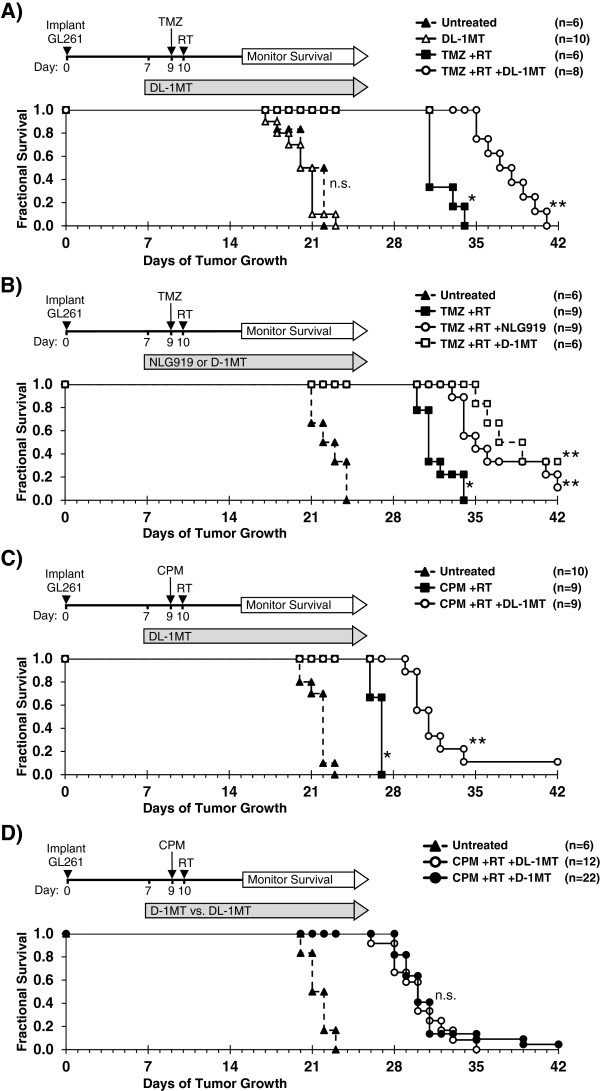
Mice bearing large established B16F10 tumor In vivo DOX treatment and tumor measurement[2] To compare the inhibition of tumor growth by DOX, mouse breast tumor model was established by injecting normal 4T1 cells as above described, and DOX was injected into them at the dose of 5.0 mg/kg for 5 times at intervals of 3 days each, when the tumor volume reach about 50 mm3. Tumor volume was measured after every 4 days using an electronic caliper, and the mean tumor volumes (mm3) of the DOX and control groups were used to plot tumor growth curves. In vivo application of DOX and NLG919 combination[2] BALB/c mice bearing 4T1 tumors of ~50 mm3 volume were administered vehicle control, DOX, NLG919, or a combination of DOX and NLG919 after every 3 days for 5 times; the administration dosage of DOX and NLG919 were 5.0 mg/kg (i.v.) and 20 mg/kg (orally), respectively. The tumor volume in each mouse group was measured as described earlier after every 3 days up to 21 days. The Kyn (nM)/Trp (µM) ratios in the tumor homogenates and plasma of mice in each group were assessed. Then, the tumor tissues were subjected to the hematoxylin and eosin stain (H&E) and proliferating cell nuclear antigen (PCNA) staining and analyzed by IHC. |
| References | |
| Additional Infomation |
Indoleamine 2,3-dioxygenase 1 (IDO1) has attracted much attention as a drug target in the field of tumor immunology because it is highly expressed in cancer cells and participates in the tumor immune editing process. Although a variety of IDO1 inhibitors have been reported in the literature and patent applications, only a few compounds have shown the best pharmacological properties in preclinical studies, thus enabling them to enter clinical trials. Therefore, finding new structural classes of IDO1 inhibitors remains an urgent problem to be solved. This article reports a fragment-based screening method that combines aqueous LOGSY NMR experiments and microscale thermophoresis to identify fragments that may help develop new IDO1 inhibitors as therapeutic drugs for tumor immune diseases. [1]
Objective: Breast cancer has become a major public health threat in today's society. The anthracycline drug doxorubicin (DOX) is a widely used drug in breast cancer chemotherapy. This study aims to investigate the immunogenic death of breast cancer cells induced by doxorubicin (DOX) and to test the efficacy of DOX combined with small molecule inhibitors in a mouse model of tumor transplantation. [2] Methods: We used 4T1 breast cancer cells to investigate the immunogenic death of breast tumor cells mediated by the anthracycline drug DOX by detecting calreticulin exposure, adenosine triphosphate (ATP) release, and high-mobility group box 1 (HMGB1). Using a 4T1 tumor cell transplantation mouse model, we also detected the expression of indoleamine 2,3-dioxygenase (IDO) in tumor tissue after DOX treatment and further explored whether the specific small molecule IDO1 inhibitor NLG919 combined with DOX could better treat breast cancer. [2] Results: DOX induced immunogenic cell death in mouse 4T1 breast cancer cells and upregulated IDO1 expression. We also found that NLG919 treatment dose-dependently enhanced the inhibitory effect of kynurenine. The IDO1 inhibitor reversed the CD8+ T cell suppression mediated by IDO-expressing 4T1 mouse breast cancer cells. Compared with monotherapy or the control group, the combination of DOX and NLG919 significantly inhibited tumor growth, indicating that the two drugs have a synergistic effect. The combination therapy also increased the expression of transforming growth factor-β, while decreasing the expression of interleukin-12p70 and interferon-γ. [2] Conclusion: Compared with monotherapy, NLG919 combined with DOX showed better therapeutic effects in the 4T1 mouse breast tumor model. The inhibition of IDO by NLG919 enhanced the therapeutic effect of DOX in breast cancer, achieving a synergistic effect. [2] Mechanism of action: NLG919, as a competitive inhibitor of IDO1, binds to the active site of the enzyme and blocks the conversion of tryptophan to kynurenine. Its structure was determined by a fragment-based drug discovery method, optimizing the interaction with key amino acid residues of the active site of IDO1. [1] Target selectivity: NLG919 showed high selectivity for IDO1, superior to related enzymes such as tryptophan 2,3-dioxygenase (TDO) and other heme-dependent enzymes, thereby minimizing potential off-target effects. [1] |
| Molecular Formula |
C18H22N2O
|
|
|---|---|---|
| Molecular Weight |
282.38
|
|
| Exact Mass |
282.173
|
|
| Elemental Analysis |
C, 76.56; H, 7.85; N, 9.92; O, 5.67
|
|
| CAS # |
1402836-58-1
|
|
| Related CAS # |
|
|
| PubChem CID |
66558287
|
|
| Appearance |
White to khaki solid powder
|
|
| Density |
1.3±0.1 g/cm3
|
|
| Boiling Point |
524.6±33.0 °C at 760 mmHg
|
|
| Flash Point |
271.1±25.4 °C
|
|
| Vapour Pressure |
0.0±1.4 mmHg at 25°C
|
|
| Index of Refraction |
1.676
|
|
| LogP |
3.28
|
|
| Hydrogen Bond Donor Count |
1
|
|
| Hydrogen Bond Acceptor Count |
2
|
|
| Rotatable Bond Count |
3
|
|
| Heavy Atom Count |
21
|
|
| Complexity |
355
|
|
| Defined Atom Stereocenter Count |
0
|
|
| SMILES |
OC(C1CCCCC1)CC(C2=C3C=CC=C2)N4C3=CN=C4
|
|
| InChi Key |
YTRRAUACYORZLX-UHFFFAOYSA-N
|
|
| InChi Code |
InChI=1S/C18H22N2O/c21-18(13-6-2-1-3-7-13)10-16-14-8-4-5-9-15(14)17-11-19-12-20(16)17/h4-5,8-9,11-13,16,18,21H,1-3,6-7,10H2
|
|
| Chemical Name |
1-cyclohexyl-2-(5H-imidazo[5,1-a]isoindol-5-yl)ethanol
|
|
| Synonyms |
|
|
| HS Tariff Code |
2934.99.9001
|
|
| Storage |
Powder -20°C 3 years 4°C 2 years In solvent -80°C 6 months -20°C 1 month |
|
| Shipping Condition |
Room temperature (This product is stable at ambient temperature for a few days during ordinary shipping and time spent in Customs)
|
| Solubility (In Vitro) |
DMSO: 15 mg/mL warmed (53.1 mM)
Water: 1 mg/mL (Insoluble) Ethanol: 30 mg/mL (106.2 mM) |
|---|---|
| Solubility (In Vivo) |
Solubility in Formulation 1: ≥ 2.5 mg/mL (8.85 mM) (saturation unknown) in 10% DMSO + 40% PEG300 + 5% Tween80 + 45% Saline (add these co-solvents sequentially from left to right, and one by one), clear solution.
For example, if 1 mL of working solution is to be prepared, you can add 100 μL of 25.0 mg/mL clear DMSO stock solution to 400 μL PEG300 and mix evenly; then add 50 μL Tween-80 to the above solution and mix evenly; then add 450 μL normal saline to adjust the volume to 1 mL. Preparation of saline: Dissolve 0.9 g of sodium chloride in 100 mL ddH₂ O to obtain a clear solution. Solubility in Formulation 2: ≥ 2.5 mg/mL (8.85 mM) (saturation unknown) in 10% DMSO + 90% (20% SBE-β-CD in Saline) (add these co-solvents sequentially from left to right, and one by one), clear solution. For example, if 1 mL of working solution is to be prepared, you can add 100 μL of 25.0 mg/mL clear DMSO stock solution to 900 μL of 20% SBE-β-CD physiological saline solution and mix evenly. Preparation of 20% SBE-β-CD in Saline (4°C,1 week): Dissolve 2 g SBE-β-CD in 10 mL saline to obtain a clear solution. View More
Solubility in Formulation 3: ≥ 2.5 mg/mL (8.85 mM) (saturation unknown) in 10% DMSO + 90% Corn Oil (add these co-solvents sequentially from left to right, and one by one), clear solution. Solubility in Formulation 4: ≥ 2.5 mg/mL (8.85 mM) (saturation unknown) in 10% EtOH + 40% PEG300 + 5% Tween80 + 45% Saline (add these co-solvents sequentially from left to right, and one by one), clear solution. For example, if 1 mL of working solution is to be prepared, you can add 100 μL of 25.0 mg/mL clear EtOH stock solution to 400 μL of PEG300 and mix evenly; then add 50 μL of Tween-80 to the above solution and mix evenly; then add 450 μL of normal saline to adjust the volume to 1 mL. Preparation of saline: Dissolve 0.9 g of sodium chloride in 100 mL ddH₂ O to obtain a clear solution. Solubility in Formulation 5: ≥ 2.5 mg/mL (8.85 mM) (saturation unknown) in 10% EtOH + 90% (20% SBE-β-CD in Saline) (add these co-solvents sequentially from left to right, and one by one), clear solution. For example, if 1 mL of working solution is to be prepared, you can add 100 μL of 25.0 mg/mL clear EtOH stock solution to 900 μL of 20% SBE-β-CD physiological saline solution and mix evenly. Preparation of 20% SBE-β-CD in Saline (4°C,1 week): Dissolve 2 g SBE-β-CD in 10 mL saline to obtain a clear solution. Solubility in Formulation 6: ≥ 2.5 mg/mL (8.85 mM) (saturation unknown) in 10% EtOH + 90% Corn Oil (add these co-solvents sequentially from left to right, and one by one), clear solution. For example, if 1 mL of working solution is to be prepared, you can add 100 μL of 25.0 mg/mL clear EtOH stock solution to 900 μL of corn oil and mix evenly. |
| Preparing Stock Solutions | 1 mg | 5 mg | 10 mg | |
| 1 mM | 3.5413 mL | 17.7066 mL | 35.4133 mL | |
| 5 mM | 0.7083 mL | 3.5413 mL | 7.0827 mL | |
| 10 mM | 0.3541 mL | 1.7707 mL | 3.5413 mL |
*Note: Please select an appropriate solvent for the preparation of stock solution based on your experiment needs. For most products, DMSO can be used for preparing stock solutions (e.g. 5 mM, 10 mM, or 20 mM concentration); some products with high aqueous solubility may be dissolved in water directly. Solubility information is available at the above Solubility Data section. Once the stock solution is prepared, aliquot it to routine usage volumes and store at -20°C or -80°C. Avoid repeated freeze and thaw cycles.
Calculation results
Working concentration: mg/mL;
Method for preparing DMSO stock solution: mg drug pre-dissolved in μL DMSO (stock solution concentration mg/mL). Please contact us first if the concentration exceeds the DMSO solubility of the batch of drug.
Method for preparing in vivo formulation::Take μL DMSO stock solution, next add μL PEG300, mix and clarify, next addμL Tween 80, mix and clarify, next add μL ddH2O,mix and clarify.
(1) Please be sure that the solution is clear before the addition of next solvent. Dissolution methods like vortex, ultrasound or warming and heat may be used to aid dissolving.
(2) Be sure to add the solvent(s) in order.
| NCT Number | Recruitment | interventions | Conditions | Sponsor/Collaborators | Start Date | Phases |
| NCT05469490 | Withdrawn | Radiation: Stereotactic Body Radiotherapy (SBRT) |
Advanced Solid Tumors | Luke, Jason, MD | October 2022 | Phase 1 |
 |
|---|
|
|
|
|---|
|
|
Products are for research use only; We do not sell to patients
Copyright 2020 InvivoChem LLC | All Rights Reserved


 COA
COA