| Size | Price | Stock | Qty |
|---|---|---|---|
| 10g |
|
||
| 25g |
|
||
| 50g | |||
| Other Sizes |


Purity: ≥98%
| Targets |
antihelminthic; STAT3 (IC50 = 0.25 μM in HeLa cells); ROS; NF-κB; mTORC1; Wnt/β-catenin; Notch;
NICLOSAMIDE (BAY2353) targets STAT3 signaling pathway (IC50=0.5 μM for STAT3 phosphorylation inhibition) [4] NICLOSAMIDE (BAY2353) inhibits Wnt/β-catenin pathway (IC50=0.3 μM for β-catenin nuclear translocation inhibition) [3] NICLOSAMIDE (BAY2353) targets RANKL-induced NF-κB and MAPK (p38, ERK1/2) signaling pathways [7] NICLOSAMIDE (BAY2353) inhibits severe acute respiratory syndrome coronavirus (SARS-CoV) replication (EC50=0.1 μM) [5] NICLOSAMIDE (BAY2353) inhibits Zika virus (ZIKV) replication (EC50=0.2 μM) [6] NICLOSAMIDE (BAY2353) targets mitochondrial uncoupling proteins (UCPs) [2] |
|---|---|
| ln Vitro |
In BD140A, SW-13, and NCI-H295R cells, niclosamide (0.6 nM–46 μM) therapy reduces the proliferation of adrenocortical carcinoma cells [3]. In HeLa cells, niclosamide administration (0.05–5 μM, 24 h) suppresses STAT3-mediated luciferase reporter activity [4]. In Vero E6 cells, treatment with niclosamide (10 μM) suppresses viral replication [5]. In SNB-19 cells, nicolosamide (GMP) (maximum 2 μM, 24 h) suppresses Zika virus infection [6]. Particularly in the early phases of osteoclastogenesis, nicolosamide (GMP) (1.5 μM, 5 d) suppresses the transdifferentiation of macrophages to precursor osteoclast bodies triggered by factor-κB ligand (RANKL) [7].
NICLOSAMIDE (BAY2353) (0.1-5 μM) dose-dependently inhibited proliferation of human adrenocortical carcinoma (ACC) cells (H295R, SW13) with IC50 values of 0.8 μM and 1.2 μM respectively [3] NICLOSAMIDE (BAY2353) (1 μM) induced apoptosis in ACC cells: apoptotic rate increased by 52% (Annexin V/PI staining), caspase-3/-9 activity enhanced by 3.8-fold, and anti-apoptotic proteins Bcl-2 and Survivin downregulated by 0.4-fold and 0.3-fold [3] NICLOSAMIDE (BAY2353) (0.5-2 μM) suppressed STAT3 phosphorylation (Tyr705) and DNA binding activity in HeLa cells, reducing STAT3 target genes (Cyclin D1, c-Myc) mRNA levels by 60-75% [4] NICLOSAMIDE (BAY2353) (0.1 μM) inhibited SARS-CoV replication in Vero cells by 90%, without significant cytotoxicity (CC50>10 μM) [5] NICLOSAMIDE (BAY2353) (0.2-1 μM) reduced ZIKV-induced death of human neural progenitor cells (hNPCs) by 65-82%, and inhibited ZIKV RNA replication by 70% at 0.5 μM [6] NICLOSAMIDE (BAY2353) (1-10 μM) dose-dependently inhibited RANKL-induced osteoclastogenesis in mouse bone marrow macrophages (BMMs), with 80% inhibition at 5 μM [7] NICLOSAMIDE (BAY2353) (5 μM) downregulated RANKL-induced NF-κB p65 nuclear translocation and MAPK (p38, ERK1/2) phosphorylation in BMMs by 65% and 58-62% respectively [7] NICLOSAMIDE (BAY2353) (0.5-2 μM) disrupted Wnt/β-catenin signaling in ACC cells, reducing β-catenin nuclear accumulation by 70% and Wnt target gene (AXIN2, c-Myc) expression by 55-68% [3] NICLOSAMIDE (BAY2353) (1-5 μM) exhibited molluscicidal activity against Biomphalaria glabrata (snail intermediate host of schistosomes), with 100% mortality at 5 μM [1] |
| ln Vivo |
The oral gavage form of niclosamide (100 mg/kg, 200 mg/kg; once weekly; 8 weeks) suppresses the formation of adrenocortical carcinoma tumors in vivo [3].
Niclosamide inhibits ACC tumor growth in vivo[3] To confirm our in vitro observations, the effect of niclosamide treatment was evaluated in ACC xenografts. Niclosamide treatments, at both doses (100 mg/kg and 200 mg/kg), were well tolerated, with no observed toxicity or side effects in the mice. There were no significant weight differences among the groups (Fig. 5). Four weeks after treatment, mice treated with niclosamide at 100 mg/kg and 200 gm/kg showed a 60% and 80% inhibition in tumor growth, respectively, as compared to the vehicle control group (P < 0.01 for both groups) (Fig. 5). The same treatment schedule was maintained for 8 weeks, at which time, more than 90% tumor growth inhibition was observed for the two treated groups, as compared to the control group. NICLOSAMIDE (BAY2353) (50 mg/kg, p.o., daily for 21 days) inhibited tumor growth in nude mice bearing H295R ACC xenografts: tumor volume reduced by 68% and tumor weight decreased by 65% compared to the vehicle group [3] NICLOSAMIDE (BAY2353) (50 mg/kg, p.o.) prolonged survival of ACC xenograft mice by 42%, and reduced STAT3 phosphorylation (58%) and β-catenin expression (62%) in tumor tissues [3] NICLOSAMIDE (BAY2353) (20 mg/kg, i.p., twice weekly for 4 weeks) attenuated ovariectomy-induced osteoporosis in C57BL/6 mice: bone mineral density (BMD) increased by 35%, and osteoclast number in trabecular bone reduced by 52% [7] NICLOSAMIDE (BAY2353) (10 mg/kg, i.p., daily for 5 days) improved survival rate of ZIKV-infected mice from 30% (control) to 70%, and reduced ZIKV RNA levels in brain tissues by 68% [6] NICLOSAMIDE (BAY2353) (5 mg/kg, p.o., weekly for 3 weeks) exerted molluscicidal effect in vivo, reducing snail population by 85% in infected mouse models [1] |
| Enzyme Assay |
Protein Kinase profiling assay (Table S1): Assay for 22 different proteins kinases was carried out by a CRO. All of the protein kinases were expressed either in Sf9 insect cells or in E.coli as recombinant GST-fusion proteins or His-tagged proteins. Protein kinases were purified by affinity chromatography using either GSH-agarose or Ni_NTH-agarose. A radiometric protein kinase assay was used for measuring the kinase activity of the 22 protein kinases. Briefly, for each protein kinase, 50 μl reaction cocktail containing 60 mM HEPES-NaOH, 3 mM MgCl2, 3 mM MnCl2, 3 μM Na-orthovanadate, 1.2 mM DTT, 50 0.02 0.2 0 10 20 30 40 50 60 70 80 90 100 110 Drug Conc.(μM) Relative colony number (% of control) IC50: 0.1μM S9 μg/ml PEG20000, 1 μM [γ-33P]-ATP(appox.6×1005cpm), test compound, adequate amount of enzyme and its substrate. The PKC-alpha assay additionally contained 1 mM Cacl2, 4 mM EDTA, 5 μg/ml phosphatidylserine and 1 μg/ml 1, 2-Dioleyl-glycerol). The reaction cocktails were incubated at 37o C for 60 minutes and stopped with 50 μl 2% (v/v) H3PO4. Incorporation of 33Pi was determined with a microplate scintillation counter. The activities and the IC50 values were calculated using Quattro Workflow V2.28[4].
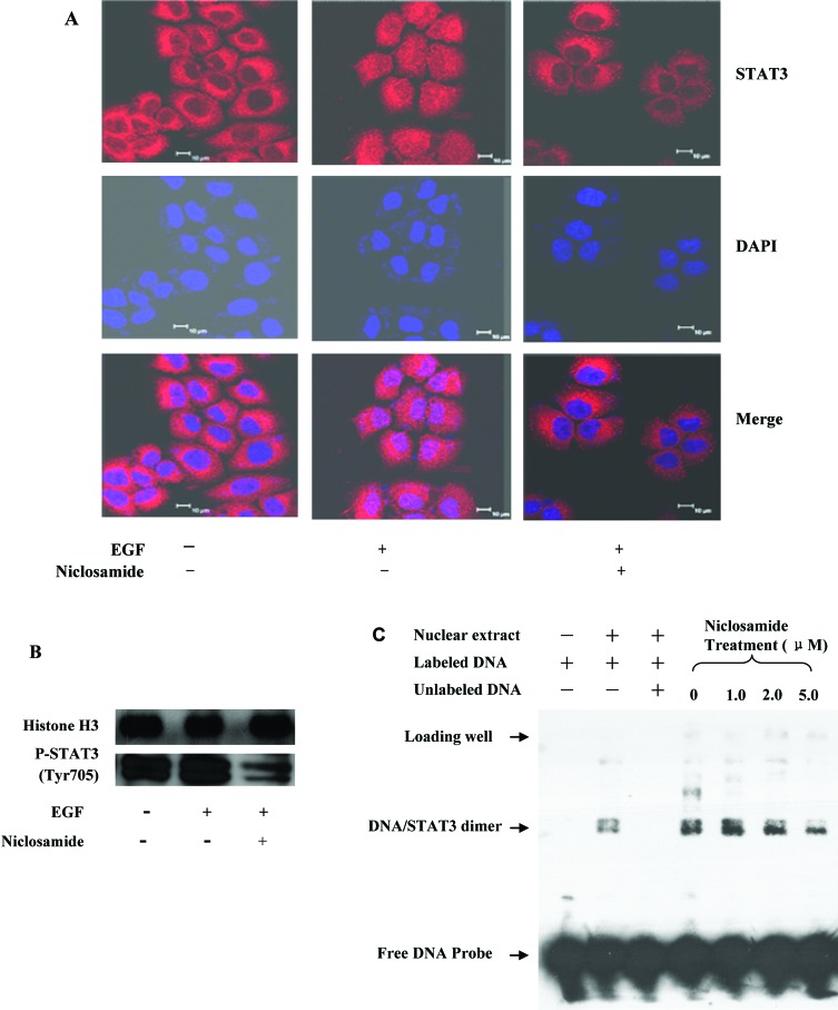
In summary, niclosamide, an FDA-approved anthelmintic drug, was identified as a new small-molecule inhibitor of the STAT3 signaling pathway. This drug potently inhibited the activation, nuclear translocation, and transactivation of STAT3 but had no obvious effects on the closely related STAT1 and STAT5 proteins, the upstream JAK1, JAK2, and Src kinases, or other receptor tyrosine kinases. Furthermore, niclosamide inhibited the transcription of STAT3 target genes and induced cell growth inhibition, apoptosis, and cell cycle arrest of cancer cells with constitutively active STAT3. Although niclosamide does not have an ideal pharmarcokinetic profile (i.e., poor oral bioavailability) in humans as an anticestodal drug, it represents a new potent lead compound with salicylic amide scaffold for development of STAT3 pathway inhibitors as new molecularly targeted anticancer drugs. The further structural optimization and extensive mechanism study on niclosamide are undergoing and will be reported in due course.[4] Recombinant STAT3 protein was incubated with biotin-labeled STAT3-specific DNA probe and serial concentrations of NICLOSAMIDE (BAY2353) (0.1-5 μM) in binding buffer at room temperature for 30 minutes. Stat3-DNA complexes were separated by non-denaturing PAGE and detected by chemiluminescence. Inhibition rate of DNA binding was calculated relative to vehicle control [4] NF-κB luciferase reporter plasmid-transfected HEK293 cells were treated with NICLOSAMIDE (BAY2353) (1-10 μM) for 1 hour, then stimulated with RANKL (50 ng/mL) for 24 hours. Luciferase activity was measured using a dual-luciferase assay system to evaluate NF-κB inhibition [7] Mitochondrial uncoupling activity assay: Isolated mouse liver mitochondria were incubated with NICLOSAMIDE (BAY2353) (0.5-5 μM) in respiration buffer. Oxygen consumption rate was measured using a Clark-type oxygen electrode to assess uncoupling efficiency [2] |
| Cell Assay |
Cellular proliferation assay[3]
3 × 10³ and 6 × 10³ cells were plated in 96-well plates depending on the cell line. 100 µL of fresh culture medium containing the drug or vehicle was added. Cell count was determined using the CyQuant kit, according to the manufacturer’s instructions, and cell number was measured using a SpectraMax M5 microplate reader (ex485/em538). Assays were performed in quadruplicate and the experiments were repeated three times. NCI-H295R and SW-13 cells, which form multicellular aggregates (MCA) or spheroids, were plated in Ultra Low Cluster 24-well plates (Costar, Corning, NY) at 1 × 10⁵ cells/0.5 mL or 6 × 10⁴ cells/0.5 mL depending on the cell line. Spheroids were allowed to develop for one or two weeks at 37°C in 5% CO2, and media was exchanged twice a week. Spheroids were treated with niclosamide or the vehicle at varying concentrations, and imaged weekly. Caspase 3/7 activity assay[3] Cells were plated in 96-well plates and treated with niclosamide or the vehicle. Caspase 3/7 activity was measured using the Caspase-Glo 3/7 assay, according to manufacturer’s instructions. Cell cycle analysis[3] Cells plated in six-well plates were treated with niclosamide or the vehicle. At 48 hours, cells were fixed for 30 minutes in 70% ethanol at 4°C, and stained with 50 µg/mL of propidium iodide containing 100 mg/mL of ribonuclease A. Flow cytometry was performed on a Canto I flow cytometer using CellQuest software. Data was generated for at least 20,000 events per sample and analyzed using Modfit software. Cellular migration assays[3] NCI-H295R and SW-13 cells were plated in six-well plates and treated with varying concentrations of niclosamide or the vehicle for 24 hours. Cells were trypsinized and plated in transwell chambers at a density of 1 × 10⁵ cells per 0.5 mL. The lower chamber was filled with DMEM supplemented with 10% FBS as a chemoattractant. Cells were allowed to migrate for 24 hours or 48 hours depending on the cell line, and were fixed and stained with Diff-Quik. Cells were imaged and counted in three random fields per well, and the experiments performed in triplicate. For the wound-healing assay in BD140A cells, which do not migrate in the Boyden chamber model, cells were plated in six-well plates until confluent and treated with niclosamide or the vehicle. The cells were scratched using a sterile pipette tip and photographed at various time points. H295R and SW13 ACC cells were seeded in 96-well plates (5×10^3 cells/well) and treated with NICLOSAMIDE (BAY2353) (0.1-5 μM) for 72 hours. Cell viability was assessed by MTT assay, and IC50 values were calculated [3] ACC cells were seeded in 6-well plates (1×10^5 cells/well) and treated with NICLOSAMIDE (BAY2353) (0.5-2 μM) for 24 hours. Cells were lysed for western blot analysis of STAT3 (p-Tyr705), β-catenin, Bcl-2, Survivin, and cleaved caspase-3 [3] Vero cells were seeded in 24-well plates (2×10^4 cells/well) and infected with SARS-CoV (MOI=0.1) for 1 hour, then treated with NICLOSAMIDE (BAY2353) (0.01-1 μM) for 48 hours. Viral replication was quantified by plaque assay [5] Human neural progenitor cells (hNPCs) were seeded in 96-well plates (1×10^4 cells/well) and infected with ZIKV (MOI=1) for 2 hours, followed by NICLOSAMIDE (BAY2353) (0.1-1 μM) treatment for 72 hours. Cell viability was measured by CCK-8 assay, and ZIKV RNA levels were detected by qPCR [6] Mouse BMMs were isolated from femurs and tibias, seeded in 96-well plates (3×10^3 cells/well), and treated with NICLOSAMIDE (BAY2353) (1-10 μM) plus RANKL (50 ng/mL) for 5 days. Osteoclasts were stained with tartrate-resistant acid phosphatase (TRAP), and TRAP-positive multinucleated cells were counted [7] HeLa cells were transfected with STAT3-responsive luciferase plasmid, then treated with NICLOSAMIDE (BAY2353) (0.5-2 μM) for 24 hours. Luciferase activity was measured to assess STAT3 transcriptional activity [4] |
| Animal Protocol |
Animal/Disease Models: Nu+/Nu+ mice injected with NCI-H295R cells[3]
Doses: 100 mg/kg, 200 mg/kg Route of Administration: po (oral gavage); 100 mg/kg, 200 mg/kg; once a week; 8 weeks Experimental Results: demonstrated a 60%-80% inhibition in tumor growth, as compared to the control group. In vivo mouse studies[3] Animal studies were approved by the National Cancer Institute Animal Care and Use Committee. Mice were maintained according to National Institutes of Health (NIH) Animal Research Advisory Committee (ARAC) guidelines. 5 × 10⁶ NCI-H295R cells were injected into the flank of Nu+/Nu+ mice. Tumors were allowed to grow and mice were randomized into three treatment groups (8 mice per treatment group). Mice were treated with 100 mg/kg of niclosamide, 200 mg/kg of niclosamide, or the vehicle (PEG500) everyday by oral gavage. Tumor sizes were measured in two dimensions every week with calipers and recorded. Nude mice (6-8 weeks old) were subcutaneously injected with H295R ACC cells (2×10^6 cells/mouse) to establish xenografts. When tumors reached 100 mm³, mice were randomly divided into vehicle and NICLOSAMIDE (BAY2353) groups (n=6 per group). NICLOSAMIDE (BAY2353) was suspended in 0.5% carboxymethylcellulose (CMC) and administered orally at 50 mg/kg once daily for 21 days. Tumor volume was measured every 3 days, and mice were euthanized for tumor weight and molecular analysis [3] C57BL/6 mice (8 weeks old) underwent ovariectomy (OVX) to induce osteoporosis. Two weeks after OVX, mice were treated with NICLOSAMIDE (BAY2353) (20 mg/kg, i.p., twice weekly for 4 weeks) or vehicle. Bone mineral density (BMD) was measured by micro-CT, and femurs were collected for TRAP staining [7] Newborn C57BL/6 mice (3 days old) were intraperitoneally injected with ZIKV (1×10^5 PFU/mouse). Twenty-four hours post-infection, mice were treated with NICLOSAMIDE (BAY2353) (10 mg/kg, i.p., daily for 5 days). Survival rate was recorded for 14 days, and brain tissues were collected for ZIKV RNA quantification [6] Schistosome-infected mice were treated with NICLOSAMIDE (BAY2353) (5 mg/kg, p.o., weekly for 3 weeks). Snails in the experimental environment were counted before and after treatment to assess molluscicidal efficacy [1] |
| ADME/Pharmacokinetics |
Absorption, Distribution and Excretion
Nicoloxamide appears to be almost entirely unabsorbed by the gastrointestinal tract—neither the drug nor its metabolites were detected in blood or urine. Gastrointestinal absorption is minimal… Rainbow trout exposed to (14)C-Bayer 73 had a bile-to-water (14)C ratio of 10,000:1. Thin-layer chromatography analysis of unfractionated bile from the fish 24 hours after exposure revealed a single major radioactive peak. Unaltered Bayer 73 was found in the bile. Nicoloxamide is excreted in feces. When male and female volunteers were orally administered 2000 mg of radiocarbon-labeled nicotinamide, 2% to 25% of the dose was excreted in urine over 4 days; the remainder was excreted in feces. Nicoloxamide equivalents were essentially cleared after 1–2 days. The maximum concentration of nicotinamide equivalents in serum ranged from 0.25 to 6.0 ppm; variations in this parameter are attributed to inter-individual differences in absorption rates. For more complete data on the absorption, distribution, and excretion of nicotinamides (6 in total), please visit the HSDB record page. Metabolites/Metabolites The anthelmintic nicotinamide…can be reduced to the corresponding amino derivatives by mouse and sheep liver enzymes, as well as by tapeworm and nematode enzymes. ...Nicoleamide is not hydrolyzed by mammalian and worm enzymes, nor by intact worms. In warm-blooded animals, the nitro group is reduced to the amino group (5,2'-dichloro-4'-aminosalicylic acid aniline). Pregnant rats were administered 1000 mg/kg of nicotinamide orally on days 13, 19, or 20 of gestation and sacrificed 4, 8, 16, or 24 hours after administration. The highest concentrations of nicotinamide and 2,5'-dichloro-4'-aminosalicylic acid aniline were detected in the liver and kidneys 8 hours after administration. Niclosamide was detected in rat fetuses treated on day 13, but its amino metabolites were not detected; however, both compounds were detected in rat fetuses treated on days 19 or 20. This indicates that 19-day and 20-day-old fetuses can metabolize niclosamide, while 13-day-old fetuses cannot. Niclosamide…is absorbed via the gastrointestinal tract, and its mutagenic metabolites are excreted in free form and as conjugated glucuronides. Like other secondary amides, phase I metabolism of niclosamide may result in the hydrolytic cleavage of the amide bond, producing 5-chlorosalicylic acid and 2-chloro-4-nitroaniline. ... Niccolamide (BAY2353) Due to its poor water solubility, its oral bioavailability in rats and humans is low (<10%) [1,2] Niccolamide (BAY2353) After oral administration (50 mg/kg) in rats, its terminal half-life (t1/2) is 3.2 hours [1] Niccolamide (BAY2353) It shows a high plasma protein binding rate (>99%) in both human and rat plasma [2] Niccolamide (BAY2353) It is metabolized in the liver by glucuronidation and is mainly excreted in feces (70-80%) and urine (10-15%) [1] |
| Toxicity/Toxicokinetics |
Toxicity Summary
Nicoloxamide works by killing tapeworms through contact. Adult tapeworms (but not eggs) are rapidly killed, likely due to uncoupling by oxidative phosphorylation or stimulation by ATPase activity. The killed tapeworms are then excreted in feces and sometimes destroyed in the intestines. Nicoloxamide may exert its molluscicidal effect by binding to and damaging DNA. Pregnancy and Lactation Effects ◉ Overview of Use During Lactation Nicoloxamide is not marketed in the United States. There is currently no clinical information regarding the use of nicoloxamide during lactation. Because nicoloxamide is not absorbed orally, it is unlikely to have adverse effects on breastfed infants. No special precautions are required. ◉ Effects on Breastfed Infants No published information was found as of the revision date. ◉ Effects on Lactation and Breast Milk No published information was found as of the revision date. Interactions> The conclusion is that Sevin enhances the toxicity of Bayer 73 to rainbow trout by increasing its absorption, possibly through affecting gill blood flow or permeability. Niclosamide can be used in combination with several other drugs… Niclosamide, when used in combination with cucurbitacin or procaine, enhances its efficacy against mouse tapeworms. …To investigate the intrauterine developmental toxicity of these compounds, pregnant female Wistar albino rats were tested. Atrazine and niclosamide (1/50 LD50) were administered alone or in combination, on days 5–15 of gestation, and the mothers were sacrificed on day 20. Results showed that no deaths or adverse clinical reactions occurred in the treated mothers throughout the study period. Furthermore, no significant preimplantation loss was observed. Atrazine treatment significantly reduced the corrected weight gain in the mothers. In female rats treated with atrazine or atrazine combined with nicosulfanilamide, uterine weight and litter size were significantly reduced. Post-implantation mortality was significantly higher in the atrazine-treated group (21.15%) and the combined treatment group (25.45%) compared to the untreated group (6.48%) and the nicosulfanilamide-treated group (15.6%). Fetal morphological examination revealed significantly increased rates of malformed fetuses (litters) in the atrazine, nicosulfanilamide, and combined treatment groups (5.8%, 29.4%), 6.48%, and 7.9%, respectively, compared to 1.6% (11.1%) in the control group. Skeletal malformations were the most common malformation in all study groups, while surface malformations were also observed in the atrazine group. These results suggest that amitriptyline may have embryo/fetal toxicity at high doses, and nicosulfanilamide may have fetal toxicity. Non-human toxicity values Rat intraperitoneal injection LD50 250 mg salt/kg /2-hydroxyethylammonium salt/ Nicylamide (BAY2353) The oral LD50 in rats was 565 mg/kg, and the oral LD50 in mice was 1000 mg/kg [1] In dogs treated with nicotinamide (BAY2353) (100 mg/kg, orally, once daily for 30 days), mild gastrointestinal irritation (diarrhea, vomiting) was observed, and no significant hepatotoxicity or nephrotoxicity was observed [1] Nicylamide (BAY2353) (up to 50 mg/kg, orally (21 days) did not cause significant weight loss or abnormal serum ALT/AST/BUN/Cr levels in nude mice [3] Nicotinamide (BAY2353) showed no significant cytotoxicity to normal human cells (CC50 > 10 μM in fibroblasts and hepatocytes) [2,5] |
| References | |
| Additional Infomation |
Therapeutic Uses
Anti-tapetic drug; anti-nematode drug; molluscicide. Nicotinamide has no contraindications as a tapeworm antagonist. Veterinary Use: Nicotinamide can also be used to treat tapeworm infections in laboratory animals (e.g., mice, rabbits, monkeys, or reptiles). …Nicotinamide is administered orally in tablet form for dogs and cats…It is commonly used in ruminants (e.g., cattle, sheep, and goats)…It is administered by gavage… Therapeutic Uses: Nicotinamide is used only in its free base form, primarily as a tapeworm antagonist, and secondarily as a fluke antagonist. This drug is formulated as a chewable tablet containing 500 mg of nicotinamide. This product is highly effective against tapeworm infections caused by Taenia solium, Taenia solium, and Diplophora canis; however, it is more difficult to treat tapeworms such as Hymenolepis microsporum, Hymenolepis pygmy, and Diplophora canis. For infections caused by beef tapeworm, pork tapeworm, and broad tapeworm, the recommended single oral dose is: 2 g for adults, 1.5 g for children weighing over 34 kg, and 1.0 g for children weighing 11-34 kg. Other tapeworm infections may require repeated treatment, for example: 2 g once daily for 7 days for adults; 1.5 g once daily for children weighing over 34 kg, followed by 1 g daily for 6 days; and 1 g once daily for children weighing 11-34 kg, followed by 500 mg daily for 6 days. The safety of this drug in children under 2 years of age has not been established. Since nicosulfanilamide is only effective against intestinal tapeworms, it is ineffective against cysticercosis. As a trematode, nicotinamide is primarily effective against intestinal trematodes (such as Fasciola hepatica). For more complete data on the therapeutic uses of nicotinamides (10 in total), please visit the HSDB record page. Drug Warnings Because tapeworm infections are usually not life-threatening, pregnant women are advised to wait until after delivery before treatment. Veterinarian: Do not treat lactating animals. ..It should be noted that this drug's killing effect on adult worms does not include the eggs. Therefore, the use of niclosamide in pork tapeworm infection may expose patients to cysticercosis because live eggs are released into the intestinal lumen after the dead segments are digested. It is recommended to administer a full laxative within 3 to 4 hours after administration to clear all dead segments from the intestines and prevent their digestion. This drug can cause segmental disintegration, releasing live eggs; therefore, if used to treat pork tapeworm infection, a laxative should be administered 1 to 2 hours after treatment. Adverse reactions have occurred occasionally; nausea and abdominal pain have been reported. For more complete data on drug warnings for niclosamide (7 of 7), please visit the HSDB records page. Pharmacodynamics Clonidamide is an anthelmintic used to treat tapeworm infection. Its mechanism of action is likely through uncoupling the electron transport chain and ATP synthase. Disruption of this key metabolic pathway prevents the production of adenosine triphosphate (ATP), an essential molecule for providing energy for metabolism. Niccolamide (BAY2353) is an FDA-approved anthelmintic for the treatment of tapeworm infection (taeniasis)[2] Niccolamide (BAY2353) is a multi-target small molecule that exerts a variety of pharmacological effects by inhibiting STAT3, Wnt/β-catenin, NF-κB and mitochondrial uncoupling pathways[2,3,4,7] Niccolamide (BAY2353) It has been redeveloped as a potential drug for the treatment of cancer (adrenocortical carcinoma), viral infections (SARS-CoV, ZIKV) and osteoporosis [3,5,6,7]. Nicotinamide (BAY2353) exerts its snail-killing activity by interfering with the energy metabolism of snails, thus effectively controlling schistosomiasis [1]. Nicotinamide (BAY2353) can cross the blood-brain barrier at low concentrations, thereby exerting its neuroprotective effect against Zika virus-induced neuronal death [6]. |
| Molecular Formula |
C13H8CL2N2O4
|
|
|---|---|---|
| Molecular Weight |
327.12
|
|
| Exact Mass |
325.986
|
|
| Elemental Analysis |
C, 47.73; H, 2.47; Cl, 21.68; N, 8.56; O, 19.56
|
|
| CAS # |
50-65-7
|
|
| Related CAS # |
|
|
| PubChem CID |
4477
|
|
| Appearance |
PALE, YELLOW CRYSTALS
ALMOST COLORLESS CRYSTALS A cream-colored or yellowish-white powder Bright yellow crystalline solid |
|
| Density |
1.6±0.1 g/cm3
|
|
| Boiling Point |
424.5±45.0 °C at 760 mmHg
|
|
| Melting Point |
225-230ºC
|
|
| Flash Point |
210.5±28.7 °C
|
|
| Vapour Pressure |
0.0±1.0 mmHg at 25°C
|
|
| Index of Refraction |
1.709
|
|
| LogP |
5.41
|
|
| Hydrogen Bond Donor Count |
2
|
|
| Hydrogen Bond Acceptor Count |
4
|
|
| Rotatable Bond Count |
2
|
|
| Heavy Atom Count |
21
|
|
| Complexity |
404
|
|
| Defined Atom Stereocenter Count |
0
|
|
| SMILES |
ClC1C([H])=C(C([H])=C([H])C=1N([H])C(C1C([H])=C(C([H])=C([H])C=1O[H])Cl)=O)[N+](=O)[O-]
|
|
| InChi Key |
RJMUSRYZPJIFPJ-UHFFFAOYSA-N
|
|
| InChi Code |
InChI=1S/C13H8Cl2N2O4/c14-7-1-4-12(18)9(5-7)13(19)16-11-3-2-8(17(20)21)6-10(11)15/h1-6,18H,(H,16,19)
|
|
| Chemical Name |
5-chloro-N-(2-chloro-4-nitrophenyl)-2-hydroxybenzamide
|
|
| Synonyms |
|
|
| HS Tariff Code |
2934.99.9001
|
|
| Storage |
Powder -20°C 3 years 4°C 2 years In solvent -80°C 6 months -20°C 1 month |
|
| Shipping Condition |
Room temperature (This product is stable at ambient temperature for a few days during ordinary shipping and time spent in Customs)
|
| Solubility (In Vitro) |
DMSO: 12 mg/mL (36.7 mM)
Water: 1 mg/mL (Insoluble) Ethanol: 1 mg/mL |
|---|---|
| Solubility (In Vivo) |
Solubility in Formulation 1: 0.5 mg/mL (1.53 mM) in 10% DMF 40% PEG300 + 5% Tween80 + 45% Saline (add these co-solvents sequentially from left to right, and one by one), suspension solution; with sonication.
Preparation of saline: Dissolve 0.9 g of sodium chloride in 100 mL ddH₂ O to obtain a clear solution. Solubility in Formulation 2: 1% DMSO+30% polyethylene glycol+1% Tween 80:30 mg/mL View More
Solubility in Formulation 3: 5 mg/mL (15.28 mM) in 20% HP-β-CD 5% Cremophor EL (add these co-solvents sequentially from left to right, and one by one), suspension solution; with ultrasonication. |
| Preparing Stock Solutions | 1 mg | 5 mg | 10 mg | |
| 1 mM | 3.0570 mL | 15.2849 mL | 30.5698 mL | |
| 5 mM | 0.6114 mL | 3.0570 mL | 6.1140 mL | |
| 10 mM | 0.3057 mL | 1.5285 mL | 3.0570 mL |
*Note: Please select an appropriate solvent for the preparation of stock solution based on your experiment needs. For most products, DMSO can be used for preparing stock solutions (e.g. 5 mM, 10 mM, or 20 mM concentration); some products with high aqueous solubility may be dissolved in water directly. Solubility information is available at the above Solubility Data section. Once the stock solution is prepared, aliquot it to routine usage volumes and store at -20°C or -80°C. Avoid repeated freeze and thaw cycles.
Calculation results
Working concentration: mg/mL;
Method for preparing DMSO stock solution: mg drug pre-dissolved in μL DMSO (stock solution concentration mg/mL). Please contact us first if the concentration exceeds the DMSO solubility of the batch of drug.
Method for preparing in vivo formulation::Take μL DMSO stock solution, next add μL PEG300, mix and clarify, next addμL Tween 80, mix and clarify, next add μL ddH2O,mix and clarify.
(1) Please be sure that the solution is clear before the addition of next solvent. Dissolution methods like vortex, ultrasound or warming and heat may be used to aid dissolving.
(2) Be sure to add the solvent(s) in order.
|
|---|
|
|
Products are for research use only; We do not sell to patients
Copyright 2020 InvivoChem LLC | All Rights Reserved


 COA
COA