| Size | Price | |
|---|---|---|
| Other Sizes |


| ADME/Pharmacokinetics |
Absorption, Distribution and Excretion
This study determined the absorption, distribution, and excretion of radioactive material 6 hours after topical application of 0.5 mg/kg body weight of ring-labeled 14C-muscon (dissolved in a mixture of phenylethanol and ethanol) to the shaved back of 21 male rats (16 CD Sprague-Dawley rats and 5 Long-Evans rats). The dose was uniformly applied over an area of 9 square centimeters. The application rate was 0.01 mg/cm²… In CD rats and Long-Evans rats, 14.6–26.3% and 13.3% of the radiolabeled dose, respectively, were absorbed from the shaved back within 6 hours. After removal of 14C-muscon 6 hours after application, 16% of the dose remained on the skin and continued to be absorbed. Data from subsequently euthanized animals also supported this conclusion. The data showed that the amount of residual substance on the treated skin exhibited a steady decreasing trend, from 7.2%–9.85% of the dose at 8 hours to 3.1%–4.1% at 24 hours, and further to 2.0%–3.4% at 48 hours. The actual absorption rate was approximately 19% at 8 hours, 25.1%–28.3% at 24 hours, and 26.2%–32.7% at 48 hours. After 48 hours, absorption essentially ceased, with approximately 3% of the dose remaining unabsorbed through the skin. In Long-Evans rats, the distribution of radioactive material was similar, with the absorption rate of ¹⁴C-muscone increasing to 29.3%–40.2% of the dose between 48 and 120 hours. After 24 hours, the average percentage of the dose remaining on the treated skin was approximately 3%. A ring-labeled 14C-musconone (dissolved in a mixture of phenylethanol and ethanol) at a dose of 0.5 mg/kg body weight was topically applied to the shaved backs of 21 male rats (16 CD Sprague-Dawley strain and 5 Long-Evans strain) for 6 hours. Radioactive absorption, distribution, and excretion were measured. The dose was uniformly applied over an area of 9 square centimeters. The application rate was 0.01 mg/cm²… After 6 hours of application, the dressing and foil were removed, and any residual dose in the treated area was wiped away… In CD strain rats, 7.3% and 17.2% of the applied dose were excreted in urine and feces, respectively, after 120 hours. In Long-Evans rats, the elimination rates of radioactive material were comparable, with 10.8% and 27.2% of the dose excreted in urine and feces, respectively, within 5 days. Most of the radioactive material was eliminated within the first 48 hours after administration. No radioactive material was detected in exhaled breath. A ring-labeled 14C-musconone (dissolved in a mixture of phenylethanol and ethanol) at a dose of 0.5 mg/kg body weight was applied topically to the shaved backs of 21 male rats (16 CD Sprague-Dawley rats and 5 Long-Evans rats) for 6 hours, and the absorption, distribution, and excretion of radioactive material were measured. The dose was evenly applied over an area of 9 square centimeters. The administered dose was 0.01 mg/cm²… Six hours after administration, the dressing and foil were removed, and any residual dose in the treated area was wiped away… In cannulated CD rats, only 1.8% of the dose was excreted in the urine within 48 hours, while 25.3% accumulated in the bile (of which 15.8% was excreted within 24 hours). In uncannulated rats, 8% of the dose was excreted in the urine within 48 hours. These results indicate that (14)C-musconone is primarily excreted via bile; therefore, most of the radioactive material in the urine of intubated animals originates from substances reabsorbed from the gastrointestinal tract. In bile, at least six drug-related components are present as β-glucuronic acid conjugates, which apparently debind in the gastrointestinal tract and are further metabolized into other, more polar components, some of which are at least partially reabsorbed, resulting in a complex urinary metabolite profile. Radioactivity was detected in almost all tissues of animals euthanized within 1–120 hours of administration. Radioactivity concentrations in all tissues peaked approximately 6 hours after administration. Between 8 and 120 hours, radioactivity concentrations in all tissues steadily decreased, and thus, at 120 hours post-administration, radioactivity concentrations in each tissue were typically less than 20% of their peak value. Throughout the study, the highest concentrations of radioactive substances were observed in the gastrointestinal tract, liver, adipose tissue, adrenal glands, thyroid gland, and kidneys of CD rats. Six hours after administration, the average muscone equivalents in these tissues were 0.645 μg/g, 0.32 μg/g, 0.19 μg/g, 0.12 μg/g, 0.10 μg/g, and 0.08 μg/g, respectively. The radioactivity distribution in Long-Evans rats was similar; peak radioactivity concentrations were observed in the gastrointestinal tract (0.47 μg muscone equivalent/g), liver (0.26 μg/g), adrenal glands (0.1 μg/g), thyroid gland (0.18 μg/g), and adipose tissue (0.16 μg/g) six hours after administration. A ring-labeled 14C-musconone (dissolved in phenylethanol and ethanol) was applied in a closed-cell manner to the shaved backs (approximately 9 square centimeters) of 10 male Sprague-Dawley CD rats once daily for 24 hours at a dose of 0.5 mg/kg body weight for 14 consecutive days. The skin was not rinsed between applications. Two rats were sacrificed for whole-body autoradiography: one 24 hours after the first administration and the other 24 hours after the 14th administration. Urine and fecal samples were collected from the remaining eight rats at multiple time points, and blood, processed skin, brain, kidney, liver, thyroid, and adipose tissue samples were collected at sacrifice. Whole-body autoradiography results showed that the radioactive material was not widely distributed in the body 24 hours after the first administration. Relatively high radioactivity concentrations were observed at the administration site, in the cecal contents, large intestine contents, and bile ducts. Lower radioactivity concentrations were observed in the small intestine contents and liver. Rats euthanized 24 hours after the 14th dose generally showed high levels of radioactive material in their tissues, but the highest concentrations remained concentrated at the administration site and in the gastrointestinal tract, while lower concentrations were observed in the liver, blood, and thyroid gland. Therefore, the absorption of radioactive material was incomplete, as a significant amount of radioactive material remained at the application site. Within 24 hours of the first dose, an average of 1.48 and 2.34 micrograms of muscone equivalent were excreted in urine and feces, respectively. Within 24 hours of the 14th dose, the average excretion rate in urine increased to 6.54 micrograms/day. Within 24 hours of the 12th and 14th doses, the average excretion rate in feces increased to a maximum of approximately 14.8 micrograms/day. At euthanasia, the radioactive concentration in treated skin was high, while the total radioactivity in blood and selected tissues represented only a small fraction of the total 14 doses (0.22–0.37% in the liver, and even lower in fat, blood, kidneys, brain, and thyroid gland). For more complete data on the absorption, distribution, and excretion of muscone (9 doses), please visit the HSDB record. [Page hr] Metabolites/Metabolites Synthetic nitromuses are well-documented in the literature. This study used selected ion monitoring gas chromatography-mass spectrometry (GC-SIM-MS) to analyze muscone (MX) and muscone (MK)-protein adducts in trout liver in vivo. The biotransformation, dose-response, and toxicokinetics of the metabolites 2-amino-MX (2-AMX), 2-amino-MK (2-AMK), and 4-amino-MX (4-AMX) generated from the enzymatic nitro reduction of MX and MK are described. These metabolites are covalently bound to cysteine residues in fish liver proteins. Trout were exposed to 0.010, 0.030, 0.10, and 0.30 mg/g of MX and/or MK, respectively. Forty-two liver samples were collected from fish in the exposed and control groups, with exposure times of 1, 3, and 7 days, respectively, and were mixed according to the exposure protocol and time. Alkaline hydrolysis was used to release bound metabolites from the mixed liver samples of the exposed group, followed by extraction with n-hexane, concentration, and GC-SIM-MS analysis. The presence of metabolites in the liver extracts was confirmed by consistency with standard mass spectrometry characteristics and retention times. In the dose-response study, after 1 day of exposure, the maximum adduct formation of 2-AMX, 2-AMK, and 4-AMX in the livers of fish exposed to 0.03 mg/g MX and MK was 492.0 ng/g, 505.5 ng/g, and 12588.5 ng/g, respectively. In the toxicokinetic study, the peak concentration of the target metabolites was the same as observed in the dose-response study, reaching its peak concentration within 1 day after exposure to 0.03 mg/g MX and MK. Based on first-order kinetic assumptions, the half-life of the metabolites is estimated to be 2–9 days. The average recovery exceeded 95%, with a relative standard deviation (RSD) of approximately 9%, and a limit of detection (LOD) ranging from 0.91 to 3.8 ng/g based on a signal-to-noise ratio of 10 (S/N=10). No metabolites were detected in either the control group or the exposed unhydrolyzed liver extract. This is the first report on the dose-response and toxicokinetics of nitromuscon-cysteine-protein adducts in fish liver. Two healthy male volunteers underwent a uniform application of 2.2 mg of ring-labeled 14C-musconone (dissolved in a mixture of phenylethanol and ethanol) to uncoiled skin in the upper left quadrant of their chest over 6 hours. The dose was uniformly applied over an area of 100 cm². The application rate was 0.02 mg/cm². …When urine samples were treated with β-glucuronidase and extracted with ethyl acetate, the recovery increased by approximately 5-fold, indicating that the majority of musconone metabolites in human urine are present as glucuronide conjugates. Human urine extracts treated with β-glucuronidase contain a major (unidentified) metabolite that may also be present as a small component in rat bile extracts. |
|---|---|
| Toxicity/Toxicokinetics |
Toxicity Summary
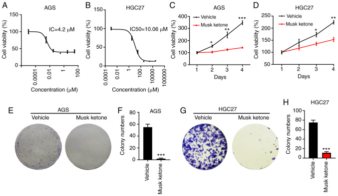
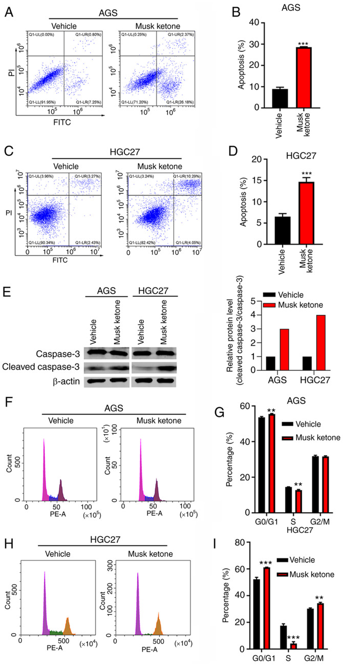
Identification and Uses: Muscone forms yellow crystals and is a fragrance ingredient. It is widely used as a fixative in floral and dreamy perfumes. Human Studies: In maximal studies on human volunteers, muscone did not induce sensitization at 48 and 72 hours. A photopatch test in one patient with chronic actinic dermatitis showed a response to both muscone and thymol (both present in their aftershave water). Muscone at concentrations of 0.068 to 68 μM did not induce sister chromatid exchange in human lymphocytes under either metabolically activated or non-metabolic conditions. In in vitro micronucleus assays, concentrations of muscone up to 136 μM and 250 μM did not increase the frequency of micronuclei in human lymphocytes or the Hep G2 hepatocellular carcinoma cell line. Animal Studies: Muscone has no skin irritation or systemic toxicity in rabbits, but it has a slight eye irritant effect. Muscone does not cause contact sensitization in guinea pigs. In mice, administration of muscone resulted in a dose-dependent increase in relative liver weight at a dose of 50 mg/kg body weight, with an increase of up to 50% at a dose of 500 mg/kg body weight. Muscone also caused histological changes in the liver, primarily manifested as hypertrophy of central lobular hepatocytes, with panlobular hepatocyte hypertrophy observed at the highest dose. Pregnant rats were administered muscone orally via gavage at doses of 0, 60, 200, 600, or 2000 mg/kg body weight/day from days 7 to 17 of gestation. Post-cesarean section observations showed that at doses of 200 mg/kg body weight and above, fetal weight, litter size, and live birth count decreased, while early and late embryonic resorption rates and the percentage of embryonic resorption increased. No obvious external fetal abnormalities were observed. Muscone negatively impacted reproduction and early life stage survival in zebrafish. Muscone was tested in five strains of Salmonella Typhimurium, and the results showed negative toxicity regardless of activation. Ecotoxicity Study: Cytochrome P450 may be a potential sensitive target for the synthesis of musk substances in fish. Interactions ... Thirteen patients suspected of having photosensitive contact dermatitis underwent questionnaires and clinical examination. After a pre-test for UV skin sensitivity, nitromuses (dosage and carrier not specified) were applied to the skin and protected with occlusive patches (two sites for each substance). Twenty-four hours after exposure, the patches were removed under dim light, and the skin was examined for signs of contact dermatitis. Half of the contact sites were re-covered, and the other half were irradiated with UV light. All subjects showed photosensitivity to thymol. One person also showed photosensitivity to muscone. No reaction was observed in unirradiated areas. This study could not determine whether the patient had cross-sensitivity to thymol and muscone, or whether there was independent photosensitivity to both nitromuses simultaneously. This study also could not determine the prevalence of this condition. ...No synergistic effect was detected when cells were treated simultaneously with MK (5-5000 ng/mL) and 0.2 ug/mL benzo[a]pyrene; benzo[a]pyrene (B(a)P) itself increased the number of micronuclei (MNs) by 1.5 times compared to the spontaneous background frequency (60 MNs/1000 binucleated cells vs. 39 MNs/1000 binucleated cells). ... In in vitro micronucleus assays using metabolically healthy human hepatocellular carcinoma cells (Hep G2 cell line), the results showed that when cells were pretreated with muscon for 28 hours before exposure to B(a)P, a synergistic toxic effect of muscon was observed; however, this effect was not observed when cells were treated simultaneously with muscon and B(a)P. Muskcon pretreatment significantly increased the number of B(a)P-induced micronuclei. When muscone concentrations were 500-5,000 ng/mL, enhanced genotoxicity of benzo[a]pyrene (B(a)P) was observed. This concentration effectively induced CYP1A activity (as shown by EROD assays in Hep G2 cells), an enzyme that plays a crucial role in B(a)P activation. Non-human toxicity values Oral LD50 in rats >10 g/kg Dermal LD50 in rabbits >10 g/kg |
| References |
|
| Additional Infomation |
Musk ketone is a pale yellow crystalline solid, insoluble in water. (NTP, 1992)
4'-tert-butyl-2',6'-dimethyl-3',5'-dinitroacetophenone is an aromatic ketone. Muscone has been found in plants of the genus Muscaria, and relevant data are available. See also: ...View more... |
| Molecular Formula |
C14H18N2O5
|
|---|---|
| Molecular Weight |
294.31
|
| Exact Mass |
294.121
|
| CAS # |
81-14-1
|
| PubChem CID |
6669
|
| Appearance |
White to off-white solid powder
|
| Density |
1.2±0.1 g/cm3
|
| Boiling Point |
369.0±42.0 °C at 760 mmHg
|
| Melting Point |
135-139 °C(lit.)
|
| Flash Point |
153.2±20.7 °C
|
| Vapour Pressure |
0.0±0.8 mmHg at 25°C
|
| Index of Refraction |
1.548
|
| LogP |
3.86
|
| Hydrogen Bond Donor Count |
0
|
| Hydrogen Bond Acceptor Count |
5
|
| Rotatable Bond Count |
2
|
| Heavy Atom Count |
21
|
| Complexity |
418
|
| Defined Atom Stereocenter Count |
0
|
| InChi Key |
WXCMHFPAUCOJIG-UHFFFAOYSA-N
|
| InChi Code |
InChI=1S/C14H18N2O5/c1-7-10(9(3)17)8(2)13(16(20)21)11(14(4,5)6)12(7)15(18)19/h1-6H3
|
| Chemical Name |
1-(4-tert-butyl-2,6-dimethyl-3,5-dinitrophenyl)ethanone
|
| Synonyms |
NSC-15339; NSC 15339; Musk ketone
|
| HS Tariff Code |
2934.99.9001
|
| Storage |
Powder -20°C 3 years 4°C 2 years In solvent -80°C 6 months -20°C 1 month Note: Please store this product in a sealed and protected environment (e.g. under nitrogen), avoid exposure to moisture and light. |
| Shipping Condition |
Room temperature (This product is stable at ambient temperature for a few days during ordinary shipping and time spent in Customs)
|
| Solubility (In Vitro) |
DMSO : ≥ 125 mg/mL (~424.74 mM)
|
|---|---|
| Solubility (In Vivo) |
Solubility in Formulation 1: ≥ 2.08 mg/mL (7.07 mM) (saturation unknown) in 10% DMSO + 90% Corn Oil (add these co-solvents sequentially from left to right, and one by one), clear solution.
For example, if 1 mL of working solution is to be prepared, you can add 100 μL of 20.8 mg/mL clear DMSO stock solution to 900 μL of corn oil and mix evenly. (Please use freshly prepared in vivo formulations for optimal results.) |
| Preparing Stock Solutions | 1 mg | 5 mg | 10 mg | |
| 1 mM | 3.3978 mL | 16.9889 mL | 33.9778 mL | |
| 5 mM | 0.6796 mL | 3.3978 mL | 6.7956 mL | |
| 10 mM | 0.3398 mL | 1.6989 mL | 3.3978 mL |
*Note: Please select an appropriate solvent for the preparation of stock solution based on your experiment needs. For most products, DMSO can be used for preparing stock solutions (e.g. 5 mM, 10 mM, or 20 mM concentration); some products with high aqueous solubility may be dissolved in water directly. Solubility information is available at the above Solubility Data section. Once the stock solution is prepared, aliquot it to routine usage volumes and store at -20°C or -80°C. Avoid repeated freeze and thaw cycles.
Calculation results
Working concentration: mg/mL;
Method for preparing DMSO stock solution: mg drug pre-dissolved in μL DMSO (stock solution concentration mg/mL). Please contact us first if the concentration exceeds the DMSO solubility of the batch of drug.
Method for preparing in vivo formulation::Take μL DMSO stock solution, next add μL PEG300, mix and clarify, next addμL Tween 80, mix and clarify, next add μL ddH2O,mix and clarify.
(1) Please be sure that the solution is clear before the addition of next solvent. Dissolution methods like vortex, ultrasound or warming and heat may be used to aid dissolving.
(2) Be sure to add the solvent(s) in order.
|
|
|
Products are for research use only; We do not sell to patients
Copyright 2020 InvivoChem LLC | All Rights Reserved