| Size | Price | Stock | Qty |
|---|---|---|---|
| 25mg |
|
||
| 50mg |
|
||
| 100mg |
|
||
| 250mg |
|
||
| 500mg |
|
||
| 1g |
|
||
| Other Sizes |
Purity: ≥98%
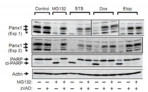
MG-132, a peptide aldehyde, is a novel, specific, potent, reversible, and cell-permeable proteasome inhibitor with an IC50 of 100 nM in a cell-free assay. It also has an IC50 of 1.2 μM for calpain inhibition.
| Targets |
Proteasome (IC50 = 100 nM); Calpain (IC50 = 1.2 μM)
|
|---|---|
| ln Vitro |
MG-132 exhibits over 1000 times greater activity than ZLLal in suppressing the 20S proteasome's ZLLL-MCA-degrading activity, with an IC50 of 100 nM as opposed to 110 μM. With an IC50 of 1.2 μM, MG-132 also inhibits calpain. At an ideal concentration of 20 nM, MG-132 exhibits 500 times greater potency than ZLLal in inducing neurite outgrowth in PC12 cells.[1] MG-132 (10 μM) inhibits the proteasome-mediated degradation of IκBα in A549 cells, thereby potently inhibiting TNF-α-induced NF-κB activation, IL-8 gene transcription, and IL-8 protein release.[2] MG-132 treatment strongly inhibits 26S proteasome, thereby inducing p53-dependent apoptosis in KIM-2 cells.[3] Multiple myeloma cells (U266 and OPM-2) are more susceptible to MG-132-induced apoptosis than BzLLLCOCHO, but MG-132 treatment results in weak inhibition of the chymotrypsinlike (CT-L) and peptidylglutamyl peptide hydrolysing (PGPH) activities of the 26S proteasome, in contrast to BzLLLCOCHO or PS-341.[4] By activating AP-1 family members c-Fos and c-Jun, which in turn repress the antiapoptotic molecule c-FLIP(L), MG-132 (1 μM) sensitizes TRAIL-resistant prostate cancer cells.[5] In the PC3 and DU145 androgen-independent prostate cancer (AIPCa) cell lines, MG-132 dramatically increases inositol hexakisphosphate's (IP6) capacity to lower cellular metabolic activity.[6]
- Induces apoptosis in human promyelocytic leukemia HL - 60 cells. Flow cytometry shows that MG - 132 treatment leads to an increase in the sub - G1 phase cell population, indicating apoptosis. Western blot analysis reveals that MG - 132 up - regulates the expression of p53 and p21 proteins, and down - regulates the expression of Bcl - 2 protein, which are related to the apoptosis pathway[3] - Inhibits the proliferation of ovarian carcinoma SKOV3 cells. MTT assay shows that MG - 132 at concentrations of 0.5, 1.5, 2.5, 3.5, and 5.0 μg/ml for 24, 48, and 72 hours can inhibit cell growth in a dose - and time - dependent manner. When combined with cisplatin, the inhibitory effect on cell growth and the promotion of apoptosis are more significant than those of cisplatin alone. Flow cytometry shows that the apoptosis rate of the combination group is higher, and Western blot and RT - PCR detect that the relative contents of caspase 3 and beclin 1 in the combination group are higher[4] |
| ln Vivo |
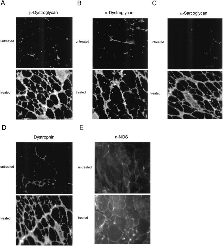
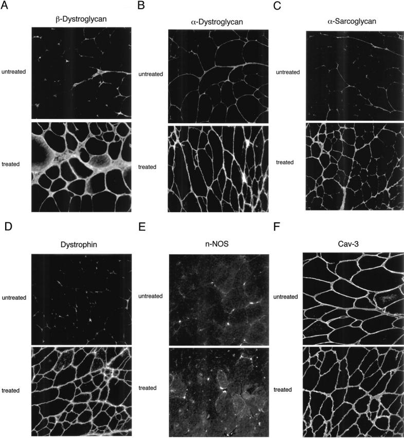
In skeletal muscle fibers from mdx mice, MG-132 administration efficiently restores the expression levels and plasma membrane localization of dystrophin, β-dystroglycan, α-bdystroglycan, and α-sarcoglycan, minimizes damage to muscle membranes, and improves the histopathological symptoms of muscular dystrophy.[8] By downregulating the muscle-specific ubiquitin ligases atrogin-1/MAFbx and MuRF-1 mRNA, MG-132 treatment dramatically reduces immobilization-induced skeletal muscle atrophy in mice.[8]
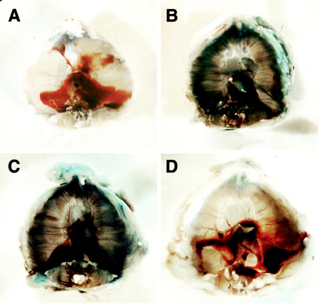
Dystrophin, the protein product of the Duchenne muscular dystrophy (DMD) gene, is absent in the skeletal muscle of DMD patients and mdx mice. At the plasma membrane of skeletal muscle fibers, dystrophin associates with a multimeric protein complex, termed the dystrophin-glycoprotein complex (DGC). Protein members of this complex are normally absent or greatly reduced in dystrophin-deficient skeletal muscle fibers, and are thought to undergo degradation through an unknown pathway. As such, we reasoned that inhibition of the proteasomal degradation pathway might rescue the expression and subcellular localization of dystrophin-associated proteins. To test this hypothesis, we treated mdx mice with the well-characterized proteasomal inhibitor MG-132. First, we locally injected MG-132 into the gastrocnemius muscle, and observed the outcome after 24 hours. Next, we performed systemic treatment using an osmotic pump that allowed us to deliver different concentrations of the proteasomal inhibitor, over an 8-day period. By immunofluorescence and Western blot analysis, we show that administration of the proteasomal inhibitor MG-132 effectively rescues the expression levels and plasma membrane localization of dystrophin, beta-dystroglycan, alpha-dystroglycan, and alpha-sarcoglycan in skeletal muscle fibers from mdx mice. Furthermore, we show that systemic treatment with the proteasomal inhibitor 1) reduces muscle membrane damage, as revealed by vital staining (with Evans blue dye) of the diaphragm and gastrocnemius muscle isolated from treated mdx mice, and 2) ameliorates the histopathological signs of muscular dystrophy, as judged by hematoxylin and eosin staining of muscle biopsies taken from treated mdx mice. Thus, the current study opens new and important avenues in our understanding of the pathogenesis of DMD. Most importantly, these new findings may have clinical implications for the pharmacological treatment of patients with DMD. [7] In the present study, we showed that the proteasome inhibitor MG132 significantly inhibited IκBα degradation thus preventing NFκB activation in vitro. MG132 preserved muscle and myofiber cross-sectional area by downregulating the muscle-specific ubiquitin ligases atrogin-1/MAFbx and MuRF-1 mRNA in vivo. This effect resulted in a diminished rehabilitation period. Conclusion: These finding demonstrate that proteasome inhibitors show potential for the development of pharmacological therapies to prevent muscle atrophy and thus favor muscle rehabilitation [8]. Treatment of recurrent or advanced cervical cancer is still limited, and new therapeutic choices are needed for improving prognosis and quality of life of patients. Because human papilloma virus (HPV) infection is critical in cervical carcinogenesis, with the E6 and E7 oncogenes of HPV degrading tumor suppressor proteins through the ubiquitin proteasome system, the inhibition of the ubiquitin proteasome system appears to be an ideal target to suppress the growth of cervical tumors. Herein, we focused on the ubiquitin proteasome inhibitor MG132 (carbobenzoxy-Leu-Leu-leucinal) as an anticancer agent against cervical cancer cells, and physically incorporated it into micellar nanomedicines for achieving selective delivery to solid tumors and improving its in vivo efficacy. These MG132-loaded polymeric micelles (MG132/m) showed strong tumor inhibitory in vivo effect against HPV-positive tumors from HeLa and CaSki cells, and even in HPV-negative tumors from C33A cells. Repeated injection of MG132/m showed no significant toxicity to mice under analysis by weight change or histopathology. Moreover, the tumors treated with MG132/m showed higher levels of tumor suppressing proteins, hScrib and p53, as well as apoptotic degree, than tumors treated with free MG132. This enhanced efficacy of MG132/m was attributed to their prolonged circulation in the bloodstream, which allowed their gradual extravasation and penetration within the tumor tissue, as determined by intravital microscopy. These results support the use of MG132 incorporated into polymeric micelles as a safe and effective therapeutic strategy against cervical tumors[10]. Enhances the sensitivity of ovarian carcinoma cells to cisplatin in nude mice. Twenty 4 - 6 - week - old female Balb/c nude mice were divided into four groups: control group, MG - 132 group, cisplatin group, and combination group. Oral administration of MG - 132 combined with cisplatin can more effectively inhibit the growth of ovarian carcinoma xenografts in nude mice than cisplatin alone[4] |
| Enzyme Assay |
MG-132, 20S proteasome, pH 7.0, 0.1 M Tris-acetate, and 25 μM substrate dissolved in dimethyl sulfoxide in a final volume of 1 mL make up the reaction mixture for the 20S proteasome inhibitory assay. 0.1 mL of 10% SDS and 0.9 mL of 0.1M Tris acetate, pH 9.0, are added to stop the reaction after it has been incubated at 37 °C for 15 minutes. The reaction products' fluorescence is measured. Different concentrations of MG-132 are added to the assay mixture in order to calculate the IC50 against 20S proteasome.
The 26S proteasome is a multicatalytic protease responsible for regulated intracellular protein degradation. Its function is mediated by three main catalytic activities: (a) chymotrypsin-like (CT-L), (b) trypsin-like, and (c) peptidylglutamyl peptide hydrolysing (PGPH). Proteasome inhibition is an emerging therapy for many cancers and is a novel treatment for multiple myeloma. Here, we profile the contributions of the three catalytic activities in multiple myeloma cell lines and compare the specificity and cytotoxicity of the novel proteasome inhibitor BzLLLCOCHO and inhibitors PS-341 (Velcade, bortezomib) and MG-132. Using fluorogenic substrates and an active site-directed probe specific for proteasome catalytic subunits, we show differential subunit specificity for each of the inhibitors. Addition of BzLLLCOCHO strongly inhibited all three catalytic activities, treatment with PS-341 completely inhibited CT-L and PGPH activities, and treatment with MG-132 resulted in weak inhibition of the CT-L and PGPH activities. Multiple myeloma cells were more sensitive to induction of apoptosis by PS-341 and MG-132 than BzLLLCOCHO. This study emphasizes the need for further investigation of the effects of these compounds on gene and protein expression in the cell to allow for the development of more specific and targeted inhibitors [4]. |
| Cell Assay |
MG-132 is added to cells at different concentrations for 24, and 48 hours. Centrifugation is used to collect the supernatant and monolayer cells, which are then preserved in 70% ethanol in PBS before being stained with acridine orange. Acridine orange (5 mg/mL in PBS) and equal volumes of cells are combined on a microscope slide, and fluorescence microscopy is used to examine the mixture. Cells are collected by centrifugation and stained with propidium iodide and annexin V for annexin V analysis. Propidium iodide (5 mg/mL) staining is done after rehydrating cells in PBS at room temperature for ten minutes in order to analyze the cell cycle. Utilizing a Coulter Epics XL flow cytometer, every sample is examined.
Cell Viability Assay[1] Cell Types: C6 glioma cells Tested Concentrations: 10, 20, 30, 40 μM Incubation Duration: 24 hrs (hours) Experimental Results: Dramatically decreased the viability of C6 glioma cells beginning at 6 h in both time- and concentration-dependent manners and showed the IC50 of 18.5 μM at 24 hours. Cell Viability Assay[1] Cell Types: A549 cells Tested Concentrations: 10 μM Incubation Duration: 1 hrs (hours) Experimental Results: Reversed the effects of TNF-α on IκB degradation and resulted in a reversal of TNF-α-induced NF-κB activation. - For HL - 60 cells, seed them in culture plates, add different concentrations of MG - 132, and incubate for a certain time. Then use flow cytometry to detect the cell cycle distribution to evaluate apoptosis, and use Western blot to detect the protein expression levels of p53, p21, and Bcl - 2[3] - For SKOV3 cells, seed them in culture plates, add MG - 132 at concentrations of 0.5, 1.5, 2.5, 3.5, and 5.0 μg/ml, and incubate for 24, 48, and 72 hours. Use MTT assay to detect cell viability. To study the combined effect, add cisplatin alone or in combination with MG - 132, incubate for 12, 24, and 36 hours, and use flow cytometry to detect apoptosis. Use Western blot and RT - PCR to detect the protein and mRNA expression levels of caspase 3 and beclin 1[4] |
| Animal Protocol |
Animal/Disease Models: Female athymic nude mice bearing EC9706 xenograft (5- to 6-weeks old) [10]
Doses: 10 mg/kg Route of Administration: i.p.; daily for 25 days starting 5 days after inoculation of EC9706 tumors Experimental Results: Dramatically suppressed tumor growth of the EC9706 xenograft without apparrent toxicity to mice. Animal/Disease Models: Female C.B-17/lcr-scid/scidJcl mice bearing HeLa tumors (5- to 6-weeks old) [10] Doses: 10 mg/kg Route of Administration: Intravenous/i.v. injection; twice a week for 4 weeks Experimental Results: The growth inhibition rates in HeLa tumors was 49% compared to the control. Dramatically suppressed tumor growth of HeLa tumors with a TGI of 49%. Dissolve MG - 132 in an appropriate solvent, and orally administer it to 4 - 6 - week - old female Balb/c nude mice bearing ovarian carcinoma xenografts. The specific dosage is not mentioned. Combine it with cisplatin, and set up a control group, a MG - 132 group, a cisplatin group, and a combination group. Observe the growth of xenografts[4] Male mdx (C57BL/10ScSn DMD mdx) mice ~10 μg/kg/day Injection |
| ADME/Pharmacokinetics |
Distribution:
1. Brain penetration: Following intraperitoneal injection (10 mg/kg) in mice, the brain concentration of MG-132 reached 0.5–1 μM within 2 hours, approximately 10% of the plasma concentration. -Metabolism: 1. Rat studies: MG-132 (10 mg/kg, intraperitoneal injection) induced proteasome-dependent proteolytic hydrolysis in the liver and kidneys, manifested as increased degradation of ubiquitinated proteins in these tissues. |
| Toxicity/Toxicokinetics |
Neurotoxicity:
1. In vivo experiments: Intrastitutive injection of MG-132 into the substantia nigra of mice resulted in dose-dependent loss of dopaminergic neurons; 0.4 μg caused 40-60% cell death. -Systemic toxicity: 1. Rat studies: Daily intraperitoneal injection of MG-132 (10 mg/kg) for 7 consecutive days did not cause significant changes in liver function (ALT, AST) or kidney function (creatinine) indicators. 2. Acute toxicity: LD₅₀ >50 mg/kg (intraperitoneal injection) in mice; no death was observed at doses ≤20 mg/kg. |
| References |
[1]. J Biochem. 1996 Mar;119(3):572-6. [2]. Am J Respir Cell Mol Biol. 1998 Aug;19(2):259-68. [3]. Cell Death Differ. 2001 Mar;8(3):210-8. [4]. Cancer Res. 2006 Jun 15;66(12):6379-86. [5]. J Med ChemCancer Res. 2007 Mar 1;67(5):2247-55. [6]. Br J Cancer. 2008 Nov 18;99(10):1613-22. [7]. Am J Pathol. 2003 Oct;163(4):1663-75. [8]. BMC Musculoskelet Disord. 2011 Aug 15:12:185. |
| Additional Infomation |
MG-132 is a highly permeable, efficient, and reversible proteasome inhibitor. It inhibits the proteasome, disrupts the normal protein degradation pathway of cells, leading to the accumulation of ubiquitinated proteins, and then induces apoptosis or inhibits cell proliferation by regulating the expression of related proteins [3][4]. N-benzyloxycarbonyl-L-leucyl-L-leucyl-L-leucine aldehyde is a tripeptide with the structure L-leucyl-L-leucyl-L-leucine, in which the C-terminal carboxyl group is reduced to the corresponding aldehyde group, and the N-terminal amino group is protected by benzyloxycarbonyl. It has the function of proteasome inhibitor. It is a tripeptide composed of an amino aldehyde and a carbamate. Z-Leu-leu-leu-al has been reported to exist in the Lamiaceae plants Tricholoma pardinum, Glycyrrhiza glabra, and Glycyrrhiza inflata, and there is relevant data. To explore inhibitors of membrane permeability synthesis that can distinguish between endogenous calpains and proteasomes, we examined the inhibitory effects of tripeptide aldehydes containing dileucine and trileucine on calpains and proteasomes in vitro and in vivo. The tripeptide aldehyde benzyloxycarbonyl-leucyl-leucine aldehyde (ZLLLal) strongly inhibited the activity of calpains and proteasomes in vitro. The concentration (IC50) required to inhibit 50% of the calpains' casein degradation activity was 1.25 μM, while the IC50s for proteasome degradation activity against succinylleucylleucylvaline tyrosine-4-methylcoumaryl-7-amide (Suc-LLVY-MCA) and benzyloxycarbonylleucylleucyl-leucine-4-methylcoumaryl-7-amide (ZLLL-MCA) were 850 nM and 100 nM, respectively. On the other hand, the synthesized dipeptide aldehyde benzyloxycarbonyl-leucyl-leucine aldehyde (ZLLal) strongly inhibited the degradation activity of calpain against casein (IC50 1.20 μM), but its inhibitory effect on the proteasome was weak (IC50 for SucLLVY-MCA and ZLLLL-MCA degradation activities were 120 μM and 110 μM, respectively). Therefore, although ZLLal and ZLLLLal had similar inhibitory concentrations against calpain, ZLLLal's inhibitory efficacy against the degradation activities of ZLLL-MCA and Suc-LLVY-MCA in the proteasome was 1100-fold and 140-fold higher than that of ZLLal. To evaluate the effectiveness of these inhibitors against the intracellular proteasome, we examined the proteasome inhibition-induced neurite growth in PC12 cells. ZLLLLal and ZLLal induced neurite growth at optimal concentrations of 20 nM and 10 μM, respectively, again demonstrating a significant difference in their effective concentrations for proteasome inhibition, consistent with in vitro experimental results. As for the effect on intracellular calpain, the concentrations of ZLLLal and ZLLal required to inhibit the autolytic activation of calpain in rabbit erythrocytes were 100 μM and 100 μM or higher, respectively. The almost identical inhibitory efficacy of ZLLLal and ZLLal was consistent with the results of calpain inhibition in vitro. The different effects of these inhibitors on calpain and proteasomes may help to elucidate the functions of calpain and proteasomes in cell physiology and pathology. [1]
The working hypothesis of the study described in this paper is that inhibition of proteasome-mediated IκB degradation can inhibit TNF-α-induced activation of nuclear factor κB (NF-κB), transcription of interleukin-8 (IL-8) gene, and release of IL-8 protein in A549 cells. Mutation analysis of the 5' flanking region of the IL-8 gene confirmed that the intact NF-κB binding site is essential for TNF-α-induced IL-8 gene transcription. The addition of TNF-α to A549 cells led to a rapid loss of IκB in the cytoplasm, while the NF-κB binding activity in the nuclear extract increased accordingly. However, pretreatment of cells with the proteasome inhibitor N-cbz-Leu-Leu-leucinal (MG-132, 10 μM) reversed the effects of TNF-α on IL-8 release (as determined by enzyme-linked immunosorbent assay [ELISA]) and IL-8 gene transcription (as determined by reporter gene assay) in A549 cells. MG-132 reversed the effect of TNF-α on IκB degradation (as determined by Western blot analysis). MG-132 did not affect IκB phosphorylation and ubiquitination, indicating that the effect of MG-132 is secondary to proteasome inhibition. MG-132 also reversed the increase in NF-κB binding in nuclear extracts from TNF-α-treated cells. These studies suggest that inhibiting proteasome-mediated IκB degradation can suppress TNF-α-induced IL-8 production in A549 cells by limiting NF-κB-mediated gene transcription. [2] We investigated the effects of 26S proteasome inhibitors on the mouse mammary cell line KIM-2 using the peptidaldehyde inhibitor MG132. These studies showed that proteasome function is essential for cell survival. In KIM-2 cells, MG132 treatment induced apoptosis, and this cell death was dependent on the active progression of the cell cycle. KIM-2 cells were constructed using a temperature-sensitive T antigen (Tag), and studies at the permissible temperature (33°C) showed that Tag-binding proteins were essential for this apoptotic response. Studies on two other cell lines (HC11, a mammary epithelial cell line carrying a mutant p53 allele; and p53-deficient ES cells) showed that p53 is essential for proteasome inhibition-induced apoptosis. These results suggest that the 26S proteasome degradation pathway plays a key role in the cell cycle progression of proliferating cells. [3] Tumor necrosis factor-associated apoptosis-inducing ligand (TRAIL) is a promising anticancer drug because it induces apoptosis in cancer cells but not in normal cells. However, some cancer cells develop resistance to TRAIL-induced apoptosis. Therefore, identifying the molecular mechanisms that differentiate TRAIL-sensitive and TRAIL-resistant tumors is of significant clinical importance. We have previously demonstrated that the long isoform of the anti-apoptotic molecule cellular FLICE inhibitor protein [c-FLIP(L)] is a necessary and sufficient condition for maintaining TRAIL-induced apoptosis resistance. We found that the transcription of c-FLIP(L) is regulated by c-Fos, a member of the activator protein-1 (AP-1) family. This article reports that the proteasome small molecule inhibitor MG-132 can sensitize TRAIL-resistant prostate cancer cells by inducing c-Fos expression and inhibiting c-FLIP(L) expression. MG-132-activated c-Fos negatively regulates c-FLIP(L) by directly binding to the putative promoter region of the c-FLIP(L) gene. In addition to activating c-Fos, MG-132 also activates another AP-1 family member, c-Jun. We found that c-Fos forms a heterodimer with c-Jun, thereby inhibiting the transcription of c-FLIP(L). Thus, MG-132 enhances the sensitivity of TRAIL-resistant prostate cancer cells by activating AP-1 family members c-Fos and c-Jun, which in turn inhibit the anti-apoptotic molecule c-FLIP(L). [5] There is currently a lack of effective treatments for androgen-independent prostate cancer (AIPCa). To address this issue, some emerging therapies, such as proteasome inhibitors, are currently undergoing clinical trials. Inositol hexaphosphate (IP6) is an orally non-toxic phytochemical with antitumor activity against a variety of cancers, including prostate cancer (PCa). We have previously demonstrated that treatment of PC3 cells with IP6 induces transcription of a subset of nuclear factor-κB (NF-κB) responsive and pro-apoptotic BCL-2 family genes. This study found that although the NF-κB subunit p50/p65 translocates to the PC3 cell nucleus upon IP6 stimulation, inhibition of NF-κB-mediated transcription using the non-degradable κB inhibitor (IκB)-α did not affect the sensitivity of PC3 cells to IP6. PUMA, BIK/NBK, and NOXA protein levels increased within 4 to 8 hours after IP6 treatment, while MCL-1 and BCL-2 protein levels decreased after 24 hours. Although blocking transcription with actinomycin D did not affect the sensitivity of PC3 cells to IP6, inhibiting protein translation with cycloheximide had a significant protective effect. Conversely, blocking proteasome-mediated protein degradation with MG-132 significantly enhanced the ability of IP6 to reduce the metabolic activity of PC3 and DU145 AIPCa cell lines. The combined treatment had a particularly significant effect on mitochondrial depolarization, and this effect was also reproduced with another proteasome inhibitor (ALLN). Cycloheximide almost completely inhibited the enhancement effect of the MG132/IP6 combination therapy, and this enhancement effect was associated with changes in the levels of BCL-2 family proteins. In summary, these results suggest that BCL-2 family proteins play a role in mediating the combined effects of IP6 and proteasome inhibitors and warrant further preclinical studies in AIPCa therapy. [6] MG-132 is a tripeptide aldehyde (Zl-leu-l-leu-l-leu-H, 2) proteasome inhibitor with antitumor activity and the ability to enhance the cell-suppressive/cytotoxic effects of chemotherapy and radiotherapy. Due to the difficulty in synthesizing tripeptides with non-natural configurations and modified amino acid side chains, only two stereoisomers of MG-132 have been reported so far. This paper proposes a new method for synthesizing tripeptide aldehydes based on the Ugi reaction. The tripeptide backbone is generated by the Ugi reaction using chiral, enantiomerically stable 2-isocyano-4-methylpentyl acetate as a substrate. The resulting product is further functionalized to synthesize tripeptide aldehydes. We synthesized all stereoisomers of MG-132 and investigated them as potential inhibitors of proteasome chymotrypsin, trypsin, and peptidyl glutamyl peptide hydrolysis activities. The study showed that the absolute configuration of the chiral aldehyde affects the cytotoxic/cytotoxic effects of the synthesized compounds, and found that only the (S,R,S)-(-)-2 stereoisomer had stronger proteasome inhibitory activity than MG-132. [9] |
| Molecular Formula |
C26H41N3O5
|
|---|---|
| Molecular Weight |
475.62
|
| Exact Mass |
475.304
|
| Elemental Analysis |
C, 65.66; H, 8.69; N, 8.83; O, 16.82
|
| CAS # |
133407-82-6
|
| Related CAS # |
(R)-MG-132;1211877-36-9;MG-132 (negative control)
|
| PubChem CID |
462382
|
| Sequence |
Z-Leu-Leu-Leu-al;
N-benzoxycarbonyl-L-leucyl-L-leucyl-L-leucinal
|
| SequenceShortening |
Cbz-Leu-Leu-Leu-al; LLL
|
| Appearance |
White to yellow solid powder
|
| Density |
1.1±0.1 g/cm3
|
| Boiling Point |
682.0±55.0 °C at 760 mmHg
|
| Melting Point |
80-84℃ (DEC.)
|
| Flash Point |
366.3±31.5 °C
|
| Vapour Pressure |
0.0±2.1 mmHg at 25°C
|
| Index of Refraction |
1.506
|
| LogP |
5.75
|
| Hydrogen Bond Donor Count |
3
|
| Hydrogen Bond Acceptor Count |
5
|
| Rotatable Bond Count |
15
|
| Heavy Atom Count |
34
|
| Complexity |
644
|
| Defined Atom Stereocenter Count |
3
|
| SMILES |
O=C([C@]([H])(C([H])([H])C([H])(C([H])([H])[H])C([H])([H])[H])N([H])C([C@]([H])(C([H])([H])C([H])(C([H])([H])[H])C([H])([H])[H])N([H])C(=O)OC([H])([H])C1C([H])=C([H])C([H])=C([H])C=1[H])=O)N([H])[C@]([H])(C([H])=O)C([H])([H])C([H])(C([H])([H])[H])C([H])([H])[H]
|
| InChi Key |
TZYWCYJVHRLUCT-VABKMULXSA-N
|
| InChi Code |
InChI=1S/C26H41N3O5/c1-17(2)12-21(15-30)27-24(31)22(13-18(3)4)28-25(32)23(14-19(5)6)29-26(33)34-16-20-10-8-7-9-11-20/h7-11,15,17-19,21-23H,12-14,16H2,1-6H3,(H,27,31)(H,28,32)(H,29,33)/t21-,22-,23-/m0/s1
|
| Chemical Name |
benzyl N-[(2S)-4-methyl-1-[[(2S)-4-methyl-1-[[(2S)-4-methyl-1-oxopentan-2-yl]amino]-1-oxopentan-2-yl]amino]-1-oxopentan-2-yl]carbamate
|
| Synonyms |
MG-132; MG 132; MG132; MGI132; MGI 132; Z-Leu-leu-leu-al; Zlllal; Z-Leu-leu-leucinal; Z-LLL-CHO; MGI-132
|
| HS Tariff Code |
2934.99.9001
|
| Storage |
Powder -20°C 3 years 4°C 2 years In solvent -80°C 6 months -20°C 1 month |
| Shipping Condition |
Room temperature (This product is stable at ambient temperature for a few days during ordinary shipping and time spent in Customs)
|
| Solubility (In Vitro) |
DMSO: ~95 mg/mL (~199.7 mM)
Water: 1 mg/mL (Insoluble) Ethanol: ~95 mg/mL (~199.7 mM) |
|---|---|
| Solubility (In Vivo) |
Solubility in Formulation 1: ≥ 1.67 mg/mL (3.51 mM) (saturation unknown) in 10% DMSO + 40% PEG300 + 5% Tween80 + 45% Saline (add these co-solvents sequentially from left to right, and one by one), clear solution.
For example, if 1 mL of working solution is to be prepared, you can add 100 μL of 16.7 mg/mL clear DMSO stock solution to 400 μL PEG300 and mix evenly; then add 50 μL Tween-80 to the above solution and mix evenly; then add 450 μL normal saline to adjust the volume to 1 mL. Preparation of saline: Dissolve 0.9 g of sodium chloride in 100 mL ddH₂ O to obtain a clear solution. Solubility in Formulation 2: 1.67 mg/mL (3.51 mM) in 10% DMSO + 90% (20% SBE-β-CD in Saline) (add these co-solvents sequentially from left to right, and one by one), suspension solution; with ultrasonication. For example, if 1 mL of working solution is to be prepared, you can add 100 μL of 16.7 mg/mL clear DMSO stock solution to 900 μL of 20% SBE-β-CD physiological saline solution and mix evenly. Preparation of 20% SBE-β-CD in Saline (4°C,1 week): Dissolve 2 g SBE-β-CD in 10 mL saline to obtain a clear solution. View More
Solubility in Formulation 3: ≥ 1.67 mg/mL (3.51 mM) (saturation unknown) in 10% DMSO + 90% Corn Oil (add these co-solvents sequentially from left to right, and one by one), clear solution. Solubility in Formulation 4: 4% DMSO+30% PEG 300+20% propylene glycol+ddH2O: 2 mg/mL |
| Preparing Stock Solutions | 1 mg | 5 mg | 10 mg | |
| 1 mM | 2.1025 mL | 10.5126 mL | 21.0252 mL | |
| 5 mM | 0.4205 mL | 2.1025 mL | 4.2050 mL | |
| 10 mM | 0.2103 mL | 1.0513 mL | 2.1025 mL |
*Note: Please select an appropriate solvent for the preparation of stock solution based on your experiment needs. For most products, DMSO can be used for preparing stock solutions (e.g. 5 mM, 10 mM, or 20 mM concentration); some products with high aqueous solubility may be dissolved in water directly. Solubility information is available at the above Solubility Data section. Once the stock solution is prepared, aliquot it to routine usage volumes and store at -20°C or -80°C. Avoid repeated freeze and thaw cycles.
Calculation results
Working concentration: mg/mL;
Method for preparing DMSO stock solution: mg drug pre-dissolved in μL DMSO (stock solution concentration mg/mL). Please contact us first if the concentration exceeds the DMSO solubility of the batch of drug.
Method for preparing in vivo formulation::Take μL DMSO stock solution, next add μL PEG300, mix and clarify, next addμL Tween 80, mix and clarify, next add μL ddH2O,mix and clarify.
(1) Please be sure that the solution is clear before the addition of next solvent. Dissolution methods like vortex, ultrasound or warming and heat may be used to aid dissolving.
(2) Be sure to add the solvent(s) in order.
 |
|---|
 |
 |
|
|---|
 |
 |
Products are for research use only; We do not sell to patients
Copyright 2020 InvivoChem LLC | All Rights Reserved


 COA
COA