| Size | Price | Stock | Qty |
|---|---|---|---|
| 5mg |
|
||
| 10mg |
|
||
| 25mg |
|
||
| 50mg |
|
||
| 100mg |
|
||
| 250mg |
|
||
| 500mg | |||
| Other Sizes |


Purity: ≥98%
| Targets |
ATM ( IC50 = 6.3 nM ); DNA-PKcs ( IC50 = 1.7 μM )
KU-60019 targets ataxia telangiectasia mutated (ATM) kinase (IC50 = 6.3 nM) [1] KU-60019 targets ATM kinase (IC50 = 5 nM; selectivity over ATR: IC50 > 10 μM, DNA-PKcs: IC50 > 10 μM) [2] |
|---|---|
| ln Vitro |
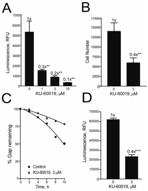
In vitro activity: In contrast to KU-55933, KU-60019 exhibits similar target selectivity but is a better ATM kinase inhibitor. KU-60019 exhibits negligible efficacy against 229 protein kinases, including PI3K, mTOR, and mTOR/FKBP12, and DNA-PKcs and ATR, with IC50 values of 1.7 μM and >10 μM, respectively. In human glioma U87 and U1242 cells, KU-60019 exhibits 3- to 10-fold greater potency than KU-55933 at inhibiting radiation-induced phosphorylation of important ATM protein targets, including p53, γ-H2AX, and CHK2. This is because 1 μM of KU-60019 significantly reduces p53 (S15) phosphorylation by >70%, a reduction that requires ~10 μM of KU-55933 to attain. In addition to radiosensitizing human glioma cells, KU-60019 also radiosensitizes normal fibroblasts but not A-T fibroblasts, with dose-enhancement ratios of 1.7 and 4.4 at 1 μM and 10 μM, respectively. Treatment with KU-60019 (3 μM) completely reduces radiation-induced AKT phosphorylation below the control level and blocks 70% of basal and ~50% of insulin-induced AKT S473 phosphorylation, respectively. The phosphorylase inhibitor okadaic acid significantly blocks the effect of KU-60019 on AKT S473 phosphorylation in glioma cell lines and normal fibroblasts, but not in A-T (h-TERT) cells. This suggests a critical role for ATM kinase in regulating AKT phosphorylation via unknown phosphatase. KU-60019 at 3 μM significantly inhibits the migration and invasion of human glioma U87 cells by >70% and ~60%, respectively, and U1242 cells by >50% and ~60%, respectively, which is consistent with the inhibition of prosurvival AKT signaling. [1]
In human glioma cell lines (U87MG, U251MG, T98G), KU-60019 (0.5–10 μM) alone inhibits cell proliferation with IC50 values ranging from 3.2 to 5.8 μM. When combined with ionizing radiation (IR, 2–8 Gy), it acts as a radiosensitizer: the survival fraction of U87MG cells after 4 Gy IR + 5 μM KU-60019 is reduced by ~70% compared to IR alone [1] - KU-60019 inhibits ATM kinase activity in glioma cells, reducing phosphorylation of ATM (p-ATM) and downstream substrates (p-Chk2, p-p53) (Western blot). It also compromises prosurvival signaling: downregulates insulin receptor β (IRβ) phosphorylation, and inhibits AKT and ERK activation (reduced p-AKT, p-ERK levels) [1] - In glioma cells, KU-60019 (2.5–10 μM) inhibits cell migration and invasion: transwell assay shows ~50% reduction in migration and ~60% reduction in invasion at 5 μM compared to control. It downregulates matrix metalloproteinase-2 (MMP-2) and MMP-9 expression (Western blot and zymography) [1] - In PTEN-deficient tumor cell lines (PC3, DU145, U87MG), KU-60019 (1–5 μM) selectively inhibits cell viability (IC50 = 2.3–3.7 μM) compared to PTEN-proficient cells (IC50 > 10 μM). It induces accumulation of double-strand DNA breaks (DSBs) (elevated γ-H2AX foci) and G2/M cell cycle arrest (flow cytometry) [2] - In PTEN-deficient cells, KU-60019 (3 μM) enhances apoptosis: Annexin V-FITC/PI staining shows apoptotic rate increased from ~8% (control) to ~45% (treatment). This is associated with activation of caspase-3 and PARP cleavage (Western blot) [2] |
| ln Vivo |
Radiation plus KU-60019 greatly increases mouse survival in orthotopic glioma U1242/luc-GFP xenograft models compared to radiation alone, KU-60019 alone, or no treatment at all. Furthermore, p53-mutant gliomas respond far more strongly to KU-60019 radiosensitization than do gliomas of the wild type.[2]
In vivo efficacy of ATM inhibition in PTEN-deficient xenografts To investigate the in vivo efficacy of ATM specific inhibition in PTEN-deficient cells, we used the PC3 PTEN Tetracycline inducible cell line model in a subcutaneous xenograft setting. Firstly, we established that the Tetracycline derivative Doxycycline was efficient at inducing PTEN expression in vitro (Supplementary Fig. 4A) and in vivo (Supplementary Fig. 4B). Calliper measurements of tumour volumes showed that induction of PTEN using Doxycycline, led to a slowing of tumour growth from 72 hours onwards (Supplementary Fig. 4C). Next, we investigated the selective toxicity of the ATM specific kinase inhibitor KU-60019 as a single modality in the PC3-PTEN inducible model. This inhibitor was chosen, as it is a potent ATM inhibitor and unlike KU-55933 is active in animal systems. Despite PTEN-deficient control tumours reaching a 4-fold increase in size before PTENwild-typecontrols, KU-60019 treated PTEN-deficient tumours displayed a statistically significant slowing in growth (Fig. 4A). This growth inhibition was especially evident at the start of the experiment (days 5-12) just after KU-60019 was administered (day 1-5). There were no significant changes in the mean relative body weights of each treatment groups (Fig. 4B). Inducible PTEN expression in vivo was analysed in resected tumours by immunofluorescence [2]. In a subcutaneous xenograft model of PTEN-deficient PC3 prostate cancer (nude mice), intraperitoneal administration of KU-60019 (25 mg/kg/day) for 21 days inhibits tumor growth by ~65% compared to vehicle control. Tumor tissues show reduced p-ATM, p-Chk2, and γ-H2AX levels (immunohistochemistry), confirming ATM inhibition and DSB accumulation [2] - In the U87MG glioma xenograft model (nude mice), combination treatment with KU-60019 (20 mg/kg/day, i.p.) and IR (4 Gy, once weekly for 3 weeks) inhibits tumor growth by ~80%, significantly higher than either agent alone (~30% for IR alone, ~45% for KU-60019 alone). The combination also prolongs mouse survival (median survival increased from 32 days to 56 days) [1] |
| Enzyme Assay |
Recombinant human ATM kinase was incubated with a peptide substrate (derived from Chk2) and ATP in kinase buffer. KU-60019 was added at concentrations ranging from 0.1 nM to 10 μM, and the mixture was incubated at 30°C for 45 minutes. The phosphorylated peptide was detected using a time-resolved fluorescence resonance energy transfer (TR-FRET) assay. The inhibition rate was calculated relative to the vehicle control, and the IC50 value was determined by nonlinear regression [1]
- ATM kinase activity assay with purified ATM protein: The reaction mixture contained ATM, [γ-32P]ATP, and histone H2AX substrate. KU-60019 (0.01–50 nM) was added, and the mixture was incubated at 37°C for 30 minutes. The reaction was terminated by adding SDS sample buffer, and proteins were separated by SDS-PAGE. Radioactivity of phosphorylated H2AX was quantified by autoradiography, and the IC50 was calculated [2] |
| Cell Assay |
Cell growth was determined by AlamarBlue®. U1242 cells were serially diluted, allowedto attach for 6 h and then exposed to KU-60019 at 3 μM. At days 1, 3 and 5 after seeding, AlamarBlue® was added to the medium to the recommended final concentration. Plates were incubated for 1 h at 37°C and fluorescence determined on a FluoroCount plate reader excitation 530 nm, emission 590 nm) and values taken as a measure of cell growth.[1]
KU-60019 is exposed to cells for 1, 3, and 5 days. AlamarBlue is the measure of cell growth. The medium is supplemented with AlamarBlue up to the suggested final concentration. The Fluoro-Count plate reader (excitation 530 nm, emission 590 nm) is used to measure fluorescence on the plates after an hour of incubation at 37 °C. The values obtained represent the growth of the cells. The trypan blue/fluorescence activated cell sorting (FACS) assay is used to assess cell survival. Radiosensitization assay: Glioma cells (1×10⁴ per well) were seeded in 6-well plates, treated with KU-60019 (2.5–10 μM) for 1 hour, then exposed to IR (2–8 Gy). After 14 days of culture, colonies were stained with crystal violet and counted. The survival fraction was calculated relative to unirradiated control cells [1] - Cell viability and apoptosis assay: PTEN-deficient/proficient cells (5×10³ per well) were seeded in 96-well plates, treated with KU-60019 (0.1–20 μM) for 48 hours. Cell viability was measured by CCK-8 assay (absorbance at 450 nm) to determine IC50. For apoptosis, cells were stained with Annexin V-FITC/PI and analyzed by flow cytometry [2] - Migration and invasion assay: Glioma cells (2×10⁴ per well) were seeded in transwell inserts (uncoated for migration, Matrigel-coated for invasion) with KU-60019 (2.5–10 μM) in the upper chamber. After 24 hours (migration) or 48 hours (invasion), cells that migrated/invaded to the lower chamber were stained with crystal violet and counted under a microscope [1] - Western blot analysis: Cells treated with KU-60019 (1–10 μM) for 24–48 hours were lysed to extract total protein. Equal amounts of protein were subjected to SDS-PAGE, transferred to PVDF membranes, and probed with antibodies against p-ATM, ATM, p-Chk2, Chk2, p-AKT, AKT, p-ERK, ERK, MMP-2, MMP-9, γ-H2AX, caspase-3, cleaved caspase-3, PARP, cleaved PARP, or GAPDH (loading control). Protein bands were visualized by chemiluminescence [1,2] - Cell cycle analysis: PTEN-deficient cells were treated with KU-60019 (3 μM) for 24 hours, fixed with 70% ethanol, stained with PI/RNase solution, and analyzed by flow cytometry to determine cell cycle distribution (G0/G1, S, G2/M phases) [2] - Immunofluorescence staining for γ-H2AX foci: PC3 cells were seeded on coverslips, treated with KU-60019 (2 μM) for 12 hours, fixed with paraformaldehyde, permeabilized with Triton X-100, and stained with anti-γ-H2AX antibody (FITC-conjugated) and DAPI. The number of γ-H2AX foci per cell was counted by confocal microscopy (n ≥ 50 cells per group) [2] |
| Animal Protocol |
Mice: Male Fox Chase Severe Combined Immunodeficiency (SCID) mice are implanted with cells (3×10⁷). Doxycycline is given every 48 hours until the animal is removed from the experiment, beginning when the tumor reaches a volume of 100 mm3. Animals are given 100 mg/kg of KU-60019 for five days in a row following PTEN induction, and their volume is measured every day until the animals reach the 400 mm3 target. Every three days, the tumor volume and body weight were measured with calipers.
PC3 prostate cancer xenograft model: Nude mice (4-week-old, male) were subcutaneously injected with PC3 cells (5×10⁶ cells/mouse) into the right flank. When tumors reached ~100 mm3, mice were randomly divided into control (n = 6) and KU-60019 treatment (n = 6) groups. KU-60019 was dissolved in DMSO (5%) + saline (95%), administered via intraperitoneal injection at 25 mg/kg once daily for 21 days. Tumor volume was measured every 3 days (volume = length × width2 / 2), and mice were euthanized at the end of treatment for tumor weight measurement and immunohistochemical analysis [2] - U87MG glioma xenograft model: Nude mice (4-week-old, male) were subcutaneously implanted with U87MG cells (1×10⁷ cells/mouse). When tumors reached ~120 mm3, mice were divided into four groups (n = 6 per group): control, IR alone (4 Gy, intravenous irradiation once weekly for 3 weeks), KU-60019 alone (20 mg/kg/day, i.p. for 21 days), and combination group. KU-60019 was dissolved in 0.9% saline containing 3% DMSO. Tumor volume and mouse survival were recorded, and tumor tissues were collected for Western blot analysis [1] |
| Toxicity/Toxicokinetics |
In vitro toxicity: KU-60019 showed low cytotoxicity to normal human astrocytes (NHA) and prostate epithelial cells (PrEC) at concentrations up to 10 μM (cell survival >80% vs. control group) [1,2] - Acute in vivo toxicity: No death or severe toxic symptoms (drowsiness, weight loss) were observed in mice after a single intraperitoneal injection of KU-60019 (up to 100 mg/kg) within 7 days [2] - Long-term in vivo toxicity: No significant changes in body weight, liver function (ALT, AST) or kidney function (BUN, creatinine) were observed in mice treated with KU-60019 (20–25 mg/kg/day, intraperitoneal injection, for 21–28 days) compared to the control group. Histological examination of the liver, kidneys and heart tissues revealed no abnormal lesions [1,2]
- Plasma protein binding rate: The plasma protein binding rate of KU-60019 in human plasma was 92%, and the plasma protein binding rate in mouse plasma was 89%, as determined by balanced dialysis [2] |
| References |
|
| Additional Infomation |
The ataxia-telangiectasia (AT) mutant gene (ATM) is crucial for cell cycle checkpoints and DNA repair. Therefore, specific small-molecule inhibitors targeting ATM may be developed into highly potent radiosensitizers. Recently, an ATM kinase-specific inhibitor, KU-55933, was shown to enhance the radiosensitivity of human cancer cells. This article reports a modified analogue of KU-55933 (KU-60019) with only half the Ki and IC50 values of KU-55933. KU-60019's ability to block radiation-induced phosphorylation of key ATM targets in human glioma cells is 10 times greater than that of KU-55933. As expected, KU-60019 is a highly potent radiosensitizer for human glioma cells. AT fibroblasts were not radiosensitized by KU-60019, strongly suggesting that ATM kinase is its specific target. Furthermore, KU-60019 reduced basal phosphorylation levels at the AKT S473 site, suggesting that ATM kinase may regulate a protein phosphatase acting on AKT. Consistent with this finding, low concentrations of the phosphatase inhibitor okadaic acid antagonized the effect of KU-60019 on AKT phosphorylation, and AT cells showed impaired AKT phosphorylation at the S473 site under radiation and insulin stimulation, showing no response to KU-60019. We also found that KU-60019 inhibited glioma cell migration and invasion in vitro, suggesting that glioma growth and migration may be regulated by ATM through AKT. MEK and AKT inhibitors did not further enhance the radiosensitivity of KU-60019-treated cells, supporting the view that KU-60019 interferes with pro-survival signaling pathways rather than having a radiosensitizing effect. In conclusion, KU-60019 inhibits DNA damage responses, reduces AKT phosphorylation and pro-survival signaling, inhibits cell migration and invasion, and effectively enhances the radiosensitivity of human glioma cells. [1]
Ataxia-telangiectasia mutant gene (ATM) is an important signaling molecule in the DNA damage response (DDR). Loss of ATM function can combine with tumor-associated mutations in the FA/BRCA pathway components to produce a synthetic lethal phenotype. In this study, we used an siRNA screening strategy to identify other tumor suppressor factors that, when inhibited, also enhance the sensitivity of cells to ATM inhibitors in a similar manner. Using this approach, we found that PTEN and ATM are synthetically lethal when inhibited in combination. PTEN-deficient cells exhibited elevated levels of reactive oxygen species, increased endogenous DNA damage, and persistent ATM activation. Compared with wild-type cells, ATM inhibitors specifically induced catastrophic DNA damage, mitotic cell cycle arrest, and apoptosis in PTEN-deficient cells. Antioxidants eliminated the increased DNA damage and ATM activation in PTEN-deficient cells, indicating that oxidative DNA damage is a necessary condition for cell death mechanisms. Finally, the ATM inhibitor KU-60019 exhibited specific toxicity to PTEN-mutant cancer cells in a tumor xenograft model, and this toxicity could be reversed by reintroducing wild-type PTEN. Our findings collectively provide a mechanistic justification for the clinical evaluation of ATM inhibitors in PTEN-deficient tumors. [2] KU-60019 is a modified analogue of the first-generation ATM inhibitor KU-55933, with higher potency (lower IC50) and better pharmacokinetic properties. [1] - Its mechanism of action involves binding to the ATP-binding pocket of ATM, inhibiting ATM-mediated DNA damage repair. This leads to the accumulation of DNA double-strand breaks (DSBs), cell cycle arrest, and apoptosis in cancer cells, especially PTEN-deficient cancer cells (which rely on ATM for survival) [2] - In glioma cells, KU-60019 exerts multiple anticancer effects: radiosensitization, inhibition of the pro-survival AKT/ERK signaling pathway, and inhibition of cell migration/invasion, making it a potential therapeutic for gliomas (which are usually radiation resistant) [1] - KU-60019 has higher selectivity for ATM than other PIKKs (ATR, DNA-PKcs), thereby reducing off-target effects and improving safety [1,2] |
| Molecular Formula |
C30H33N3O5S
|
|
|---|---|---|
| Molecular Weight |
547.67
|
|
| Exact Mass |
547.214
|
|
| CAS # |
925701-49-1
|
|
| Related CAS # |
|
|
| PubChem CID |
15953870
|
|
| Appearance |
Light yellow to khaki solid powder
|
|
| Density |
1.3±0.1 g/cm3
|
|
| Boiling Point |
786.6±60.0 °C at 760 mmHg
|
|
| Flash Point |
429.5±32.9 °C
|
|
| Vapour Pressure |
0.0±2.7 mmHg at 25°C
|
|
| Index of Refraction |
1.645
|
|
| LogP |
5.9
|
|
| Hydrogen Bond Donor Count |
1
|
|
| Hydrogen Bond Acceptor Count |
8
|
|
| Rotatable Bond Count |
5
|
|
| Heavy Atom Count |
39
|
|
| Complexity |
972
|
|
| Defined Atom Stereocenter Count |
2
|
|
| SMILES |
C[C@@H]1CN(C[C@@H](O1)C)CC(=O)NC2=CC3=C(C=C2)SC4=C(C3)C=CC=C4C5=CC(=O)C=C(O5)N6CCOCC6
|
|
| InChi Key |
SCELLOWTHJGVIC-BGYRXZFFSA-N
|
|
| InChi Code |
InChI=1S/C30H33N3O5S/c1-19-16-32(17-20(2)37-19)18-28(35)31-23-6-7-27-22(13-23)12-21-4-3-5-25(30(21)39-27)26-14-24(34)15-29(38-26)33-8-10-36-11-9-33/h3-7,13-15,19-20H,8-12,16-18H2,1-2H3,(H,31,35)/t19-,20+
|
|
| Chemical Name |
2-[(2S,6R)-2,6-dimethylmorpholin-4-yl]-N-[5-(6-morpholin-4-yl-4-oxopyran-2-yl)-9H-thioxanthen-2-yl]acetamide
|
|
| Synonyms |
|
|
| HS Tariff Code |
2934.99.9001
|
|
| Storage |
Powder -20°C 3 years 4°C 2 years In solvent -80°C 6 months -20°C 1 month |
|
| Shipping Condition |
Room temperature (This product is stable at ambient temperature for a few days during ordinary shipping and time spent in Customs)
|
| Solubility (In Vitro) |
DMSO: 18~100 mg/mL (32.9~182.6 mM)
Water: 1 mg/mL (Insoluble) Ethanol: 1 mg/mL |
|---|---|
| Solubility (In Vivo) |
5%DMSO+ 40%PEG300+5%Tween 80+50%ddH2O: 5.0mg/ml (9.13mM) (Please use freshly prepared in vivo formulations for optimal results.)
|
| Preparing Stock Solutions | 1 mg | 5 mg | 10 mg | |
| 1 mM | 1.8259 mL | 9.1296 mL | 18.2592 mL | |
| 5 mM | 0.3652 mL | 1.8259 mL | 3.6518 mL | |
| 10 mM | 0.1826 mL | 0.9130 mL | 1.8259 mL |
*Note: Please select an appropriate solvent for the preparation of stock solution based on your experiment needs. For most products, DMSO can be used for preparing stock solutions (e.g. 5 mM, 10 mM, or 20 mM concentration); some products with high aqueous solubility may be dissolved in water directly. Solubility information is available at the above Solubility Data section. Once the stock solution is prepared, aliquot it to routine usage volumes and store at -20°C or -80°C. Avoid repeated freeze and thaw cycles.
Calculation results
Working concentration: mg/mL;
Method for preparing DMSO stock solution: mg drug pre-dissolved in μL DMSO (stock solution concentration mg/mL). Please contact us first if the concentration exceeds the DMSO solubility of the batch of drug.
Method for preparing in vivo formulation::Take μL DMSO stock solution, next add μL PEG300, mix and clarify, next addμL Tween 80, mix and clarify, next add μL ddH2O,mix and clarify.
(1) Please be sure that the solution is clear before the addition of next solvent. Dissolution methods like vortex, ultrasound or warming and heat may be used to aid dissolving.
(2) Be sure to add the solvent(s) in order.
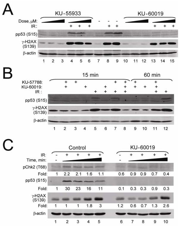 KU-60019 is a more effective inhibitor of the ATM kinase than KU-55933.Mol Cancer Ther.2009 Oct;8(10):2894-902. |
|---|
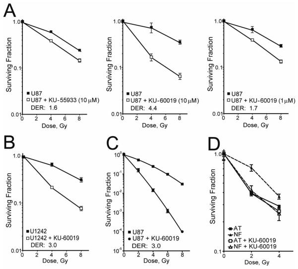 KU-60019 radiosensitizes U87 and U1242 human glioma cells and normal but not A-T fibroblasts.Mol Cancer Ther.2009 Oct;8(10):2894-902. |
KU-60019 inhibits migration and invasion of human glioma U87 and U1242 cells in vitro.Mol Cancer Ther.2009 Oct;8(10):2894-902. |
Products are for research use only; We do not sell to patients
Copyright 2020 InvivoChem LLC | All Rights Reserved


 COA
COA