| Size | Price | Stock | Qty |
|---|---|---|---|
| 5mg |
|
||
| 10mg |
|
||
| 50mg |
|
||
| 100mg |
|
||
| 250mg | |||
| 500mg | |||
| Other Sizes |


| Targets |
INDY targets the dual-specificity tyrosine phosphorylation-regulated kinases DYRK1A and DYRK1B. It acts as an ATP-competitive inhibitor, binding within the ATP pocket of the enzyme. The compound exhibits an IC50 value of 0.24 μM for DYRK1A and 0.23 μM for DYRK1B, with a Ki value of 0.18 μM for DYRK1A. At a high concentration of 10 µM, INDY shows >90% inhibition of CLK1. DYRK1A and DYRK1B are involved in various cellular processes, including cell cycle regulation, neuronal development, and differentiation. Inhibition of these kinases is being explored as a therapeutic strategy for cancer and neurodegenerative diseases.
|
|---|---|
| ln Vitro |
At 3 μM and 30 μM, INDY (0.3-30 μM; 20 hours) virtually totally blocks tau phosphorylation [1]. Nuclear factor of activated T cells, or NFAT, signaling is blocked when acid-(Y)-phosphorylation regulator 1A is overexpressed [1].
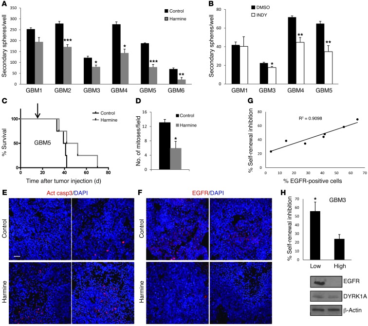
In vitro, INDY potently inhibits DYRK1A and DYRK1B kinase activity with IC50 values of 0.24 μM and 0.23 μM, respectively. The compound binds to the ATP pocket of the enzyme with a Ki of 0.18 μM for DYRK1A. INDY significantly reduces the self-renewal capacity of both normal and tumorigenic cells in primary glioblastoma (GBM) cell lines and neural progenitor cells. It is selective for DYRK1A over MAO-A and MAO-B at concentrations up to 100 μM. At 10 µM, INDY inhibits CLK1 by more than 90%, indicating some cross-reactivity at higher concentrations. INDY has also been reported to inhibit SLC13A5, a sodium-coupled citrate transporter. |
| ln Vivo |
ProINDY (2.5 μM) significantly restores synchronized normal development in Xenopus laevis [1].
In vivo studies of INDY are limited, as the compound is primarily used as a research tool for in vitro kinase inhibition studies. However, the compound's ability to diminish the self-renewal capacity of GBM cells and neural progenitor cells suggests potential in vivo applications in cancer research. INDY has been cited in biomedical literature, including a publication in Nature Medicine, indicating its relevance in preclinical research. Further in vivo studies are needed to fully characterize its pharmacokinetic profile, efficacy in animal models of disease, and potential therapeutic applications in oncology and neurology. |
| Enzyme Assay |
For in vitro kinase assays with INDY, recombinant DYRK1A or DYRK1B enzyme is incubated with a suitable peptide substrate and ATP in a kinase buffer. INDY is typically dissolved in DMSO and added to the reaction at various concentrations. The reaction is allowed to proceed for a specified time at room temperature or 30°C. Kinase activity is measured using a radiometric assay with [γ-33P]ATP, or using a luminescent ADP detection method such as the ADP-Glo™ Kinase Assay. The IC50 value is determined by plotting the percentage of remaining kinase activity against the log of inhibitor concentration and fitting the data to a sigmoidal dose-response curve.
|
| Cell Assay |
Western Blot Analysis[1]
Cell Types: COS7 cells transfected with EGFP-Dyrk1A and EGFP-tau Tested Concentrations: 0.3, 1, 3, 10, 30 μM Incubation Duration: 20 hrs (hours) Experimental Results: Mild inhibition of tau phosphorylation at 3 μM , almost complete inhibition at 30 μM. For in vitro cell-based studies, primary glioblastoma (GBM) cell lines or neural progenitor cells are cultured in appropriate growth media. INDY is dissolved in DMSO and diluted in cell culture medium to achieve the desired concentrations, with the final DMSO concentration kept below 0.1% to avoid solvent toxicity. Cells are treated with INDY for a defined period, typically 24-72 hours. The effect of INDY on cell self-renewal capacity is assessed using neurosphere formation assays or colony formation assays. Cell viability is measured using MTT, CellTiter-Glo, or similar assays. The effect on signaling pathways can be evaluated by Western blot analysis of DYRK1A substrates or downstream effectors. |
| Animal Protocol |
For in vivo animal studies, INDY is typically formulated for oral administration as a homogeneous suspension in CMC-Na at a concentration of ≥5 mg/mL. For injection, it can be prepared as a clear solution in 5% DMSO, 40% PEG300, 5% Tween 80, and 50% ddH2O at a concentration of 1.150 mg/mL (4.89 mM). Alternatively, it can be formulated in 5% DMSO and 95% corn oil. Animal studies typically involve xenograft mouse models for efficacy evaluation, where tumor-bearing mice are treated with INDY via oral gavage or intravenous administration. Tumor growth inhibition is measured over time, and pharmacokinetic parameters such as half-life, Cmax, and AUC are determined from plasma samples.
|
| ADME/Pharmacokinetics |
The pharmacokinetic properties of INDY have not been extensively characterized in the available literature. INDY is a small molecule with a molecular weight of 235.3 g/mol, which is favorable for oral bioavailability. It is soluble in DMSO at 47 mg/mL (199.74 mM) and in ethanol at 4 mg/mL, but is insoluble in water. For in vivo administration, INDY can be formulated as a homogeneous suspension in CMC-Na for oral administration or as a clear solution using DMSO, PEG300, Tween 80, and water for injection. The compound is stable when stored as a powder at -20°C. Detailed PK parameters such as half-life, bioavailability, and volume of distribution have not been reported and require further investigation.
|
| Toxicity/Toxicokinetics |
Toxicological data for INDY are limited, as the compound is a research chemical not intended for human therapeutic use. The compound is typically handled with standard laboratory safety precautions. INDY is selective for DYRK1A over MAO-A and MAO-B at concentrations up to 100 μM, suggesting a favorable off-target safety profile. However, comprehensive toxicology studies, including acute and chronic toxicity, genotoxicity, and cardiotoxicity assessments, have not been reported. The compound's safety in animal models and potential for clinical development would require further toxicological evaluation.
|
| References | |
| Additional Infomation |
Indigoxin (INDY) belongs to the benzothiazole class of compounds, with the structure 2,3-dihydro-1,3-benzothiazole, substituted at positions 2, 3, and 5 with 2-oxopropylidene, ethyl, and hydroxyl groups, respectively. It is an ATP-competitive inhibitor of Dyrk1A and Dyrk1B (IC50 values of 0.24 μM and 0.23 μM, respectively). It possesses antitumor activity, is a drug metabolite, and is also an EC 2.7.12.1 (bispecific kinase) inhibitor. Indigoxin belongs to the benzothiazole class, organic hydroxyl compounds, and enones.
INDY is a research compound and is not an FDA-approved drug. It is primarily used as a chemical probe to study the biological functions of DYRK1A and DYRK1B in neurological disorders and cancer. The compound has been cited in scientific literature, including a publication in Nature Medicine, indicating its significance in biomedical research. The benzothiazole scaffold of INDY is a common pharmacophore in drug discovery, and the compound serves as a lead for the development of more potent and selective DYRK inhibitors. INDY has also been reported as an inhibitor of SLC13A5, a sodium-coupled citrate transporter, suggesting additional research applications in metabolic diseases. |
| Molecular Formula |
C12H13NO2S
|
|---|---|
| Molecular Weight |
235.302
|
| Exact Mass |
235.067
|
| CAS # |
1169755-45-6
|
| Related CAS # |
1169755-45-6;
|
| PubChem CID |
44136031
|
| Appearance |
Off-white to light yellow solid powder
|
| LogP |
2.078
|
| Hydrogen Bond Donor Count |
1
|
| Hydrogen Bond Acceptor Count |
4
|
| Rotatable Bond Count |
2
|
| Heavy Atom Count |
16
|
| Complexity |
316
|
| Defined Atom Stereocenter Count |
0
|
| SMILES |
CCN\1C2=C(C=CC(=C2)O)S/C1=C\C(=O)C
|
| InChi Key |
GCSZJMUFYOAHFY-SDQBBNPISA-N
|
| InChi Code |
InChI=1S/C12H13NO2S/c1-3-13-10-7-9(15)4-5-11(10)16-12(13)6-8(2)14/h4-7,15H,3H2,1-2H3/b12-6-
|
| Chemical Name |
(1Z)-1-(3-ethyl-5-hydroxy-1,3-benzothiazol-2-ylidene)propan-2-one
|
| HS Tariff Code |
2934.99.9001
|
| Storage |
Powder -20°C 3 years 4°C 2 years In solvent -80°C 6 months -20°C 1 month |
| Shipping Condition |
Room temperature (This product is stable at ambient temperature for a few days during ordinary shipping and time spent in Customs)
|
| Solubility (In Vitro) |
DMSO : ≥ 33.33 mg/mL (~141.65 mM)
|
|---|---|
| Solubility (In Vivo) |
Solubility in Formulation 1: ≥ 2.5 mg/mL (10.62 mM) (saturation unknown) in 10% DMSO + 90% Corn Oil (add these co-solvents sequentially from left to right, and one by one), clear solution.
For example, if 1 mL of working solution is to be prepared, you can add 100 μL of 25.0 mg/mL clear DMSO stock solution to 900 μL of corn oil and mix evenly. (Please use freshly prepared in vivo formulations for optimal results.) |
| Preparing Stock Solutions | 1 mg | 5 mg | 10 mg | |
| 1 mM | 4.2499 mL | 21.2495 mL | 42.4989 mL | |
| 5 mM | 0.8500 mL | 4.2499 mL | 8.4998 mL | |
| 10 mM | 0.4250 mL | 2.1249 mL | 4.2499 mL |
*Note: Please select an appropriate solvent for the preparation of stock solution based on your experiment needs. For most products, DMSO can be used for preparing stock solutions (e.g. 5 mM, 10 mM, or 20 mM concentration); some products with high aqueous solubility may be dissolved in water directly. Solubility information is available at the above Solubility Data section. Once the stock solution is prepared, aliquot it to routine usage volumes and store at -20°C or -80°C. Avoid repeated freeze and thaw cycles.
Calculation results
Working concentration: mg/mL;
Method for preparing DMSO stock solution: mg drug pre-dissolved in μL DMSO (stock solution concentration mg/mL). Please contact us first if the concentration exceeds the DMSO solubility of the batch of drug.
Method for preparing in vivo formulation::Take μL DMSO stock solution, next add μL PEG300, mix and clarify, next addμL Tween 80, mix and clarify, next add μL ddH2O,mix and clarify.
(1) Please be sure that the solution is clear before the addition of next solvent. Dissolution methods like vortex, ultrasound or warming and heat may be used to aid dissolving.
(2) Be sure to add the solvent(s) in order.
| NCT Number | Recruitment | interventions | Conditions | Sponsor/Collaborators | Start Date | Phases |
| NCT02159833 | Completed | Other: Intranasal challenge with active substance (food protein) Other: Intranasal challenge with placebo |
Food Allergy | Imperial College London | 2014-10 | Not Applicable |
| NCT04681781 | Enrolling by invitation | 17p13.1 Deletions Confined to SLC13A5 Gene
SLC13A5 Deficiency Citrate Transporter Deficiency Citrate Transporter Disorder |
TESS Research Foundation | 2021-03-01 | ||
| NCT06144957 | Recruiting | SLC13A5 Deficiency Citrate Transporter Deficiency Citrate Transporter Disorder DEE25 |
2021-12-01 | TESS Research Foundation | ||
| NCT02500082 | No longer available | Drug: triheptanoin | SLC13A5 Gene Mutation Citrate Transporter Deficiency |
Irina A Anselm | ||
| NCT03964831 | Completed | Device: P200TxE Device: P200DTx |
Retinal Disease | Optos, PLC | 2019-06-18 | Not Applicable |
|
Products are for research use only; We do not sell to patients
Copyright 2020 InvivoChem LLC | All Rights Reserved