| Size | Price | Stock | Qty |
|---|---|---|---|
| 1mg |
|
||
| 5mg |
|
||
| 25mg | |||
| 50mg | |||
| 100mg | |||
| Other Sizes |


IM-54 (IM54) is a novel and potent inhibitor of necrosis induced by oxidative stress (e.g. H2O2-induced necrosis). IM-54 has potential cardioprotective activity. Oxidative stress-induced necrosis plays an important role in ischemia-reperfusion injury, such as stroke and heart attack.
| Targets |
Necrosis
|
|---|---|
| ln Vitro |
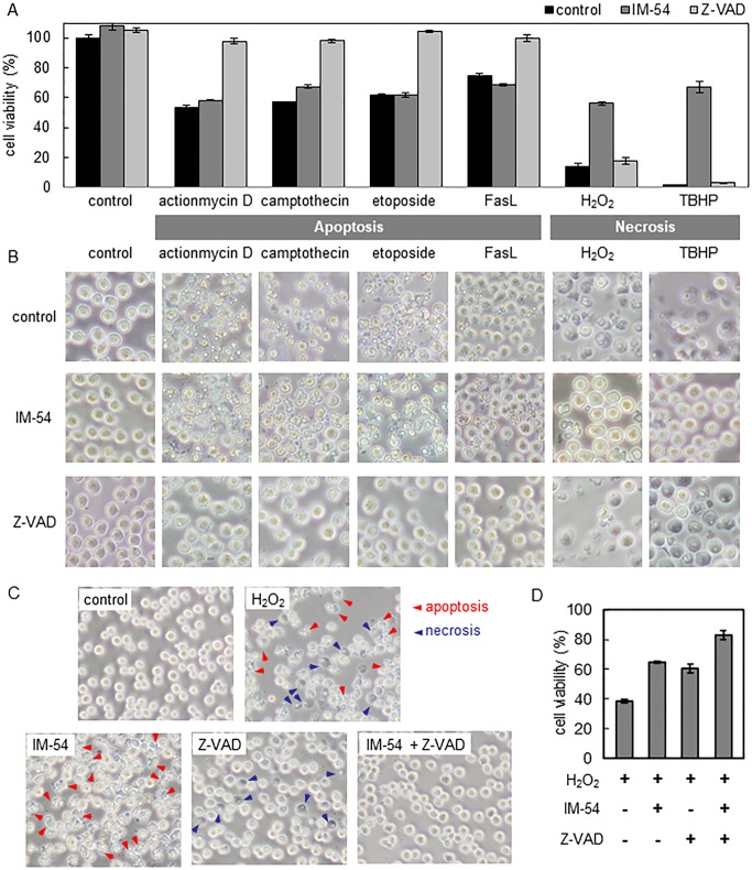
While IM-54 inhibits H2O2-induced cellular crossover, it is unable to inhibit crossover or cellular crossover brought on by other anticancer medications or Fas ligands [1]. In HL-60 cells, the IC50 is 0.25 μM[1].
|
| ln Vivo |
In vivo study of IM-54 was planned, but its water-solubility was too low. To overcome this problem, researchers designed and synthesized more water-soluble IM derivatives. [1]
|
| Enzyme Assay |
KinaseProfiler assay of IM-54 [1]
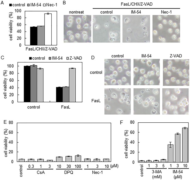
As IM-54 was originally developed from ATP-competitive PKC inhibitor bisindolylmaleimide I (BM I) 5 , we previously analyzed its kinase-inhibitory activities against all PKC subtypes and confirmed that no PKC was inhibited by IM-541 . We also evaluated its inhibitory activities against various kinases related to cell-death signaling pathways. Protein kinase inhibition assays were performed using the KinaseProfiler service. Briefly, protein kinases were assayed for their ability to phosphorylate the appropriate peptide/protein substrates in the presence of 50 µM IM-54 and 100 µM or 10 µM ATP. Activities are given as mean percentages of those in control incubations (averages of duplicate determinations). IM-54 did not inhibit more than half of any kinase activity, indicating that IC50 values against all tested kinases were over 50 µM. KINOMEscan screening of IM-17 and IM-54 [1] To examine the broad pattern of kinase inhibitory activities, KINOMEscan screening was applied for IM-54 and IM-17 at 10 µM. KINOMEscan evaluated the binding affinity to kinases, which was determined based on the ATP site-dependent competition for beads-immobilized kinase inhibitors6 . Activities are given as percentages of control values of bound kinases to beads-immobilized kinase inhibitors (averages of duplicate determinations). Less than 35 % of control at 10 µM indicates S17 significant inhibition of kinase. As shown in Table S2, no significant inhibition of 467 kinases by IM-17 or IM-54 was observed, supporting the view that these compounds do not have kinase-inhibitory activity. Analysis of stability to liver metabolism [1] To compare the drug-like properties of IM derivatives, stabilities of IM-12, IM-54, and IM-17 to liver metabolism were examined. Each compound in DMSO was mixed with 1 mL of rat liver S9 fraction containing cofactors for a final concentration of 5 µM with 0.1% DMSO. The mixture was incubated at 37 °C for 0 min, 5 min, 15 min and 30 min. After the incubation, the mixture was moved to ice quickly. Then ethyl acetate (250 µL) was added to the mixture and the compound was extracted. The extraction was repeated 5 times. The organic layer was combined and concentrated in vacuo. The residue was diluted with MeOH (30 µL) and analyzed by HPLC. Residual compound was quantified based on the peak area detected by UV (n = 3, three independent experiments). As shown in figure S1, IM-12 and IM-54 were decreased rapidly and were not detected after 5 min incubation with S9 fraction. In contract, IM-17 was detected even after 15 min incubation with 40% residual rate. These results demonstrated the higher stability of IM-17 to liver metabolism than IM-12 and IM-54. |
| Cell Assay |
AlamarBlue assay [1]
HL-60 cells (3 x 10⁴ cells/well for Fas ligand (FasL) and 4 x 10⁴ cells/well for others) or Jurkat cells (2 x 10⁴ cells/well for FasL/CHX/Z-VAD treatment or 3 x 10⁴ cells/well for FasL treatment) were suspended in fresh medium in a 96-well plate. After 2 h incubation, the cells were treated with test compounds (DMSO solution, 0.5 µL/well) for 1 h and then cell death inducer (in medium, 4 µL/well) was added (final volume 100 µL/well). In all experiments, the final DMSO concentration was the same (0.5%). At 3 h later, 10 µL of AlamarBlue was added to each well. The cell viability was determined based on the increase of fluorescence (excitation 560 nm/emission 590 nm) during 3-4 h incubation. Data are presented as mean ± S.D. (n = 4). LDH assay [1] Lactate dehydrogenase (LDH) that leaked into the culture medium was measured with a Cytotoxicity detection kitPLUS according to the manufacturer’s protocol. HL-60 cells were treated with test compounds and H2O2 (100 µM) in a 96-well plate according to the same method as described for AlamarBlue assay (triplicate). H9c2 cells (1 x 10⁴ cells/well) were treated with test compounds and TBHP (300 µM) for 4 h (triplicate). The change in absorbance at 490 nm was then measured to calculate the percentage of LDH release. IC50 values were calculated by Origin 8.0 software, and data are presented as mean ± S.D. (n = 3, three independent experiments). |
| Animal Protocol |
Langendorff isolated rat heart model [1]
The isolated rat heart preparations used in this study have been described previously. Briefly, male Sprague–Dawley rats (270–320 g) were anesthetized with sodium pentobarbital (30 mg/kg, i.p.) and heparinized (1000 IU/kg, i.v.). The hearts were rapidly removed and mounted on a Langendorff perfusion system. The environmental temperature was maintained at 37 °C with a heated glass water-bath throughout the experiments. Through the cannulated aorta, the hearts were perfused with warmed (37 °C) and gassed (95% O2 and 5% CO2, pO2>600 mmHg) Krebs–Henseleit solution containing (in mM) NaCl 120, KCl 4.7, CaCl2 1.25, MgSO4 1.2, KH2PO4 1.2, NaHCO3 25, glucose 11 at a constant perfusion pressure (70 ± 5 mm Hg). To measure the coronary flow (CF) continuously, a cannulating-type flow probe connected to an electro-magnetic blood flowmeter was inserted into a perfusion line connected to the heart. The left ventricular pressure was measured through a water-filled latex balloon inserted into the left ventricle via the left atrium, with a pressure transducer connected to an amplifier. Left ventricular end-diastolic pressure (LVEDP) was adjusted to about 5 mm Hg by adjusting the volume of the balloon, and the left ventricular developed pressure (LVDP) was obtained by deducting LVEDP from left ventricular systolic pressure. The heart rate was measured by using a cardio-tachometer triggered by the pulse of left ventricular pressure. All hemodynamic parameters and CF were continuously recorded on a multi-channel recorder. The hearts were paced at 300 beats/min using an electric stimulator, and an isolator unit with the bipolar electrode placed on the left atrium. After a 15-min period of S14 equilibration, vehicle, IM-12 (0.3 µM) or IM-17 (3 µM) was infused for 10 min with an infusion pump through a drug infusion line connected to the main perfusion line of the Langendorff system at a flow rate of 1/100 of the CF rate. Subsequent to drug treatment for 5 min, hearts were subjected to 30 min of global ischemia and 60 min of reperfusion. Global ischemia was induced by completely stopping the flow. LVDP, LVEDP and CF were measured before and 10 min after infusion of the drug, 10, 20 and 30 min after the induction of global ischemia, and 10, 20 and 30 min after reperfusion. Data are presented as mean ± S.D. (n = 3) Ischemia-reperfusion-induced arrhythmia model[1] Male Sprague-Dawley rats were anesthetized with sodium pentobarbital. The femoral vein was cannulated for intravenous test drug administration. Heart rate was measured by a cardiotachometer. A left thoracotomy at the fifth intercostal space and pericardiotomy were performed and an ELP 5-0 nylon ligature was placed around the left coronary artery about 2-3 mm from its origin. a b S15 Thereafter, both ends of the nylon ligature were passed through a small polyethylene tube to make a coronary snare. The standard limb lead II electrocardiogram (ECG) was monitored by a cardiograph. ECG, blood pressure and heart rate data were collected by an ECG processor and stored on the MO disk for further data processing. Myocardial ischemia was initiated by tightening the coronary snare and successful ischemia was confirmed by typical elevation of the ST segment in the ECG. At 5 min after the start of ischemia, reperfusion was initiated by releasing the snare. Ventricular fibrillation (Vf) after reperfusion was evaluated by ECG analysis based on the reported guideline26. Total duration of Vf was calculated as the sum of the duration of episodes that occurred within 10 min after reperfusion. 0.9% saline was used as a control. IM-17 was intravenously injected (1 ml/kg, over 1 min) at 5 min before ischemia (pre-ischemia treatment, 1 to 3 mg/kg) or at 1 min before reperfusion (post-ischemia treatment, 3 mg/kg). |
| References | |
| Additional Infomation |
IM-54 is an organic nitrogen and organic oxygen compound whose function is related to α-amino acids.
|
| Molecular Formula |
C19H23N3O2
|
|---|---|
| Molecular Weight |
325.40482
|
| Exact Mass |
325.179
|
| Elemental Analysis |
C, 70.13; H, 7.12; N, 12.91; O, 9.83
|
| CAS # |
861891-50-1
|
| PubChem CID |
16760577
|
| Appearance |
Light yellow to orange solid powder
|
| Density |
1.2±0.1 g/cm3
|
| Boiling Point |
511.5±50.0 °C at 760 mmHg
|
| Flash Point |
263.1±30.1 °C
|
| Vapour Pressure |
0.0±1.3 mmHg at 25°C
|
| Index of Refraction |
1.615
|
| LogP |
3.81
|
| Hydrogen Bond Donor Count |
1
|
| Hydrogen Bond Acceptor Count |
3
|
| Rotatable Bond Count |
6
|
| Heavy Atom Count |
24
|
| Complexity |
543
|
| Defined Atom Stereocenter Count |
0
|
| InChi Key |
SGLOMINNEBLJFF-UHFFFAOYSA-N
|
| InChi Code |
InChI=1S/C19H23N3O2/c1-4-5-8-11-20-17-16(18(23)22(3)19(17)24)14-12-21(2)15-10-7-6-9-13(14)15/h6-7,9-10,12,20H,4-5,8,11H2,1-3H3
|
| Chemical Name |
1-methyl-3-(1-methylindol-3-yl)-4-(pentylamino)pyrrole-2,5-dione
|
| Synonyms |
IM-54; Necrosis Inhibitor, IM-54; 1-Methyl-3-(1-methyl-1H-indol-3-yl)-4-(pentylamino)-1H-pyrrole-2,5-dione; 1-methyl-3-(1-methylindol-3-yl)-4-(pentylamino)pyrrole-2,5-dione; IM 54; MFCD18382109; CHEMBL365337;
|
| HS Tariff Code |
2934.99.9001
|
| Storage |
Powder -20°C 3 years 4°C 2 years In solvent -80°C 6 months -20°C 1 month Note: Please store this product in a sealed and protected environment (e.g. under nitrogen), avoid exposure to moisture. |
| Shipping Condition |
Room temperature (This product is stable at ambient temperature for a few days during ordinary shipping and time spent in Customs)
|
| Solubility (In Vitro) |
DMSO : ~50 mg/mL (~153.66 mM)
|
|---|---|
| Solubility (In Vivo) |
Solubility in Formulation 1: ≥ 2.5 mg/mL (7.68 mM) (saturation unknown) in 10% DMSO + 40% PEG300 +5% Tween-80 + 45% Saline (add these co-solvents sequentially from left to right, and one by one), clear solution.
For example, if 1 mL of working solution is to be prepared, you can add 100 μL of 25.0 mg/mL clear DMSO stock solution to 400 μL PEG300 and mix evenly; then add 50 μL Tween-80 + to the above solution and mix evenly; then add 450 μL normal saline to adjust the volume to 1 mL. Preparation of saline: Dissolve 0.9 g of sodium chloride in 100 mL ddH₂ O to obtain a clear solution. (Please use freshly prepared in vivo formulations for optimal results.) |
| Preparing Stock Solutions | 1 mg | 5 mg | 10 mg | |
| 1 mM | 3.0731 mL | 15.3657 mL | 30.7314 mL | |
| 5 mM | 0.6146 mL | 3.0731 mL | 6.1463 mL | |
| 10 mM | 0.3073 mL | 1.5366 mL | 3.0731 mL |
*Note: Please select an appropriate solvent for the preparation of stock solution based on your experiment needs. For most products, DMSO can be used for preparing stock solutions (e.g. 5 mM, 10 mM, or 20 mM concentration); some products with high aqueous solubility may be dissolved in water directly. Solubility information is available at the above Solubility Data section. Once the stock solution is prepared, aliquot it to routine usage volumes and store at -20°C or -80°C. Avoid repeated freeze and thaw cycles.
Calculation results
Working concentration: mg/mL;
Method for preparing DMSO stock solution: mg drug pre-dissolved in μL DMSO (stock solution concentration mg/mL). Please contact us first if the concentration exceeds the DMSO solubility of the batch of drug.
Method for preparing in vivo formulation::Take μL DMSO stock solution, next add μL PEG300, mix and clarify, next addμL Tween 80, mix and clarify, next add μL ddH2O,mix and clarify.
(1) Please be sure that the solution is clear before the addition of next solvent. Dissolution methods like vortex, ultrasound or warming and heat may be used to aid dissolving.
(2) Be sure to add the solvent(s) in order.
|
|
Products are for research use only; We do not sell to patients
Copyright 2020 InvivoChem LLC | All Rights Reserved