| Size | Price | Stock | Qty |
|---|---|---|---|
| 10mg |
|
||
| 25mg |
|
||
| 50mg |
|
||
| 100mg |
|
||
| 250mg |
|
||
| Other Sizes |


| Targets |
Natural flavonoid from safflower
|
|---|---|
| ln Vitro |
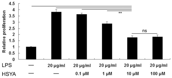
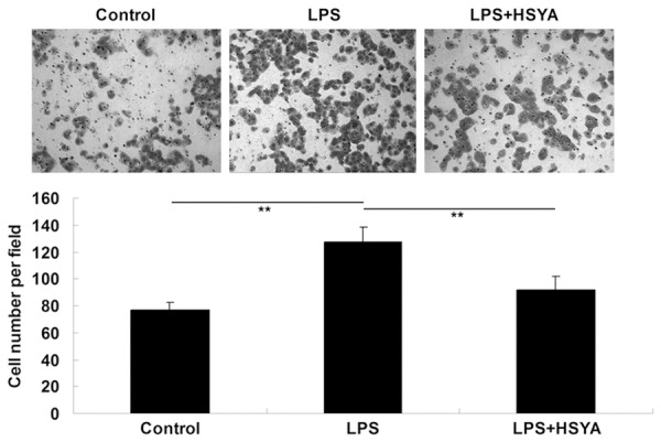
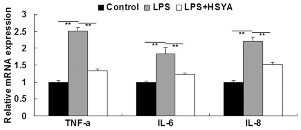
Abnormal proliferation and migration of vascular smooth muscle cells (VSMCs) is closely associated with early vascular hyperplasic lesions. Toll-like receptor (TLR)-4 is a pathogen pattern recognition receptor expressed on VSMCs, and can be activated by lipopolysaccharide. Activated TLR-4 plays a promoting role in VSMCs proliferation and migration through the downstream signaling pathways including Rac1/Akt. Hydroxysafflor yellow A (HSYA) is the main component of the safflower yellow pigments, which has long been used for the treatment of cardiovascular diseases in traditional Chinese medicine. However, the effect of HSYA on VSMC proliferation and migration remains unknown. In the present study, we showed that HYSA could inhibit LPS-induced VSMCs proliferation and migration, accompanied by the downregulated levels of several key pro-inflammatory cytokines, including TNF-α, IL-6, and IL-8. We further showed that HYSA inhibited LPS-induced upregulation of TLR-4 expression as well as the activation of Rac1/Akt pathway, suggesting that HSYA inhibits LPS-induced VSMCs proliferation and migration, partly at least, via inhibition of TLR-4/Rac1/Akt pathway. Accordingly, HSYA may be used as a promising agent for prevention and treatment of vascular hyperplasic disorders. [1]
Hydroxysafflor yellow A (HSYA), a main component of safflor yellow, has been demonstrated to prevent steroid-induced avascular necrosis of femoral head by inhibiting primary bone marrow-derived mesenchymal stromal cells adipogenic differentiation induced by steroid. In this study, we investigate the effect of HSYA on the proliferation and adipogenesis of mouse 3T3-L1 preadipocytes. The effects of HSYA on proliferation and differentiation of 3T3-L1 cells and its possible mechanism were studied by 3-(4,5-dimethylthiazol-2-yl) 2,5-diphenyl tetrazolium bromide spectrophotometry, Oil Red O staining, intracellular triglyceride assays, real-time quantitative RT-PCR, transient transfection and dual luciferase reporter gene methods. HSYA inhibited the proliferation of 3T3-L1 preadipocytes and cell viability greatly decreased in a dose and time dependent manner. HSYA (1 mg/l) notably reduced the amount of intracellular lipid and triglyceride content in adipocytes by 21.3 % (2.13 ± 0.36 vs 2.71 ± 0.40, P < 0.01) and 22.6 % (1.33 ± 0.07 vs 1.72 ± 0.07, P < 0.01) on days 8 following the differentiation, respectively. HSYA (1 mg/l) significantly increased hormone-sensitive lipase (HSL) mRNA expression and promoter activities by 2.4- and 1.55-fold, respectively (P < 0.01), in differentiated 3T3-L1 adipocytes. HSYA inhibits the proliferation and adipogenesis of 3T3-L1 preadipocytes. The inhibitory action of HYSA on adipogenesis may be due to the promotion of lipolytic-specific enzyme HSL expression by increasing HSL promoter activity. [2] HSYA/Hydroxysafflor yellow A protected EC viability against LPS-induced injury (P <0.05). LPS-induced NF-κB p65 subunit DNA binding (P <0.01) and nuclear factor of kappa light polypeptide gene enhancer in B-cells inhibitor α (IκBα) phosphorylation was inhibited by HSYA. HSYA attenuated LPS triggered ICAM-1 and E-selectin mRNA levels elevation and phosphorylation of p38 MAPK or c-Jun N-terminal kinase MAPK. HSYA also inhibited LPS-induced cell surface ICAM-1 protein expression P <0.01) and leukocyte adhesion to EC (P <0.05). Conclusion: HSYA is effective to protect LPS-induced high expression of endothelium adhesive molecule and inflammatory signal transduction [4]. |
| ln Vivo |
Hydroxysafflor yellow A (HSYA), the main active natural constituent extracted from Carthamus tinctorius L., has been widely used for the treatment of cerebrovascular and cardiovascular diseases. The aim of this study is to explore the effect of HSYA on alcohol-induced liver injury and the underlying mechanism. Male Sprague-Dawley rats were used to establish the liver injury model induced by alcohol. HSYA treatment ameliorated serum biochemical indicators by reducing the levels of alanine aminotransferase (ALT), aspartate aminotransferase (AST), hyaluronan (HA), laminin (LN), and type III precollagen (III-C) in rats. Hydroxysafflor yellow A/HSYA efficiently increased the activity and messenger RNA (mRNA) of superoxide dismutase (SOD) and glutathione peroxidase (GPx) in rat liver tissue compared with those of model group, which was obviously reduced by alcohol. HSYA also apparently decreased the levels of reactive oxygen species (ROS) and malondialdehyde (MDA) in rat liver tissue compared with those of model group, which was obviously enhanced by alcohol. Histological studies demonstrated that HSYA substantially reduced the number of macro- and micro-vesicular steatosis, suppressed hepatic fibrogenesis and shrunk ballooning degeneration areas, ameliorated the severity of liver damage induced by long-term drinking, and finally improved the liver architecture. In addition, immunohistochemistry study indicated that the activation of transforming growth factor β1 (TGF-β1) stimulated by alcohol in rat liver tissue was significantly blocked by HSYA. Collectively, these data demonstrated that HSYA can effectively protect the liver of rats from long-term alcohol injury, which relates with the enhanced antioxidant capacity of liver tissues and inhibition of TGF-β1 expression [3].
|
| Cell Assay |
MTT assay [1]
VSMCs were cultured to 70% confluence and serum-starved for 24 in 96-well plate. VSMCs in LPS group were treated with LPS (20 μg/ml) for 24 h. In LPS + Hydroxysafflor yellow A/HSYA group, VSMCs were treated with LPS (20 μg/ml) and HSYA (0.1 μM, 1 μM, 10 μM and 100 μM) for 24 h, respectively. After culture for 24 h, MTT (0.5 μg/mL) was added into the medium and then incubated for 2 h. After removing the medium, 100 µL of DMSO was added to dissolve the precipitation. The absorbance at 570 nm was determined using the microplate reader. Cell migration assay [1] A 24-well modified Boyden chamber containing polycarbonate membranes was used to examine cell migration in VSMCs treated with LPS (20 μg/ml) alone, or LPS (20 μg/mL) and HYSA (10 μM) for 48 h. In brief, the lower wells in each group were filled with DMEM with or without LPS (20 μg/mL) in the presence or absence of Hydroxysafflor yellow A/HSYA (10 μM). After 24 h incubation at 37°C with 5% CO2, VSMCs on the upper side of the membrane were removed. VSMCs on the lower side were stained with Hoechst 33342, and counted in five randomly selected squares per well. Western blot analysis [1] Western blot analysis was performed to examine the protein expression in VSMCs treated with LPS (20 μg/ml) in the presence or absence of Hydroxysafflor yellow A/HSYA (10 μM). VSMCs in each group were lysed, and the protein concentration was determined by using the BCA Protein Assay Kit (Thermo Fisher), in accordance with the manufacture’s instruction. After that, protein was separated with 12% SDS-PAGE, and transferred to a PVDF membrane, which was then blocked in 5% nonfat dried milk in PBS for 4 h. After that, the PVDF membrane was incubated with primary antibodies for 2 h, and then was incubated with the appropriate secondary antibody, after washed by PBS for three times. Determination of proliferation of 3T3-L1 preadipocytes by cell counting and MTT assay [2] Cell counting and 3-(4,5-dimethylthiazol-2-yl) 2,5-diphenyl tetrazolium bromide (MTT) assay were used to evaluate cell proliferation. For cell counting assay, cells were seeded in 24-well plates with a density of 2 × 10⁴/ml in DMEM supplemented with 10 % FBS. After 24 h, cells were washed with phosphate-buffered saline and then exposed to various concentration of Hydroxysafflor yellow A/HSYA. Four, 8, 24, 48, 72, 96 h later, Trypan Blue dye and the hemacytometer were used to calculate cell number and viability. For MTT assay, 3T3-L1 preadipocytes were plated in 96-well plate and treated using similar process as cell counting. Cell growth rate was determined by MTT assay by adding MTT solution into each well with a final concentration of 1 mg/ml for 4 h. Then 100 μl triple solution (SDS 10 g, iso-butanol 5 ml and 10 N HCL 0.1 ml dissolved in double distilled water to 100 ml) was added and cells were incubated overnight. Finally, the optical density (OD) value was obtained by using an ELISA reader at a wavelength of 620 nm. Oil Red O staining and triglycerides contents determination in 3T3-L1 cells [2] 3T3-L1 preadipocytes were plated in 24-well plate. The differentiation was induced by treating confluent preadipocytes in 10 % FBS DMEM/F12 medium with 10 μg/ml insulin, 10 μM dexamethasone and 0.5 mM 3-isobutyl-1-methylxanthine (IBMX) (Day 0). After 3 days, the medium was replaced with 10 % FBS DMEM-F12 that contained 10 μg/ml insulin for further 2 days. The medium was then changed to 10 % FBS DMEM-F12 (Day 5) and refreshed every 1–2 days. During the differentiation, these cells were treated with 0, 0.01 and 1 mg/l Hydroxysafflor yellow A/HSYA for 4, 8, 12 days. Oil Red O staining was conducted as described previously (Zhu et al. 2006, 2013). Briefly, the cells were fixed with 4 % fresh formaldehyde for 1 h at room temperature, then stained with 0.6 % (w/v) filtered Oil red O solution for 2 h. Stained lipid droplets in cytoplasm were visualized by an inverted microscope and then photographed. Then 600 μl isopropanol was added to extract oil red O dyes, followed by removing 150 μl extracting solution to detect the OD value by using an ELISA reader at a wavelength of 490 nm. For the determination of the contents of triglycerides in adipocytes, the cells were treated with 0, 0.01, 1 and 100 mg/l HSYA for 8 days, then lysed according to the instructions of commercial triglycerides GPO-POD enzymatic assay kit (Beijing SinoPCR, Beijing, China) and OD values were obtained by an ELISA reader. The total cell protein concentration was estimated with the BCA protein assay reagent kit according to the manufacturer’s instructions. Intracellular lipid content was normalized against the protein content. Experiments were replicated at least three times. Lipolysis assay [2] Cells were cultured in a 24-well plate and treated with different concentration of Hydroxysafflor yellow A/HSYA as described in the above triglycerides determination experiments. The medium was collected and incubated at 70 °C for 10 min to inactivate residual lipases. Glycerol released into the medium was determined by the glycerol assay kit at 490 nm. The total protein concentration was estimated by the BCA method. Experiments were replicated at least three times. Lipolysis data were expressed as μmol of glycerol/mg of total protein. Real-time fluorescence quantitative RT-PCR (RT-qPCR) analysis [2] Quantitative real-time RT-PCR (qRT-PCR) was performed with SYBR green fluorescent dye using an ABI7500 PCR system as previously described (Gong et al. 2009). In brief, 3T3-L1 preadipocytes were plated in 24-well plate with a density of 2 × 10⁴/ml, then differentiated and treated with Hydroxysafflor yellow A/HSYA as above. Then total cellular RNA was isolated using an E.Z.N.A Total RNA Kit I. 0.5 μg of total RNA was used to produce cDNA using an RT-PCR system including Omniscript RT kit, Oligo (dT) primer and RNA enzyme inhibitor. The primer sequences of target gene mHSL and internal control gene m18S (NR_003278) are listed in Table 1. Total reaction volume of each well was 20 μl in 96-well plate, and each gene was repeated in duplicate. The reaction condition for PCR was initial denaturation at 95 °C for 10 min, (95 °C for 15 s, 60 °C for 1 min) × 40 cycles, and the reaction condition for dissociation curve was 95 °C for 15 s, 60 °C for 1 min and 95 °C for 15 s. Dissociation curve of every gene demonstrated specific amplification. HSL mRNA expression level in arbitrary unit were acquired from the value of the threshold cycle (Ct) of the RT-PCR as related to that of m18S using the comparative Ct method through the formula 2−ΔΔCT(Ct=Ctgeneofinterest−Ctm18S) (Livak and Schmittgen 2001). House keeping gene m18S was used as internal control to normalize the expression of target gene. The variation of the expression of 18S under various culture conditions was relative stable. Experiments were replicated at least three times. |
| Animal Protocol |
Forty male Sprague-Dawley rats ranging in weight from 200 to 250 g were randomly divided into the following five groups: control group (normal saline, 8 mL/kg,), model group, Hydroxysafflor yellow A/HSYA group (2.5 or 10 mg/kg), and positive control group (colchicine, 1 mg/kg). Except for control group rats gavaging with equivalent normal saline, all rats were gavaged with 56 % alcohol (8 mL/kg/day) for 6 weeks, with the first dosage doubled. Rats in Hydroxysafflor yellow A/HSYA groups were injected intraperitoneally daily with HSYA at dosage of 2.5 or 10 mg/kg/day for 6 weeks, respectively, and the rats were treated with intraperitoneal injection of HSYA at the beginning of alcohol administration. Rats in colchicine group were treated with intraperitoneal injection of colchicine at dosage of 1 mg/kg/day. At the end of 6 weeks, all rats were briefly anesthetized with chloral hydrate and sacrificed by bleeding from abdominal aorta. Blood samples and liver tissues were collected and immediately frozen in liquid nitrogen for the further examinations. [3]
|
| References |
|
| Additional Infomation |
Hydroxysafflor yellow A is a C-glycoside compound with the chemical name 3,4,5-trihydroxycyclohexane-2,5-dien-1-one, substituted with β-D-glucosyl groups at positions 2 and 4, and p-hydroxycinnamoyl groups at position 6. It is a major bioactive component of a traditional Chinese medicine extracted from safflower (Carthamus tinctorius). It possesses various effects including anti-inflammatory, antioxidant, platelet aggregation inhibition, antitumor, free radical scavenging, inhibition of EC 3.2.1.48 (sucrase α-glucosidase) activity, neuroprotection, and plant metabolism regulation. It is a C-glycoside compound belonging to the phenolic, enone, and enol classes. Hydroxysafflor yellow A has been reported in previous literature. In summary, our results indicate that Hydroxysafflor yellow A (HSYA) inhibits LPS-induced vascular smooth muscle cell (VSMC) proliferation and migration by suppressing TLR-4 expression and the TLR-4/Rac1/Akt pathway. Therefore, HSYA may become a novel drug for the prevention and treatment of vascular proliferative diseases. However, this study has some limitations. For example, we have not investigated the effect of HYSA on LPS-induced proliferation and migration of VSMCs in vivo. In addition, whether HYSA affects other TLR4-mediated pathways (including MyD88 or TRIF) in LPS-stimulated VSMCs has not been investigated. In summary, the specific effects of HYSA on the TLR signaling pathway network in vascular smooth muscle cells (VSMCs) still need further investigation. [1] HSYA/Hydroxysafflower yellow A is the main flavonoid component in safflower, and its structure is chalcone. Studies by Hsu and Yen (2007) have shown that antioxidants, including flavonoids and phenolic acids, can significantly inhibit the production of triglycerides in adipocytes and the activity of glycerol-3-phosphate dehydrogenase (a key enzyme in adipogenesis). Similarly, Liu et al. (2007) reported that the flavonoid naringin inhibits adipogenesis in 3T3-L1 preadipocytes by reducing the expression of PPARγ and C/EBPα. Hsieh et al. (2012) also found that chalcone derivatives have potential antidiabetic activity. Xanthohumol (chalcone from hops) significantly inhibits preadipocyte differentiation (Yang et al. 2007). These results, combined with the recent discovery that Hydroxysafflor yellow A (HSYA) may protect the heart and brain from ischemic damage through its antioxidant effects (Liu et al. 2008; Wei et al. 2005), suggest that HSYA may overcome certain metabolic disorders associated with obesity and may be a potential target for anti-obesity and anti-cardiovascular ischemic disease treatment. In summary, Hydroxysafflor yellow A/HSYA inhibits the proliferation and adipogenesis of 3T3-L1 preadipocytes. The inhibitory effect of HSYA on adipogenesis may be due to its promotion of the expression of the lipolysis-specific enzyme HSL by increasing the activity of the HSL promoter. [2] In summary, the data indicate that Hydroxysafflor yellow A/HSYA can prevent alcoholic liver injury by inhibiting TGF-β1 expression and enhancing antioxidant capacity. In subsequent studies, we will fully elucidate the antifibrotic effect of HSYA and its potential mechanism, which will provide a theoretical basis for its clinical application. [3] In summary, administration of Hydroxysafflor yellow A/HSYA can significantly reduce LPS-induced endothelial cell inflammatory damage. The effects of HSYA include inhibiting p38 MAPK phosphorylation, NF-κB activation, high expression of inflammatory factors, and leukocyte adhesion to endothelial cells. [4]
|
| Molecular Formula |
C27H32O16
|
|---|---|
| Molecular Weight |
612.5334
|
| Exact Mass |
612.169
|
| Elemental Analysis |
C, 52.94; H, 5.27; O, 41.79
|
| CAS # |
78281-02-4
|
| PubChem CID |
49798103
|
| Appearance |
Yellow to orange solid powder
|
| Density |
1.9±0.1 g/cm3
|
| Boiling Point |
1015.8±65.0 °C at 760 mmHg
|
| Flash Point |
334.0±27.8 °C
|
| Vapour Pressure |
0.0±0.3 mmHg at 25°C
|
| Index of Refraction |
1.797
|
| LogP |
2.98
|
| Hydrogen Bond Donor Count |
12
|
| Hydrogen Bond Acceptor Count |
16
|
| Rotatable Bond Count |
6
|
| Heavy Atom Count |
43
|
| Complexity |
1160
|
| Defined Atom Stereocenter Count |
10
|
| SMILES |
C1=CC(=CC=C1/C=C/C(=C/2\C(=C(C(=O)C(C2=O)([C@H]3[C@@H]([C@H]([C@@H]([C@H](O3)CO)O)O)O)O)[C@H]4[C@@H]([C@H]([C@@H]([C@H](O4)CO)O)O)O)O)/O)O
|
| InChi Key |
IAVUBSCVWHLRGE-HMGSBZAVSA-N
|
| InChi Code |
InChI=1S/C27H32O16/c28-7-12-16(32)19(35)21(37)23(42-12)15-18(34)14(11(31)6-3-9-1-4-10(30)5-2-9)24(39)27(41,25(15)40)26-22(38)20(36)17(33)13(8-29)43-26/h1-6,12-13,16-17,19-23,26,28-38,41H,7-8H2/b6-3+,14-11-/t12-,13-,16-,17-,19+,20+,21-,22-,23+,26-,27?/m1/s1
|
| Chemical Name |
(6Z)-2,5-dihydroxy-6-[(E)-1-hydroxy-3-(4-hydroxyphenyl)prop-2-enylidene]-2,4-bis[(2S,3R,4R,5S,6R)-3,4,5-trihydroxy-6-(hydroxymethyl)oxan-2-yl]cyclohex-4-ene-1,3-dione
|
| Synonyms |
Hydroxysafflor Yellow A; Safflomin A; 78281-02-4; HSYA; HI7O919OYZ; 146087-19-6; (6E)-2,5-dihydroxy-6-[(E)-1-hydroxy-3-(4-hydroxyphenyl)prop-2-enylidene]-2,4-bis[(2S,3R,4R,5S,6R)-3,4,5-trihydroxy-6-(hydroxymethyl)oxan-2-yl]cyclohex-4-ene-1,3-dione; Hydroxy safflor yellow A;
|
| HS Tariff Code |
2934.99.9001
|
| Storage |
Powder -20°C 3 years 4°C 2 years In solvent -80°C 6 months -20°C 1 month Note: This product requires protection from light (avoid light exposure) during transportation and storage. |
| Shipping Condition |
Room temperature (This product is stable at ambient temperature for a few days during ordinary shipping and time spent in Customs)
|
| Solubility (In Vitro) |
DMSO : ~250 mg/mL (~408.14 mM)
H2O : ~33.33 mg/mL (~54.41 mM) |
|---|---|
| Solubility (In Vivo) |
Solubility in Formulation 1: ≥ 2.08 mg/mL (3.40 mM) (saturation unknown) in 10% DMSO + 40% PEG300 + 5% Tween80 + 45% Saline (add these co-solvents sequentially from left to right, and one by one), clear solution.
For example, if 1 mL of working solution is to be prepared, you can add 100 μL of 20.8 mg/mL clear DMSO stock solution to 400 μL PEG300 and mix evenly; then add 50 μL Tween-80 to the above solution and mix evenly; then add 450 μL normal saline to adjust the volume to 1 mL. Preparation of saline: Dissolve 0.9 g of sodium chloride in 100 mL ddH₂ O to obtain a clear solution. Solubility in Formulation 2: ≥ 2.08 mg/mL (3.40 mM) (saturation unknown) in 10% DMSO + 90% (20% SBE-β-CD in Saline) (add these co-solvents sequentially from left to right, and one by one), clear solution. For example, if 1 mL of working solution is to be prepared, you can add 100 μL of 20.8 mg/mL clear DMSO stock solution to 900 μL of 20% SBE-β-CD physiological saline solution and mix evenly. Preparation of 20% SBE-β-CD in Saline (4°C,1 week): Dissolve 2 g SBE-β-CD in 10 mL saline to obtain a clear solution. (Please use freshly prepared in vivo formulations for optimal results.) |
| Preparing Stock Solutions | 1 mg | 5 mg | 10 mg | |
| 1 mM | 1.6326 mL | 8.1629 mL | 16.3257 mL | |
| 5 mM | 0.3265 mL | 1.6326 mL | 3.2651 mL | |
| 10 mM | 0.1633 mL | 0.8163 mL | 1.6326 mL |
*Note: Please select an appropriate solvent for the preparation of stock solution based on your experiment needs. For most products, DMSO can be used for preparing stock solutions (e.g. 5 mM, 10 mM, or 20 mM concentration); some products with high aqueous solubility may be dissolved in water directly. Solubility information is available at the above Solubility Data section. Once the stock solution is prepared, aliquot it to routine usage volumes and store at -20°C or -80°C. Avoid repeated freeze and thaw cycles.
Calculation results
Working concentration: mg/mL;
Method for preparing DMSO stock solution: mg drug pre-dissolved in μL DMSO (stock solution concentration mg/mL). Please contact us first if the concentration exceeds the DMSO solubility of the batch of drug.
Method for preparing in vivo formulation::Take μL DMSO stock solution, next add μL PEG300, mix and clarify, next addμL Tween 80, mix and clarify, next add μL ddH2O,mix and clarify.
(1) Please be sure that the solution is clear before the addition of next solvent. Dissolution methods like vortex, ultrasound or warming and heat may be used to aid dissolving.
(2) Be sure to add the solvent(s) in order.
|
|
|
Products are for research use only; We do not sell to patients
Copyright 2020 InvivoChem LLC | All Rights Reserved