
| Size | Price | Stock | Qty |
|---|---|---|---|
| 25mg |
|
||
| 50mg |
|
||
| 100mg |
|
||
| 250mg |
|
||
| 500mg |
|
||
| 1g |
|
||
| Other Sizes |


Purity: =99.81%
| Targets |
Dye reagent;DNA Stain
HOE 33342 targets the minor groove of DNA, specifically binding to adenine-thymine-rich regions. Upon binding to DNA, the fluorescence greatly increases. The compound is a non-embedding dye, meaning it does not intercalate between DNA base pairs but instead binds in the grooves of the DNA double strand. This binding specificity allows for the selective staining of nuclear DNA with low background fluorescence. |
|---|---|
| ln Vitro |
1. Preparation of Hoechst working solution
1.1: Preparation of Hoechst stock solution. Prepare 1 mg/mL Hoechst stock solution using DMSO. *Note: After aliquot, Hoechst stock solution should be stored in the dark (protect from light) at -4°C or -20°C. 1.2: Preparing working solution: Dilute the stock solution with PBS or a serum-free cell culture medium to a 10 μg/mL of Hoechst working solution. *Note: Before use, please make sure that the Hoechst working solution concentration is appropriate for your experiments, and use freshly prepared working solution for optimal results. 2. Staining (Suspended cells) 2.1: Centrifuge cells, add PBS, then wash twice for five minutes each time, or until the cell density reaches 1×10⁶/mL. 2.2: Add 1 mL of Hoechst working solution and let it settle for 3–10 minutes. 2.3: Centrifuge for 3–4 minutes at 400 g, then discard supernatants. 2.4: Wash the cells twice with PBS, five minutes each time. 2.5: Re-suspend the cells in 1 mL of PBS or serum-free media, and use a flow cytometer or fluorescence microscope to observe. 3. Staining (Adherent cell) 3.1: Grow adherent cells on sterile coverslips. 3.2: Remove the cover glass from the culture medium and remove excess culture medium. 3.3: Add 100 μ L of dye working solution, gently shake to completely cover the cells, and incubate for 3-10 minutes. 3.4: Remove the dye working solution, wash 2-3 times with culture medium for 5 minutes each time, and observe using a fluorescence microscope or flow cytometer. Note 1. Please adjust the concentration of Hoechst working solution according to the actual situation and prepare it for use. 2. This product is limited to the scientific research use of professional researchers and cannot be used for clinical diagnosis, treatment, food or medicine. For your safety and health, please wear laboratory clothes and disposable gloves when operating. In vitro, HOE 33342 is used to label nuclear DNA of cells grown in culture. It binds to adenine-thymine-rich regions of DNA in the minor groove and greatly increases fluorescence upon binding. The compound allows easy visualization of the nucleus in interphase cells and chromosomes in mitotic cells. It is used in cell cycle analysis, apoptosis studies, and chromosome analysis. The dye is cell-permeable and can be used with live cells. |
| ln Vivo |
In vivo, HOE 33342 is used as a live nuclear marker dye. It is used in various biological applications including cell tracking, cell sorting, and fluorescence imaging. The compound can be administered to living organisms to label nuclei and track cell populations. Its ability to stain DNA in live cells makes it useful for studies of cell proliferation, migration, and differentiation.
|
| Enzyme Assay |
Hoechst 33342 binds to adenine-thymine-rich regions of DNA in the minor groove. On binding to DNA, the fluorescence greatly increases. This protocol describes the use of Hoechst 33342 to label nuclear DNA of cells grown in culture. Hoechst 33342 can also be used to stain fixed cells by substituting Hoechst 33342 for DAPI.[1]
Hoechst 33342 can also be used to stain fixed cells by substituting Hoechst 33342 for DAPI in the protocol described in Labeling Nuclear DNA Using DAPI (Chazotte 2011a). Autofluorescence from endogenous cellular molecules such as the reduced forms of nicotinamide adenine dinucleotide or flavin adenine dinucleotide can interfere with imaging by reducing the signal-to-noise ratio. This occurs when the excitation and/or emission wavelengths of the probe and the autofluorescing molecules are similar, e.g., frequently with excitation wavelengths <500 nm, and particularly at ultraviolet wavelengths. Autofluorescence can be reduced by careful selection of the excitation and the emission wavelengths used, by treating fixed cells with reducing agents (e.g., a 1% solution of sodium borohydride [NaBH4] for 20 min), and by comparing the experimental images with unlabeled control slides. Avoid fixation with glutaraldehyde, because it can increase interference from cellular autofluorescence, most frequently at wavelengths <500 nm.[1] This protocol assumes that the cells of interest were grown on glass microscope coverslips immersed in small Petri dishes containing culture medium. Generally, labeling conditions vary by cell type, and it might be necessary to alter the protocol for a particular use. To mount cells labeled using the technique described here, see Mounting Live Cells onto Microscope Slides (Chazotte 2011b).[3] A number of fluorescent stains are available that label DNA and allow easy visualization of the nucleus in interphase cells and chromosomes in mitotic cells. One advantage of Hoechst 33342 is that it is membrane permeant and, thus, can stain live cells. Hoechst 33342 binds to adenine-thymine-rich regions of DNA in the minor groove. On binding to DNA, the fluorescence greatly increases. This protocol describes the use of Hoechst 33342 to label nuclear DNA of cells grown in culture. In cell-free biochemical assays, HOE 33342 is characterized for its binding properties to DNA. The compound's fluorescence enhancement upon binding to AT-rich DNA sequences is measured. Its purity and molecular weight are determined using analytical techniques. The compound's binding affinity to various DNA sequences is assessed to understand its sequence specificity. Its stability and solubility in aqueous solutions are evaluated. |
| Cell Assay |
Method[1]
Do not allow the cells to dry out at any time during the protocol. 1. Dilute the Hoechst stock solution 1:100 in H2O for use in labeling. 2. Aspirate the cell medium from cells grown on coverslips. Rinse the cells three times with PBS+. 3. Incubate the cells in the Hoechst labeling solution (from Step 1) for 10-30 min at room temperature. 4. Aspirate the labeling solution. Rinse the cells three times in PBS+. 5. Mount the coverslips as described in Mounting Live Cells onto Microscope Slides (Chazotte 2011b). For live cell staining: Cells grown on coverslips are rinsed three times with phosphate-buffered saline (PBS) containing CaCl2 and MgCl2. The Hoechst 33342 stock solution (10 mg/mL in H2O) is diluted 1:100 in H2O to prepare the labeling solution. Cells are incubated in this labeling solution for 10-30 minutes at room temperature. After incubation, the labeling solution is aspirated and cells are rinsed three times with PBS. Cells are then mounted onto microscope slides. Imaging is performed with excitation at 353 nm and emission at 483 nm. [1] For fixed cell staining: Hoechst 33342 can be used to stain fixed cells by substituting it for DAPI in the DAPI staining protocol (fixation with 3.7% formaldehyde, permeabilization with 0.2% Triton X-100, then staining with Hoechst 33342). [1] Cellular assays for HOE 33342 involve evaluating its DNA binding and staining properties in various cell lines. The compound's ability to stain nuclear DNA with low background fluorescence is assessed. Its effects on cell viability and function are evaluated to ensure compatibility with live cell imaging. The dye is used in cell cycle analysis, apoptosis detection, and chromosome staining. |
| Animal Protocol |
Animal models for HOE 33342 include its use in cell tracking and lineage tracing studies. The compound can be administered to label cells in vivo for tracking their migration and differentiation. Its use in various model organisms has been documented. The compound's pharmacokinetic properties and tissue distribution have been characterized.
|
| ADME/Pharmacokinetics |
Pharmacokinetic data for HOE 33342 show that the compound has a molecular weight of 561.94 g/mol (trihydrochloride form) with a molecular formula of C27H28N6O·3HCl·xH2O. The CAS number is 23491-52-3. The compound is soluble in water and DMSO. It is also known as Pibenzimol, bisBenzimide H 33342, and HO342. Standard handling procedures for fluorescent dyes apply.
|
| Toxicity/Toxicokinetics |
Hoechst 33342 may be harmful by inhalation, ingestion, or skin absorption. Appropriate gloves and safety glasses should be worn, and the compound should be handled in a chemical fume hood. Do not breathe the dust. [1]
UV light used for excitation is dangerous and can damage the retina; never look at an unshielded UV source with naked eyes. View only through a filter or safety glasses that absorb harmful wavelengths. UV radiation is also mutagenic and carcinogenic; ensure adequate shielding and wear protective gloves when holding materials under UV light. [1] The toxicity profile of HOE 33342 indicates that it is a DNA-binding dye that can affect cell function at high concentrations. The compound is generally used at low concentrations for staining applications to minimize toxicity. Standard safety precautions for handling fluorescent dyes apply. The compound is for research use only and not for human use. |
| References |
[1]. Chazotte B. Labeling nuclear DNA with hoechst 33342. Cold Spring Harb Protoc. 2011 Jan 1;2011(1):pdb.prot5557.
[2]. Chazotte B (2011a) Labeling nuclear DNA using DAPI. Cold Spring Harb Protoc doi:10.1101/pdb.prot5556. [3]. Chazotte B (2011b) Mounting live cells onto microscope slides. Cold Spring Harb Protoc doi:10.1101/pdb.prot5554. |
| Additional Infomation |
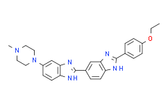
2'-(4-ethoxyphenyl)-5-(4-methylpiperazin-1-yl)-2,5'-bibenzimidazole is a bibenzimidazole and N-methylpiperazine. It can be used as a fluorescent dye. Its function is similar to that of piperbenzimidazole.
See also: Bibenzimidazole diethyl ether trihydrochloride (note moved to). Hoechst 33342 is also known as bisbenzimide. It binds to double-stranded DNA with a preference for AT-rich clusters. On binding to DNA, fluorescence increases greatly. It is membrane permeant and can stain live cells. Autofluorescence from endogenous molecules (e.g., reduced NAD, FAD) can interfere with imaging, particularly at UV wavelengths; this can be reduced by careful selection of excitation/emission wavelengths, treating fixed cells with reducing agents (e.g., 1% sodium borohydride for 20 min), and comparing with unlabeled controls. Fixation with glutaraldehyde should be avoided as it increases autofluorescence. [1] HOE 33342 (Hoechst 33342) is a non-embedding fluorescent DNA dye that binds to AT-rich regions in the minor groove. It is a live nuclear marker dye used in fluorescence microscopy, flow cytometry, and cell cycle analysis. The dye exhibits weak fluorescence in solution but becomes intensely fluorescent upon DNA binding. The CAS number is 23491-52-3. |
| Molecular Formula |
C27H28N6O
|
|---|---|
| Molecular Weight |
452.5508
|
| Exact Mass |
452.232
|
| Elemental Analysis |
C, 71.66; H, 6.24; N, 18.57; O, 3.54
|
| CAS # |
23491-52-3
|
| Related CAS # |
Hoechst 33342 trihydrochloride;875756-97-1;Hoechst 33342 analog;178481-68-0;Hoechst 33342 analog 2;106050-84-4
|
| PubChem CID |
1464
|
| Appearance |
Brown to breen solid powder
|
| Melting Point |
268ºC
|
| LogP |
7.139
|
| Hydrogen Bond Donor Count |
2
|
| Hydrogen Bond Acceptor Count |
5
|
| Rotatable Bond Count |
5
|
| Heavy Atom Count |
34
|
| Complexity |
664
|
| Defined Atom Stereocenter Count |
0
|
| SMILES |
CCOC1=CC=C(C=C1)C2=NC3=C(N2)C=C(C=C3)C4=NC5=C(N4)C=C(C=C5)N6CCN(CC6)C
|
| InChi Key |
PRDFBSVERLRRMY-UHFFFAOYSA-N
|
| InChi Code |
InChI=1S/C27H28N6O/c1-3-34-21-8-4-18(5-9-21)26-28-22-10-6-19(16-24(22)30-26)27-29-23-11-7-20(17-25(23)31-27)33-14-12-32(2)13-15-33/h4-11,16-17H,3,12-15H2,1-2H3,(H,28,30)(H,29,31)
|
| Chemical Name |
2-(4-ethoxyphenyl)-6-[6-(4-methylpiperazin-1-yl)-1H-benzimidazol-2-yl]-1H-benzimidazole
|
| Synonyms |
HO342; HOE33342; HO 342; HOE-33342; HO-342; Bisbenzimide; HOE 33342; Hoechst 33342
|
| HS Tariff Code |
2934.99.9001
|
| Storage |
Powder -20°C 3 years 4°C 2 years In solvent -80°C 6 months -20°C 1 month Note: This product requires protection from light (avoid light exposure) during transportation and storage. |
| Shipping Condition |
Room temperature (This product is stable at ambient temperature for a few days during ordinary shipping and time spent in Customs)
|
| Solubility (In Vitro) |
DMSO: ~6.25 mg/mL (~13.81 mM)
H2O: < 0.1 mg/mL (Insoluble) |
|---|---|
| Solubility (In Vivo) |
Solubility in Formulation 1: ≥ 0.5 mg/mL (1.10 mM) (saturation unknown) in 10% DMSO + 40% PEG300 + 5% Tween80 + 45% Saline (add these co-solvents sequentially from left to right, and one by one), clear solution.
For example, if 1 mL of working solution is to be prepared, you can add 100 μL of 5.0 mg/mL clear DMSO stock solution to 400 μL PEG300 and mix evenly; then add 50 μL Tween-80 to the above solution and mix evenly; then add 450 μL normal saline to adjust the volume to 1 mL. Preparation of saline: Dissolve 0.9 g of sodium chloride in 100 mL ddH₂ O to obtain a clear solution. Solubility in Formulation 2: ≥ 0.5 mg/mL (1.10 mM) (saturation unknown) in 10% DMSO + 90% (20% SBE-β-CD in Saline) (add these co-solvents sequentially from left to right, and one by one), clear solution. For example, if 1 mL of working solution is to be prepared, you can add 100 μL of 5.0 mg/mL clear DMSO stock solution to 900 μL of 20% SBE-β-CD physiological saline solution and mix evenly. Preparation of 20% SBE-β-CD in Saline (4°C,1 week): Dissolve 2 g SBE-β-CD in 10 mL saline to obtain a clear solution. View More
Solubility in Formulation 3: ≥ 0.5 mg/mL (1.10 mM) (saturation unknown) in 10% DMSO + 90% Corn Oil (add these co-solvents sequentially from left to right, and one by one), clear solution. Solubility in Formulation 4: 5 mg/mL (11.05 mM) in PBS (add these co-solvents sequentially from left to right, and one by one), clear solution; with ultrasonication (<60°C). |
| Preparing Stock Solutions | 1 mg | 5 mg | 10 mg | |
| 1 mM | 2.2097 mL | 11.0485 mL | 22.0970 mL | |
| 5 mM | 0.4419 mL | 2.2097 mL | 4.4194 mL | |
| 10 mM | 0.2210 mL | 1.1049 mL | 2.2097 mL |
*Note: Please select an appropriate solvent for the preparation of stock solution based on your experiment needs. For most products, DMSO can be used for preparing stock solutions (e.g. 5 mM, 10 mM, or 20 mM concentration); some products with high aqueous solubility may be dissolved in water directly. Solubility information is available at the above Solubility Data section. Once the stock solution is prepared, aliquot it to routine usage volumes and store at -20°C or -80°C. Avoid repeated freeze and thaw cycles.
Calculation results
Working concentration: mg/mL;
Method for preparing DMSO stock solution: mg drug pre-dissolved in μL DMSO (stock solution concentration mg/mL). Please contact us first if the concentration exceeds the DMSO solubility of the batch of drug.
Method for preparing in vivo formulation::Take μL DMSO stock solution, next add μL PEG300, mix and clarify, next addμL Tween 80, mix and clarify, next add μL ddH2O,mix and clarify.
(1) Please be sure that the solution is clear before the addition of next solvent. Dissolution methods like vortex, ultrasound or warming and heat may be used to aid dissolving.
(2) Be sure to add the solvent(s) in order.
Products are for research use only; We do not sell to patients
Copyright 2020 InvivoChem LLC | All Rights Reserved


 COA
COA