| Size | Price | Stock | Qty |
|---|---|---|---|
| 100mg |
|
||
| Other Sizes |


| ln Vitro |
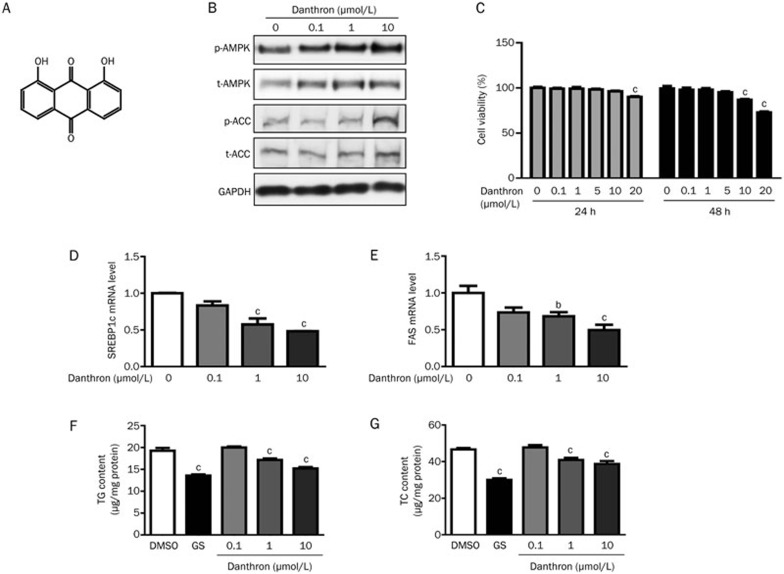
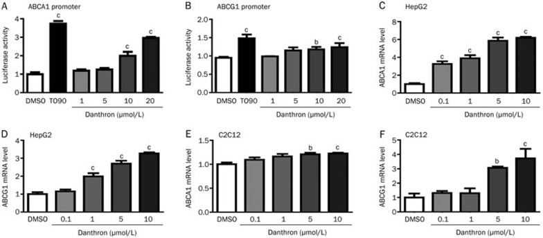
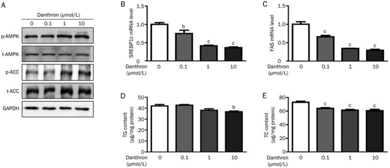
In HepG2 and C2C12 cells, dantrofrine (0.1, 1, and 10 μM) dose-dependently increased AMPK and acetyl-CoA carboxylase (ACC) phosphorylation. Simultaneously, the administration of Danthron resulted in a considerable decrease in the expression of the fatty acid synthase (FAS) and sterol regulatory element binding protein 1c (SREBP1c) genes, as well as in the levels of total cholesterol (TC) and triglycerides (TG). Furthermore, the use of Danthron effectively raises glucose consumption. Danthron activates the AMPK signaling system, which efficiently lowers intracellular lipid content and promotes in vitro glucose intake. For HepG2 cells, 10 μM Danthron/24 hours is safe. HepG2 cells were cultured with Danthron (0.1–10 μM) in FBS-free media for 8 hours after they reached 80% confluence. Cells were then extracted in preparation for the Western blot test. While there are no changes in t-AMPK protein, there is a dose-dependent rise in p-AMPK protein while using danthron [1]. With an IC50 of 0.11 μM, danthron inhibits the transactivation of retinoic acid X receptor alpha (RXRα) produced by 9-cis retinoic acid (9cRA). Using isothermal titration calorimetry (ITC) assays, the stoichiometry of Danthron binding to the RXRα-ligand binding domain (LBD) was further clarified. The ITC experiment yielded a KD value of 7.5 μM for Danthron binding to RXRα-LBD[2].
|
|---|---|
| ln Vivo |
Danthron functions as an insulin sensitizer in the body. Danthron enhances insulin sensitivity in mice with diet-induced obesity (DIO). Insulin tolerance test results demonstrated that Danthron (5 mg/kg)-treated diet-induced obese mice displayed lower blood glucose levels after insulin challenge compared with the control group [2].
|
| ADME/Pharmacokinetics |
Absorption, Distribution and Excretion
Following administration of tansine within 24 hours of labor induction in 12 women, tansine was detected in maternal urine, neonatal urine, and amniotic fluid. Most of the drug exists in the maternal and infant forms as glucuronide. Male Wistar rats were administered tansine sodium intravenously at doses of 4.8, 22, or 58 μmol/kg (1.2, 5.3, or 14 mg/kg) body weight, or via gastric tube at a dose of 12 μmol/kg (28.8 mg/kg) body weight. …After intravenous administration, approximately 80% of the tansine conjugates were excreted in bile after 1 hour; dose fractions detected after 5 hours represented approximately 20%, 30%, and 40% of the low, medium, and high dose levels, respectively. The corresponding urinary components were 16%, 12%, and 10%, with bile-to-urinary excretion ratios of 1.3, 2.7, and 4.0, respectively. Only 30–50% of the dose was explainable by the conjugates. Early studies also indicated that only 30-40% of the total dose of tansone was recovered from feces and urine after oral administration, primarily within the first 24 hours. /tansone sodium salt/ Like other anthraquinone compounds, tansone is partially absorbed from the small intestine. Rats were administered tansone via infusion (I) at doses of 0.48, 2.2, and 5.8 μmol/100 g body weight, or via gastric tube at 12 μmol/100 g body weight. Thin-layer chromatography analysis of bile and urine revealed multiple metabolites in both routes of administration. These metabolites included tansone monosulfate (II) and tansone glucuronide (III), as well as two other phase II metabolites present as corresponding diconjugates, and several phase I metabolites present as conjugates (IV). Following infusion, approximately 80% of the tansantrone conjugates were excreted in bile after 1 hour; after 5 hours, the dose fractions in the low, medium, and high-dose groups were approximately 20%, 30%, and 40%, respectively. The corresponding fractions in urine were 16%, 12%, and 10%, with bile-to-urine excretion ratios of 1.3, 2.7, and 4.0, respectively. This change in excretion pattern correlated with changes in metabolite distribution, manifested as a decreased balance between phase IV and phase I conjugates and an increased ratio of phase III to phase II conjugates. Phase IV was present in higher concentrations in bile than in urine, and its excretion was more persistent than that of tansantrone conjugates. After intragastric administration, the cumulative excretion of phase I conjugates (bile + urine) was only 6%, 8%, and 5% of the administered dose in three consecutive 6-hour timeframes (0–6 hours, 6–12 hours, and 12–18 hours post-administration), respectively. The bile-to-urine excretion ratio appeared to decrease over time, as did the III:II ratio… Metabolism/Metabolites In vitro experiments showed that Dantron can be converted into its monoglucuronide and monosulfate in the rat jejunum and colon, with monoglucuronide being the major metabolite. Male Wistar rats were administered Dantron sodium via intravenous injection at 4.8, 22, or 58 μmol/kg (1.2, 5.3, or 14 mg/kg) body weight, or via gastric infusion at 12 μmol/kg (28.8 mg/kg) body weight. Monosulfate, β-glucuronide, and other unidentified metabolites were detected in both bile and urine after both routes of administration. /Dantron Sodium/ Following intravenous infusion of Dantron in rats, its metabolism and excretion exhibited a complex dose-dependent pattern. These metabolites, especially the more polar ones, are generally excreted primarily via bile, with a small amount excreted via urine. ...This paper describes further research on a group of bile-derived metabolites that exhibited high heterogeneity. It comprises more than a dozen metabolites, which are conjugates of four different aglycones, including the parent Dantron... ...In rat jejunum and dissected colonic sacs, the serosal side (BL) was injected with Kh-Heinz solution (KH), and the mucosal side (LU) was infiltrated with KH solution containing Dantron (3-4 nmol/mL) or emodin (10 nmol/mL). After incubation at 37°C for 60 min, reversed-phase high-performance liquid chromatography (RP-HPLC) was used to analyze the parent Dantron and its metabolites in the LU and BL solutions and intestinal tissue. Reference metabolites were isolated and purified from the urine and bile of rats perfused with Dantron or emodin. Studies have shown that: (1) only a small amount of unmetabolized drug is present on the contralateral side; (2) in both tissues, dananthrone is converted into its monoglucuronide (G) and monosulfate (S); the G:S ratio in the jejunum is 6-8:1, and the ratio is higher in the colon; (3) in the jejunum, G and S are mainly secreted (LU:BL distribution ratio greater than 10:1); (4) however, in the colon, G is mainly absorbed (BL:LU ratio is 3:1), while S appears to have a small net secretion; (5) the residual amount (%) in intestinal tissue is very small; (6) the absorption and metabolism of emodin are slower, but in other respects it appears similar to dananthrone… |
| Toxicity/Toxicokinetics |
Interactions
This study investigated the regulatory effects of calendula extract on the development of 1,2-dimethylhydrazine (DMH)-induced colon and liver cancer in male ICR/CD-1 mice. Starting at 6 weeks of age, mice were divided into four groups. Two groups received weekly subcutaneous injections of DMH (20 mg/kg body weight) for 12 weeks. One week after the last DMH injection, one group was fed a basal diet for the entire study period (Group I), while the other group was fed a diet containing 0.2% calendula extract (dissolved in the basal diet) for 42 weeks (Group II). The remaining two groups received either saline injections followed by a diet containing 0.2% calendula extract for 42 weeks (Group III), or a basal diet for the entire experiment (Group IV). The incidence and number of colon tumors in Group II were significantly higher than in Group I (P < 0.05, P < 0.01). The incidence and number of hepatocellular carcinomas in Group II were also significantly higher than in Group I (P < 0.002, P < 0.02). No colonic tumors were found in the third group, but a small number of liver tumors and severe colonic inflammatory lesions were observed. Mice exposed to calendula extract showed higher ornithine decarboxylase activity in the colonic mucosa than mice not exposed to calendula extract. These results suggest that the pro-tumorigenic effect of calendula extract may be related to increased cell proliferation in target organs. DMH and calendula extract also showed a synergistic effect in liver tumorigenesis. When tannin was added to the diet and mice were simultaneously given 1,2-dimethylhydrazine, the incidence and number of colonic and hepatic adenomas significantly increased. To investigate the pro-tumorigenic activity of the anthraquinone laxative tannin, researchers administered a single subcutaneous injection of the colonic tumor inducer 1,2-dimethylhydrazine (DMH) to three groups of male rats. One week later, the animals were fed diets containing 0, 600, or 2400 ppm tannin, respectively, for 26 weeks. Two other groups of rats participated in the study; one group received no treatment, and the other group received only tannin. Nine tumors were observed in animals receiving DMA (with or without concurrent administration of danantrone). The incidence of colonic tumors was higher in animals treated with both DMH and danantrone than in those treated with DMH alone (5/60 vs. 0/30), but the difference was not statistically significant. Kidneys and mesangial lymph nodes were enlarged, exhibiting yellow-red and brown discoloration, respectively. The pigments were mostly PAS-positive, but did not contain lipids as detected by various staining methods. Current evidence suggests that the pigments are derived from the drug. Non-human toxicity values Oral LD50 in mice < 7 g/kg [Merck Index, 14th Edition (2006)] Intraperitoneal LD50 in mice 500 mg/kg |
| References | |
| Additional Infomation |
According to the International Agency for Research on Cancer (IARC) of the World Health Organization, Dantron (calendula oleracea; 1,8-dihydroxyanthraquinone) is a possible carcinogen. Dantron is an orange crystalline powder, almost tasteless and odorless. (NTP, 1992) Calendula oleracea is a dihydroxyanthraquinone, a compound in which anthracene-9,10-dione is substituted with hydroxyl groups at positions 1 and 8. It is an apoptosis inducer and a plant metabolite. Due to its genotoxicity, Dantron was withdrawn from the markets of Canada, the United States, and the United Kingdom in 1998. Dantron has been reported to be found in senna leaves, Hedyotis diffusa, and several other organisms with relevant data. Dantron is a red synthetic anthraquinone derivative. Dantron was once widely used as a laxative, but is no longer used to treat constipation; instead, it is used as an antioxidant in synthetic lubricants, in the synthesis of experimental antitumor drugs, as a bactericide, and as an intermediate in dye production. This substance is suspected of being mutagenic, and based on evidence of carcinogenicity in experimental animals, there is reason to expect it to be carcinogenic to humans. (NCI05)
Mechanism of Action This study used DNA sequencing technology to investigate the carcinogen Dantron (1,8-dihydroxyanthraquinone) and the anthraquinone-mediated metal DNA damage mechanism using 32P-tagged human DNA fragments obtained from the human c-Ha-ras-1 proto-oncogene and p53 tumor suppressor gene. In the presence of Cu(II), cytochrome P450 reductase, and the NADPH generation system, Dantron mainly caused DNA damage to guanine in the 5'-GG-3', 5'-GGGG-3', and 5'-GGGGG-3' sequences (damaged bases are underlined). Catalase and bartophenone inhibited this DNA damage, suggesting that H₂O₂ and Cu(I) are involved. With increasing Dantron concentration, the production of 8-oxo-7,8-dihydro-2'-deoxyguanosine also increased. On the other hand, the oxidative DNA damage induced by the carcinogen anthraquinone was less than that Dantron. Electron spin resonance studies showed that the P450 reductase and NADPH-mediated reduction of Dantron generated semiquinone radicals, while anthraquinone produced almost no signal. These results indicate that Dantron is more readily reduced by P450 reductase and generates reactive oxygen species through redox cycles, leading to more extensive Cu(II)-mediated DNA damage than anthraquinone. For anthraquinone, its hydroxylated metabolites have similar reactivity to Dantron and may be involved in in vivo DNA damage. In conclusion, the oxidative DNA damage induced by Dantron and anthraquinone appears to be associated with their carcinogenic expression. All three tested anthraquinone compounds—emodin, aloe-emodin, and tansacrine—exhibited the ability to inhibit the non-covalent binding of the benzimidazole dye Hoechst 33342 to isolated DNA and mouse lymphoma L5178Y cells, with effects comparable to topoisomerase II inhibitors and the intercalating agent m-amsacrine. In cell-free helicalization assays, emodin showed stronger inhibition of topoisomerase II activity than meta-amsacrine, tansacrine showed similar inhibition, while aloe-emodin showed weaker inhibition than meta-amsacrine. The chromosomal extent of DNA damage induced by these anthraquinone compounds was analyzed in mouse lymphoma L5178Y cells. Anthraquinone-induced mutant cell clones showed similar chromosomal damage compared to the topoisomerase II inhibitors etoposide and meta-amsacrine, but differed from mutants induced by the DNA alkylating agent ethyl methanesulfonate. These data support the view that inhibition of topoisomerase II catalytic activity is one of the causes of anthraquinone-induced genotoxicity and mutagenicity. Therapeutic Use Danantrolone has been widely used as a laxative since the beginning of this century. In 1987, the U.S. Food and Drug Administration (FDA) ordered the withdrawal of the drug from the market, prohibiting its use as a laxative. U.S. manufacturers also voluntarily stopped producing all human medicines containing this compound. /Previous U.S. Use/ Therapeutic Indication: Constipation in patients with advanced disease. Drug Warnings Contraindications: As with other gastrointestinal laxatives, compound danantrolone capsules should not be taken when there are acute or painful abdominal symptoms, or when constipation is suspected to be caused by intestinal obstruction. Contraindicated in patients with hypersensitivity to any component of this product. Contraindicated in patients with peanut or soy allergy. Dantrolone may cause a temporary, harmless pink or red discoloration of urine and perianal skin.Prolonged use of high doses may cause discoloration of the colonic mucosa. Combined tanthrone capsules are contraindicated in pregnant and breastfeeding women. One woman experienced severe skin discoloration after taking a large dose of a laxative containing tanthrone. Similar discoloration has been observed in other studies, primarily in older adults, mainly concentrated on the buttocks and thighs, accompanied by mild inflammation. Skin contact with feces or urine containing the drug appears to be a necessary condition for the discoloration. The inflammation may be due to the reduction of the parent compound to a diol derivative in the colon, which irritates the intestines and skin, while the parent compound does not. For more complete data on drug warnings for 1,8-dihydroxyanthraquinone (6 of 6), please visit the HSDB record page. |
| Molecular Formula |
C14H8O4
|
|---|---|
| Molecular Weight |
240.2109
|
| Exact Mass |
240.042
|
| CAS # |
117-10-2
|
| PubChem CID |
2950
|
| Appearance |
Brown to breen solid powder
|
| Density |
1.5±0.1 g/cm3
|
| Boiling Point |
452.7±35.0 °C at 760 mmHg
|
| Melting Point |
191-193 °C(lit.)
|
| Flash Point |
241.7±22.4 °C
|
| Vapour Pressure |
0.0±1.1 mmHg at 25°C
|
| Index of Refraction |
1.733
|
| LogP |
4.57
|
| Hydrogen Bond Donor Count |
2
|
| Hydrogen Bond Acceptor Count |
4
|
| Rotatable Bond Count |
0
|
| Heavy Atom Count |
18
|
| Complexity |
346
|
| Defined Atom Stereocenter Count |
0
|
| InChi Key |
QBPFLULOKWLNNW-UHFFFAOYSA-N
|
| InChi Code |
InChI=1S/C14H8O4/c15-9-5-1-3-7-11(9)14(18)12-8(13(7)17)4-2-6-10(12)16/h1-6,15-16H
|
| Chemical Name |
1,8-dihydroxyanthracene-9,10-dione
|
| HS Tariff Code |
2934.99.9001
|
| Storage |
Powder -20°C 3 years 4°C 2 years In solvent -80°C 6 months -20°C 1 month |
| Shipping Condition |
Room temperature (This product is stable at ambient temperature for a few days during ordinary shipping and time spent in Customs)
|
| Solubility (In Vitro) |
DMSO : ~5 mg/mL (~20.82 mM)
|
|---|---|
| Solubility (In Vivo) |
Solubility in Formulation 1: 10 mg/mL (41.63 mM) in 50% PEG300 +50% Saline (add these co-solvents sequentially from left to right, and one by one), suspension solution; with sonication (<60°C).
Preparation of saline: Dissolve 0.9 g of sodium chloride in 100 mL ddH₂ O to obtain a clear solution. (Please use freshly prepared in vivo formulations for optimal results.) |
| Preparing Stock Solutions | 1 mg | 5 mg | 10 mg | |
| 1 mM | 4.1630 mL | 20.8151 mL | 41.6302 mL | |
| 5 mM | 0.8326 mL | 4.1630 mL | 8.3260 mL | |
| 10 mM | 0.4163 mL | 2.0815 mL | 4.1630 mL |
*Note: Please select an appropriate solvent for the preparation of stock solution based on your experiment needs. For most products, DMSO can be used for preparing stock solutions (e.g. 5 mM, 10 mM, or 20 mM concentration); some products with high aqueous solubility may be dissolved in water directly. Solubility information is available at the above Solubility Data section. Once the stock solution is prepared, aliquot it to routine usage volumes and store at -20°C or -80°C. Avoid repeated freeze and thaw cycles.
Calculation results
Working concentration: mg/mL;
Method for preparing DMSO stock solution: mg drug pre-dissolved in μL DMSO (stock solution concentration mg/mL). Please contact us first if the concentration exceeds the DMSO solubility of the batch of drug.
Method for preparing in vivo formulation::Take μL DMSO stock solution, next add μL PEG300, mix and clarify, next addμL Tween 80, mix and clarify, next add μL ddH2O,mix and clarify.
(1) Please be sure that the solution is clear before the addition of next solvent. Dissolution methods like vortex, ultrasound or warming and heat may be used to aid dissolving.
(2) Be sure to add the solvent(s) in order.
|
|
|
Products are for research use only; We do not sell to patients
Copyright 2020 InvivoChem LLC | All Rights Reserved