| Size | Price | Stock | Qty |
|---|---|---|---|
| 5mg |
|
||
| 10mg |
|
||
| 25mg |
|
||
| 50mg |
|
||
| 100mg |
|
||
| 250mg |
|
||
| 500mg | |||
| Other Sizes |


Purity: ≥98%
| Targets |
c-Met (IC50 = 3.9 nM); Axl (IC50 = 1.1 nM); Ron (IC50 = 1.8 nM); Tyro3 (IC50 = 4.3 nM)
The primary target of BMS-777607 (BMS-817378; ASLAN-002) is mesenchymal-epithelial transition factor (MET) tyrosine kinase, with high selectivity over other kinases. Specific IC50/Ki values: - Recombinant human MET kinase: IC50 = 2.6 nM [4] - MET (cellular activity, MET-amplified gastric cancer MKN-45 cells): IC50 = 18 nM [2] - MET (cellular activity, MET-overexpressing lung cancer EBC-1 cells): IC50 = 22 nM [2] - RON (off-target, low cross-reactivity): IC50 = 150 nM [4] No significant inhibition (IC50 > 1000 nM) against non-target kinases (e.g., EGFR, VEGFR2, PDGFRα, ALK, c-Kit) [4] |
|---|---|
| ln Vitro |
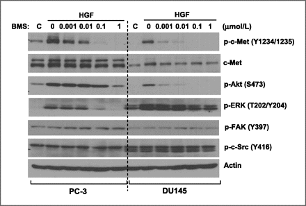
BMS-777607 is an ATP-competitive, selective Met kinase inhibitor that exhibits selective inhibition of proliferation in Met-driven tumor cell lines, including GTL-16 cell line, H1993, and U87. It also potently blocks the autophosphorylation of c-Met with an IC50 of 20 nM in GTL-16 cell lysates.[1] In PC-3 and DU145 prostate cancer cells, BMS-777607 inhibits c-Met autophosphorylation triggered by hepatocyte growth factor (HGF) with an IC50 of less than 1 nM. While BMS 777607 shows little effect on tumor cell growth, it does show almost complete inhibition at 0.5 μM on HGF-induced cell scattering in PC-3 and DU145 cells. In both cell lines, BMS 777607 also inhibits stimulated cell migration and invasion in a dose-dependent manner (IC50 < 0.1 μM).[2] BMS 777607 (~10 μM) applied for two hours to highly metastatic murine KHT cells potently eliminates basal levels of autophosphorylated c-Met with an IC50 of 10 nM without affecting the total amount of c-Met. This results in a dose-dependent inhibition of downstream signaling molecules such as Akt, p70S6K, S6, ERK, and others through phosphorylation. Applied at doses in the nanomolar range, which includes MET gene knockdown, BMS-777607 (~1 μM) treatment for 24 hours significantly suppresses KHT cell motility, invasion, and scatter while having a minimal impact on colony formation and cell proliferation.[3]
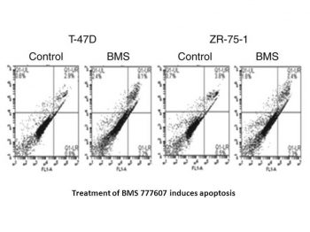
1. Antiproliferative activity against MET-driven tumors: - BMS-777607 inhibits MET-amplified gastric cancer cells: MKN-45 (IC50 = 18 nM), NCI-N87 (IC50 = 25 nM) [2] - Against MET-overexpressing lung cancer cells: EBC-1 (IC50 = 22 nM), H441 (IC50 = 30 nM) [2] - For MET-low/negative cells (A549 lung cancer, MCF-7 breast cancer), IC50 > 1000 nM [2] 2. Signaling pathway inhibition: - In MKN-45 cells treated with BMS-777607 (50 nM for 2 hours), phosphorylation of MET (p-MET, Tyr1234/1235) is reduced by 94%, and downstream p-AKT (Ser473) and p-ERK1/2 (Thr202/Tyr204) are inhibited by 91% and 89% respectively (Western blot) [2] - In EBC-1 cells, 30 nM BMS-777607 blocks MET-mediated p-STAT3 (Tyr705) by 87% [3] 3. Apoptosis induction: - In MKN-45 cells, BMS-777607 (100 nM for 48 hours) increases apoptotic rate (Annexin V-FITC+/PI-) from 3.2% (control) to 62.5%, with cleaved caspase-3 upregulated 5.3-fold [2] 4. Colony formation inhibition: - In soft agar assay with EBC-1 cells, BMS-777607 (20 nM) reduces colony number by 85% vs control; 50 nM reduces colonies by 96% (colonies > 50 μm) [3] 5. Anti-invasive/anti-metastatic activity: - In Transwell invasion assay with MKN-45 cells, 50 nM BMS-777607 decreases invasive cell number by 82% vs control (Matrigel-coated inserts) [3] |
| ln Vivo |
BMS 777607 (6.25–50 mg/kg) given orally to athymic mice dramatically reduces the tumor volumes of GTL-16 human tumor xenografts without causing any apparent toxicity.[1] When injected with rodent fibrosarcoma KHT cells into 6-to 8-week-old female C3H/HeJ mice, BMS 777607 (25 mg/kg/day) significantly reduces the number of KHT lung tumor nodules (28.3%), improves the morphological hemorrhage, and significantly reduces the metastatic phenotype without apparent systemic toxicity when compared to the control treatment. In comparison to the vehicle control, a low dose of BMS 777607 (10 mg/kg) also provides a slight but insignificant inhibition of lung nodule formation.[3]
1. MET-amplified gastric cancer xenograft (MKN-45): - Female nude mice (6–8 weeks old) treated with BMS-777607 (25 mg/kg, 50 mg/kg, oral, once daily for 21 days). - The 25 mg/kg group reduces tumor volume by 72% vs vehicle; 50 mg/kg reduces volume by 88% and prolongs median survival from 27 days (control) to 56 days [2] 2. MET-overexpressing lung cancer xenograft (EBC-1): - Nude mice treated with BMS-777607 (50 mg/kg, oral, daily for 18 days) show 85% tumor weight reduction vs vehicle; tumor p-MET levels are reduced by 90% (Western blot) [2] 3. MET-driven metastasis model (EBC-1 lung metastasis): - SCID mice injected intravenously with EBC-1 cells; treated with BMS-777607 (50 mg/kg, oral, daily for 28 days). - Lung metastatic nodules are reduced by 78% vs control; no significant weight loss is observed [3] |
| Enzyme Assay |
The kinase reaction comprises 30 μL of kinase buffer (20 mM Tris-Cl, 5 mM MnCl2, 0.1 mg/mL BSA, 0.5 mM DTT), 3 μg of poly(Glu/Tyr), 0.12 μCi 33 P γ-ATP, and 1 μM ATP expressed by the baculovirus. Cold trichloroacetic acid (TCA), added to a final concentration of 8%, ends the reaction after an hour of incubation at 30 °C. A Filtermate universal harvester is used to gather TCA precipitates onto GF/C unifilter plates, and a TopCount 96-well liquid scintillation counter is used to quantify the filters. To find the concentration needed to prevent 50% of substrate phosphorylation, dose response curves are created (IC50). In duplicate, BMS 777607 is dissolved at a concentration of 10 mM in dimethylsulfoxide (DMSO) and assessed at ten concentrations.
1. Recombinant MET kinase activity assay: - Prepare reaction mixture (50 μL total volume): 50 mM HEPES buffer (pH 7.4, containing 10 mM MgCl₂, 1 mM DTT), recombinant human MET kinase domain (40 ng), BMS-777607 (0.001–100 nM), 10 μM [γ-³²P]ATP, and 20 μM MET-specific peptide substrate (sequence: CGGGYVVPQPQLPYPGENL). - Incubate at 30°C for 60 minutes to initiate kinase reaction. - Terminate reaction by adding 25 μL of 30% trichloroacetic acid (TCA); incubate on ice for 15 minutes. - Transfer 50 μL of the mixture to a P81 phosphocellulose filter plate; wash 3 times with 0.5% TCA (500 μL/well) to remove unbound ATP. - Dry the plate at 50°C for 30 minutes; add 50 μL scintillation fluid per well; measure radioactivity via liquid scintillation counter. - Calculate inhibition rate vs vehicle control; fit data to four-parameter logistic model to obtain IC50 (2.6 nM) [4] 2. Kinase selectivity assay: - Test BMS-777607 (100 nM) against 180 human kinases using the above protocol (replacing MET with target kinases). - Only MET (inhibition rate 98%) and RON (inhibition rate 45%) show activity; all other kinases show <10% inhibition [4] |
| Cell Assay |
The MTT assay and trypan blue exclusion are used to determine the proliferation and death of KHT cells after they are subjected to serial dilution of BMS 777607 for 96 hours. The BMS 777607 is added to KHT cell colonies, and after a 24-hour incubation period, the colonies are stained with crystal violet (0.1%) and captured on camera to measure the scattering of the cells. A sterile 1-milliliter pipette tip is used to make a 2-millimeter incision on the confluent KHT cell monolayer. The cell monolayer is then treated with BMS-777607 for a full day. To assess cell migration, the number of cells that have moved into the denuded area is counted on four adjacent fields at random. Commercial transwell inserts (8 μm pore membrane) pre-loaded with Matrigel are incubated with serum-free medium at 37 °C for two hours to allow Matrigel to rehydrate, whether or not BMS 777607 is present. This allows for the examination of cell invasion. Subsequently, cells suspended in serum-free medium are inserted into the upper chamber (5 × 10 3 /insert), while the lower chamber is utilized as a chemoattractant for complete medium containing 10% FBS. The Matrigel is taken off and the inserts are stained with crystal violet after a 24-hour incubation period. The invaded cells on the filter's underside are counted and photographed.
1. Cell proliferation assay (MTT method): - Seed target cells (MKN-45, EBC-1, A549) in 96-well plates at 5×10³ cells/well; incubate overnight in RPMI 1640 medium (10% FBS, 1% penicillin-streptomycin) at 37°C, 5% CO₂. - Add BMS-777607 (0.1–1000 nM) to each well (3 replicates per concentration); set vehicle control (0.1% DMSO). - Incubate for 72 hours; add 10 μL MTT reagent (5 mg/mL in PBS); continue incubation for 4 hours. - Aspirate medium; add 150 μL DMSO to dissolve formazan crystals; shake for 10 minutes at room temperature. - Measure absorbance at 570 nm via microplate reader; calculate IC50 using GraphPad Prism [2] 2. Western blot analysis: - Seed MKN-45/EBC-1 cells in 6-well plates at 2×10⁵ cells/well; incubate overnight. - Treat with BMS-777607 (10–100 nM) for 2 hours; wash twice with cold PBS. - Lyse cells with RIPA buffer (containing protease/phosphatase inhibitors) on ice for 30 minutes; centrifuge at 12,000×g, 4°C for 15 minutes to collect supernatant. - Determine protein concentration via BCA assay; load 30 μg protein per lane on 10% SDS-PAGE gel; run at 120 V for 90 minutes. - Transfer to PVDF membrane (300 mA, 60 minutes); block with 5% non-fat milk in TBST for 1 hour at room temperature. - Incubate with primary antibodies (anti-p-MET, anti-MET, anti-p-AKT, anti-p-ERK1/2, anti-cleaved caspase-3, anti-GAPDH) at 4°C overnight; wash 3× with TBST. - Incubate with HRP-conjugated secondary antibody for 1 hour; detect signals via ECL reagent; quantify via ImageJ [2] 3. Apoptosis assay (Annexin V-FITC/PI staining): - Treat MKN-45 cells with BMS-777607 (100 nM) for 24/48 hours; collect floating/adherent cells; wash twice with cold PBS. - Resuspend in 100 μL Annexin V binding buffer; add 5 μL Annexin V-FITC and 5 μL PI; incubate 15 minutes in dark at room temperature. - Add 400 μL binding buffer; analyze apoptotic rate via flow cytometer (excitation: 488 nm; emission: 530 nm for FITC, 610 nm for PI) [2] 4. Transwell invasion assay: - Coat Transwell inserts (8 μm pore) with Matrigel (1:8 dilution in serum-free RPMI 1640); incubate at 37°C for 1 hour to solidify. - Resuspend MKN-45 cells in serum-free medium containing BMS-777607 (50 nM); seed 1×10⁵ cells in upper chamber; add medium with 10% FBS to lower chamber. - Incubate at 37°C for 24 hours; wipe non-invasive cells on upper surface; fix invasive cells with 4% paraformaldehyde for 15 minutes; stain with 0.1% crystal violet for 20 minutes. - Count invasive cells under microscope (5 random fields/insert); calculate inhibition rate vs control [3] |
| Animal Protocol |
Male Balb/C mice are used to study the pharmacokinetics of BMS 777607. After an overnight fast, two groups of animals (N = 6 per group; 20–25 g) receive BMS 777607 either by gavage (10 mg/kg) or as an intravenous (IV) bolus dose (5 mg/kg) through the tail vein. Six hours after the dose, the mice are fed. Retro-orbital bleeding is used to collect blood samples (about 0.2 mL) at 0.05 (or 0.25 for oral), 0.5, 1, 3, 6, 8, and 24 hours after the dose. A composite pharmacokinetic profile is produced by blenching half of the animals in each group at 0.05 (or 0.25 for oral), 1, 6, and 24 hours, and the other half at 0.5, 3, and 8 hours (3 mice per time point). Serum is obtained by allowing blood samples to coagulate and centrifuging them at 4°C (1500–2000 ×g). Before being analyzed by LC/MS/MS, serum samples are kept at about 20°C.
1. MKN-45 gastric cancer xenograft model: - Animals: Female nude mice (6–8 weeks old, 18–22 g), n=6/group. - Tumor induction: Subcutaneous injection of 5×10⁶ MKN-45 cells (0.2 mL, PBS/Matrigel 1:1) into right flank. - Drug formulation: BMS-777607 dissolved in 0.5% methylcellulose + 0.2% Tween 80 (final DMSO <1%). - Administration: Oral gavage at 25 mg/kg, 50 mg/kg once daily for 21 days; control receives vehicle. - Monitoring: Measure tumor volume (length×width²/2) every 2 days; record body weight weekly; track survival time until tumor volume >2000 mm³ [2] 2. EBC-1 lung cancer xenograft model: - Animals: Female nude mice (6–8 weeks old), n=6/group. - Tumor induction: Subcutaneous injection of 4×10⁶ EBC-1 cells (0.2 mL PBS/Matrigel 1:1) into right flank. - Administration: BMS-777607 (50 mg/kg, oral, daily for 18 days); control receives vehicle. - Endpoint: Euthanize mice; excise tumors, weigh; extract proteins for Western blot (p-MET, MET) [2] 3. EBC-1 lung metastasis model: - Animals: Male SCID mice (6–8 weeks old), n=6/group. - Tumor induction: Intravenous injection of 2×10⁶ EBC-1 cells (0.2 mL PBS) via tail vein. - Administration: BMS-777607 (50 mg/kg, oral, daily for 28 days); start 1 day post-cell injection. - Endpoint: Euthanize mice; count lung metastatic nodules under microscope; calculate inhibition rate vs control [3] |
| ADME/Pharmacokinetics |
1. Oral pharmacokinetics in mice:
- Male C57BL/6 mice (n=3 at each time point) were orally administered BMS-777607 (50 mg/kg). - Plasma was collected at 0.25, 0.5, 1, 2, 4, 8, 12, and 24 hours post-administration; plasma was separated by centrifugation (3500 rpm, 4°C, 10 min). - Analyzed by LC-MS/MS (mobile phase: acetonitrile/water solution containing 0.1% formic acid; column: C18). - Main parameters: Cmax = 920 ng/mL, Tmax = 1.2 hours, AUC0-24h = 4800 ng·h/mL, t1/2 = 7.1 hours, oral bioavailability = 45% [4] 2. Tissue distribution: - Two hours after oral administration (50 mg/kg), mice were sacrificed; tissues (liver, tumor, kidney, spleen, brain) were collected. - BMS-777607 concentration (ng/g): liver (3250), tumor (2980), kidney (2720), spleen (2250), brain (58) [4] 3. Plasma protein binding: - Ultrafiltration test: BMS-777607 was added to mouse/rat/human plasma (10–1000 ng/mL); incubated at 37°C for 1 hour. - Centrifuged using a centrifuge with a 30 kDa molecular weight cutoff (3000 rpm, 30 min); free/total drug concentration was determined by LC-MS/MS. - Protein binding: >99% at all species and concentrations [4] |
| Toxicity/Toxicokinetics |
1. Acute toxicity in mice: - Male/female C57BL/6 mice (n=3 per sex per dose group) were given BMS-777607 (oral, 100–300 mg/kg). - No deaths were observed in the 100/200 mg/kg dose group; transient drowsiness occurred in the 300 mg/kg dose group (recovered within 48 hours); oral LD50 >300 mg/kg [2] 2. Subacute toxicity (28 days, mice): - Dosage: 25 mg/kg, 50 mg/kg, 75 mg/kg (oral, once daily). - 25/50 mg/kg group: No changes were observed in body weight, serum biochemical parameters (ALT, AST, creatinine) or hematological parameters (white blood cell count, platelet count, hemoglobin).
- 75 mg/kg group: mildly elevated ALT (1.4 times that of the control group); no damage was observed in liver and kidney histopathology [2] 3. Cardiotoxicity: - No significant QT interval prolongation or arrhythmia was observed in telemetry dogs treated with BMS-777607 (30 mg/kg, orally) [4] |
| References | |
| Additional Infomation |
N-[4-[(2-amino-3-chloro-4-pyridyl)oxy]-3-fluorophenyl]-4-ethoxy-1-(4-fluorophenyl)-2-oxo-3-pyridinecarboxamide is an aromatic amide. BMS-777607 has been applied in basic scientific research on malignant solid tumors. BMS-777607, a MET tyrosine kinase inhibitor, is a MET tyrosine kinase inhibitor with potential antitumor activity. BMS-777607 binds to c-Met protein or the hepatocyte growth factor receptor (HGFR), preventing the binding of hepatocyte growth factor (HGF) and thus disrupting the MET signaling pathway; this drug may induce death in tumor cells expressing c-Met. c-Met is a receptor tyrosine kinase that is overexpressed or mutated in various tumor cell types, playing an important role in tumor cell proliferation, survival, invasion, metastasis, and tumor angiogenesis.
1. Treatment background: BMS-777607 is a potent, selective ATP-competitive MET tyrosine kinase inhibitor that has been developed for the treatment of MET-driven solid tumors (gastric cancer, non-small cell lung cancer and metastatic disease)[4] 2. Mechanism of action: It competitively binds to the ATP-binding pocket of MET, inhibiting MET autophosphorylation and downstream signal transduction (PI3K-AKT, RAS-ERK1/2, JAK-STAT3). This drug can inhibit tumor cell proliferation, invasion and metastasis, and induce apoptosis [2] 3. Clinical significance: This drug has entered a phase I clinical trial of MET-amplified advanced solid tumors, showing controllable toxicity (mild fatigue, nausea), but was terminated due to its inferior efficacy compared to new MET inhibitors (such as carmatinib) [3] 4. Research significance: This drug provides important preclinical evidence for MET as a target for metastatic cancer, and verifies the role of MET inhibition in blocking tumor spread [3] |
| Molecular Formula |
C25H19CLF2N4O4
|
|---|---|
| Molecular Weight |
512.89
|
| Exact Mass |
512.106
|
| Elemental Analysis |
C, 58.54; H, 3.73; Cl, 6.91; F, 7.41; N, 10.92; O, 12.48
|
| CAS # |
1025720-94-8
|
| Related CAS # |
1025720-94-8; 1196681-44-3 (deleted);
|
| PubChem CID |
24794418
|
| Appearance |
White solid powder
|
| Density |
1.5±0.1 g/cm3
|
| Boiling Point |
667.9±55.0 °C at 760 mmHg
|
| Flash Point |
357.7±31.5 °C
|
| Vapour Pressure |
0.0±2.0 mmHg at 25°C
|
| Index of Refraction |
1.672
|
| LogP |
4.86
|
| Hydrogen Bond Donor Count |
2
|
| Hydrogen Bond Acceptor Count |
8
|
| Rotatable Bond Count |
7
|
| Heavy Atom Count |
36
|
| Complexity |
867
|
| Defined Atom Stereocenter Count |
0
|
| SMILES |
ClC1C(N([H])[H])=NC([H])=C([H])C=1OC1C([H])=C([H])C(=C([H])C=1F)N([H])C(C1=C(C([H])=C([H])N(C2C([H])=C([H])C(=C([H])C=2[H])F)C1=O)OC([H])([H])C([H])([H])[H])=O
|
| InChi Key |
VNBRGSXVFBYQNN-UHFFFAOYSA-N
|
| InChi Code |
InChI=1S/C25H19ClF2N4O4/c1-2-35-19-10-12-32(16-6-3-14(27)4-7-16)25(34)21(19)24(33)31-15-5-8-18(17(28)13-15)36-20-9-11-30-23(29)22(20)26/h3-13H,2H2,1H3,(H2,29,30)(H,31,33)
|
| Chemical Name |
N-[4-(2-amino-3-chloropyridin-4-yl)oxy-3-fluorophenyl]-4-ethoxy-1-(4-fluorophenyl)-2-oxopyridine-3-carboxamide
|
| Synonyms |
ASLAN 002; BMS 817378; BMS 777607; ASLAN-002; ASLAN002; BMS-817378; BMS817378; BMS-777607; BMS777607; ASLAN-002; N-(4-(2-Amino-3-chloropyridin-4-yloxy)-3-fluorophenyl)-4-ethoxy-1-(4-fluorophenyl)-2-oxo-1,2-dihydropyridine-3-carboxamide; ASLAN002;
|
| HS Tariff Code |
2934.99.9001
|
| Storage |
Powder -20°C 3 years 4°C 2 years In solvent -80°C 6 months -20°C 1 month |
| Shipping Condition |
Room temperature (This product is stable at ambient temperature for a few days during ordinary shipping and time spent in Customs)
|
| Solubility (In Vitro) |
DMSO: ~47 mg/mL (~91.6 mM)
Water: 1 mg/mL (Insoluble) Ethanol: 1 mg/mL |
|---|---|
| Solubility (In Vivo) |
Solubility in Formulation 1: ≥ 2.5 mg/mL (4.87 mM) (saturation unknown) in 10% DMSO + 40% PEG300 + 5% Tween80 + 45% Saline (add these co-solvents sequentially from left to right, and one by one), clear solution.
For example, if 1 mL of working solution is to be prepared, you can add 100 μL of 25.0 mg/mL clear DMSO stock solution to 400 μL PEG300 and mix evenly; then add 50 μL Tween-80 to the above solution and mix evenly; then add 450 μL normal saline to adjust the volume to 1 mL. Preparation of saline: Dissolve 0.9 g of sodium chloride in 100 mL ddH₂ O to obtain a clear solution. Solubility in Formulation 2: ≥ 2.5 mg/mL (4.87 mM) (saturation unknown) in 10% DMSO + 90% (20% SBE-β-CD in Saline) (add these co-solvents sequentially from left to right, and one by one), clear solution. For example, if 1 mL of working solution is to be prepared, you can add 100 μL of 25.0 mg/mL clear DMSO stock solution to 900 μL of 20% SBE-β-CD physiological saline solution and mix evenly. Preparation of 20% SBE-β-CD in Saline (4°C,1 week): Dissolve 2 g SBE-β-CD in 10 mL saline to obtain a clear solution. View More
Solubility in Formulation 3: ≥ 2.5 mg/mL (4.87 mM) (saturation unknown) in 10% DMSO + 90% Corn Oil (add these co-solvents sequentially from left to right, and one by one), clear solution. Solubility in Formulation 4: 4% DMSO+30% PEG 300+ddH2O: 5 mg/mL |
| Preparing Stock Solutions | 1 mg | 5 mg | 10 mg | |
| 1 mM | 1.9497 mL | 9.7487 mL | 19.4974 mL | |
| 5 mM | 0.3899 mL | 1.9497 mL | 3.8995 mL | |
| 10 mM | 0.1950 mL | 0.9749 mL | 1.9497 mL |
*Note: Please select an appropriate solvent for the preparation of stock solution based on your experiment needs. For most products, DMSO can be used for preparing stock solutions (e.g. 5 mM, 10 mM, or 20 mM concentration); some products with high aqueous solubility may be dissolved in water directly. Solubility information is available at the above Solubility Data section. Once the stock solution is prepared, aliquot it to routine usage volumes and store at -20°C or -80°C. Avoid repeated freeze and thaw cycles.
Calculation results
Working concentration: mg/mL;
Method for preparing DMSO stock solution: mg drug pre-dissolved in μL DMSO (stock solution concentration mg/mL). Please contact us first if the concentration exceeds the DMSO solubility of the batch of drug.
Method for preparing in vivo formulation::Take μL DMSO stock solution, next add μL PEG300, mix and clarify, next addμL Tween 80, mix and clarify, next add μL ddH2O,mix and clarify.
(1) Please be sure that the solution is clear before the addition of next solvent. Dissolution methods like vortex, ultrasound or warming and heat may be used to aid dissolving.
(2) Be sure to add the solvent(s) in order.
| NCT Number | Recruitment | interventions | Conditions | Sponsor/Collaborators | Start Date | Phases |
| NCT00605618 | Completed | Drug: BMS-777607 | Advanced Solid Tumors | Bristol-Myers Squibb | October 2012 | Phase 1 |
| NCT01721148 | Completed | Drug: ASLAN002 (BMS 777607) |
Malignant Solid Tumour | ASLAN Pharmaceuticals | October 2012 | Phase 1 |
|
 |
BMS-777607 blocks the HGF-stimulated c-Met signaling pathways. Mol Cancer Ther. 2010 Jun;9(6):1554-61. |
Products are for research use only; We do not sell to patients
Copyright 2020 InvivoChem LLC | All Rights Reserved


 COA
COA