| Size | Price | Stock | Qty |
|---|---|---|---|
| 5mg |
|
||
| 10mg |
|
||
| 25mg |
|
||
| 50mg |
|
||
| Other Sizes |


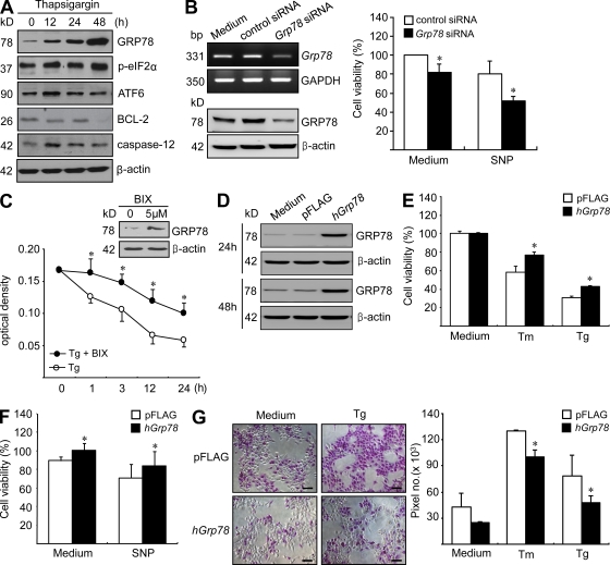
BIX (BiP Inducer X) is a novel and potent BiP (Hsp70-5) ER chaperone inducer with anticancer activity. It protects neurons from ER stress.
| ln Vitro |
BiP protein is increased by BiP inducer X (5 μM; 0–12 hours; SK–N–SH cells)[1]. BiP induces the ATF6 pathway, which in turn induces BiP[1]. Cell death caused by ER stress is decreased by BiP inducer X (5 μM; 12 hours; pretreatment with SK-N-SH cells) through suppressed activation of caspases 3/7 and 4[1].
|
|---|---|
| ln Vivo |
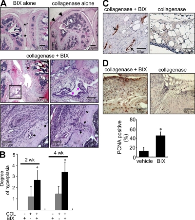
The use of BiP inducer X lessens the damage caused by cerebral infarction[1]. A 20 μg dose of BiP inducer -34 g (Japan SLC)[1] was administered intracerebroventricularly. The level of BiP protein significantly increased 24 hours following the dose, indicating that BIX treatment produces BiP protein in vivo.
|
| References |
[1]. Kudo T, et al. A molecular chaperone inducer protects neurons from ER stress. Cell Death Differ. 2008;15(2):364-375.
[2]. Yoo SA, et al. A novel pathogenic role of the ER chaperone GRP78/BiP in rheumatoid arthritis. J Exp Med. 2012;209(4):871-886. |
| Additional Infomation |
2-(3,4-Dihydroxyphenyl)-2-oxoethyl thiocyanate belongs to the thiocyanate class of compounds. Its structure is similar to 3,4-dihydroxyacetophenone, except that one methyl hydrogen atom is replaced by a sulfur atom in a cyanothiodiyl (-SC#N) group. Studies have found that this compound can induce the expression of the endoplasmic reticulum molecular chaperone protein GRP78 (a 78 kDa glucose-regulated protein, BiP, a highly conserved member of the 70 kDa heat shock protein family), thereby attenuating the unfolded protein response. It can protect neurons and retinal cells from endoplasmic reticulum stress-induced cell death. This compound belongs to the thiocyanate, aromatic ketone, and catechol classes of compounds.
|
| Molecular Formula |
C9H7NO3S
|
|---|---|
| Molecular Weight |
209.22178
|
| Exact Mass |
209.014
|
| CAS # |
101714-41-4
|
| PubChem CID |
16656807
|
| Appearance |
Brown to gray solid powder
|
| Density |
1.5±0.1 g/cm3
|
| Boiling Point |
517.1±45.0 °C at 760 mmHg
|
| Melting Point |
127 °C
|
| Flash Point |
266.5±28.7 °C
|
| Vapour Pressure |
0.0±1.4 mmHg at 25°C
|
| Index of Refraction |
1.665
|
| LogP |
1.53
|
| Hydrogen Bond Donor Count |
2
|
| Hydrogen Bond Acceptor Count |
5
|
| Rotatable Bond Count |
3
|
| Heavy Atom Count |
14
|
| Complexity |
260
|
| Defined Atom Stereocenter Count |
0
|
| SMILES |
N#CSCC(C1C=CC(O)=C(O)C=1)=O
|
| InChi Key |
SVFLBLCWKKQKDW-UHFFFAOYSA-N
|
| InChi Code |
InChI=1S/C9H7NO3S/c10-5-14-4-9(13)6-1-2-7(11)8(12)3-6/h1-3,11-12H,4H2
|
| Chemical Name |
[2-(3,4-dihydroxyphenyl)-2-oxoethyl] thiocyanate
|
| HS Tariff Code |
2934.99.9001
|
| Storage |
Powder -20°C 3 years 4°C 2 years In solvent -80°C 6 months -20°C 1 month |
| Shipping Condition |
Room temperature (This product is stable at ambient temperature for a few days during ordinary shipping and time spent in Customs)
|
| Solubility (In Vitro) |
DMSO : ~50 mg/mL (~238.98 mM)
|
|---|---|
| Solubility (In Vivo) |
Solubility in Formulation 1: ≥ 2.5 mg/mL (11.95 mM) (saturation unknown) in 10% DMSO + 90% (20% SBE-β-CD in Saline) (add these co-solvents sequentially from left to right, and one by one), clear solution.
For example, if 1 mL of working solution is to be prepared, you can add 100 μL of 25.0 mg/mL clear DMSO stock solution to 900 μL of 20% SBE-β-CD physiological saline solution and mix evenly. Preparation of 20% SBE-β-CD in Saline (4°C,1 week): Dissolve 2 g SBE-β-CD in 10 mL saline to obtain a clear solution. (Please use freshly prepared in vivo formulations for optimal results.) |
| Preparing Stock Solutions | 1 mg | 5 mg | 10 mg | |
| 1 mM | 4.7797 mL | 23.8983 mL | 47.7966 mL | |
| 5 mM | 0.9559 mL | 4.7797 mL | 9.5593 mL | |
| 10 mM | 0.4780 mL | 2.3898 mL | 4.7797 mL |
*Note: Please select an appropriate solvent for the preparation of stock solution based on your experiment needs. For most products, DMSO can be used for preparing stock solutions (e.g. 5 mM, 10 mM, or 20 mM concentration); some products with high aqueous solubility may be dissolved in water directly. Solubility information is available at the above Solubility Data section. Once the stock solution is prepared, aliquot it to routine usage volumes and store at -20°C or -80°C. Avoid repeated freeze and thaw cycles.
Calculation results
Working concentration: mg/mL;
Method for preparing DMSO stock solution: mg drug pre-dissolved in μL DMSO (stock solution concentration mg/mL). Please contact us first if the concentration exceeds the DMSO solubility of the batch of drug.
Method for preparing in vivo formulation::Take μL DMSO stock solution, next add μL PEG300, mix and clarify, next addμL Tween 80, mix and clarify, next add μL ddH2O,mix and clarify.
(1) Please be sure that the solution is clear before the addition of next solvent. Dissolution methods like vortex, ultrasound or warming and heat may be used to aid dissolving.
(2) Be sure to add the solvent(s) in order.
|
|
Products are for research use only; We do not sell to patients
Copyright 2020 InvivoChem LLC | All Rights Reserved