| Size | Price | Stock | Qty |
|---|---|---|---|
| 5mg |
|
||
| 10mg |
|
||
| 25mg |
|
||
| 50mg |
|
||
| 100mg |
|
||
| 250mg |
|
||
| 500mg | |||
| Other Sizes |


Purity: ≥98%
| Targets |
HIF-1α(hypoxia-inducible factor-1α)
BAY 87-2243 is a selective inhibitor of mitochondrial complex I (NADH:ubiquinone oxidoreductase), with an IC50 of 1.8 nM in purified bovine heart mitochondria. It shows no significant inhibition of other mitochondrial respiratory chain complexes (complex II, III, IV) even at concentrations up to 10 μM [1] |
|---|---|
| ln Vitro |
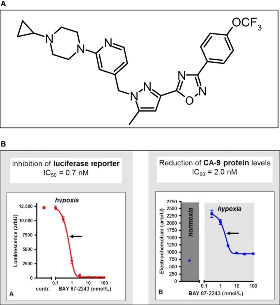
For in vitro studies, BAY 87-2243 was prepared as a 10 mmol/L stock solution in dimethyl sulfoxide (DMSO) and diluted in the relevant assay media. Luciferase activity is inhibited by BAY 87-2243, with an IC50 value of around 0.7 nM. BAY 87-2243 inhibits hypoxic expression of the HIF target gene CA9 on the protein level in HCT116luc cells, with an IC50 value of around 2 nM. With an IC50 value of less than 10 nM, BAY 87- 2243 inhibits mitochondrial oxygen consumption as determined by the oxygen-sensitive fluorescent dye LUX-MitoXpress[1]. The nuclear HIF-1α protein expression is inhibited by BAY-87-2243. In contrast to relative vascular area (RVA) and perfused vessels (PF), administration of BAY-87-2243 for approximately 18 days significantly reduces HIF-1α protein expression, necrotic fraction (NF) (mean 9% vs. 35.6%, p=0.0002), and pimonidazole hypoxic fraction (pHF) (mean 2.4% (BAY-87-2243) vs. 17.6% (carrier), p<0.0001)[2].
Antiproliferative activity across cancer cell lines: BAY 87-2243 induced dose-dependent growth inhibition in various human cancer cell lines under normoxic (21% O₂) and hypoxic (1% O₂) conditions. IC50 values (72 h, MTT assay) were: A549 (lung cancer, normoxia: 12 nM; hypoxia: 8 nM), HCT116 (colorectal cancer, normoxia: 15 nM; hypoxia: 9 nM), SK-MEL-28 (melanoma, normoxia: 10 nM; hypoxia: 7 nM), and PC-3 (prostate cancer, normoxia: 18 nM; hypoxia: 11 nM). Hypoxic conditions enhanced sensitivity to BAY 87-2243 by ~1.3–1.6-fold [1] - Inhibition of hypoxia-inducible genes: In A549 cells exposed to hypoxia (1% O₂ for 24 h), BAY 87-2243 (5–20 nM) dose-dependently reduced the expression of hypoxia-inducible factor-1α (HIF-1α) protein (by 40–85%) and its target genes, including vascular endothelial growth factor (VEGF, mRNA down 55–90%) and glucose transporter 1 (GLUT1, mRNA down 45–80%), as detected by Western blot and qPCR [1] - Mitochondrial function disruption: BAY 87-2243 (10–30 nM) treatment for 16 h in HCT116 cells decreased mitochondrial ATP production by 50–75% (luciferase-based assay) and increased reactive oxygen species (ROS) levels by 2.5–4-fold (DCFH-DA staining). It also reduced the mitochondrial membrane potential (ΔΨm) by 30–60% (JC-1 staining), indicating mitochondrial dysfunction [1] - Synergy with fractionated irradiation: In FaDu (head and neck squamous cell carcinoma, HNSCC) cells, BAY 87-2243 (3–10 nM) combined with fractionated irradiation (2 Gy per fraction, 5 fractions over 5 days) reduced cell survival compared to irradiation alone. At 5 nM, the surviving fraction (SF) after 10 Gy irradiation was 0.08 (combination) vs. 0.25 (irradiation alone), with a radiosensitization ratio (RSR) of 1.6 [2] |
| ln Vivo |
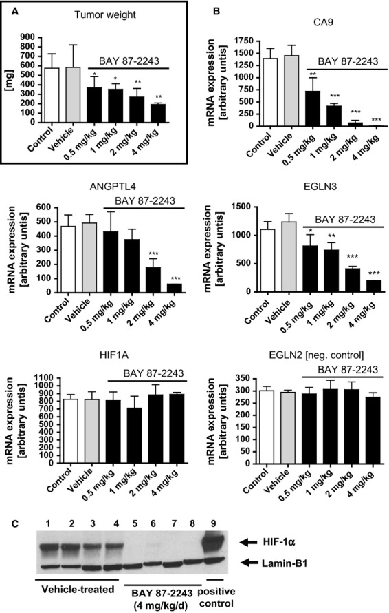
H460 cells are subcutaneously injected into naked mice, and once tumors have developed, the mice are given daily oral gavage treatments with BAY 87-2243 (0.5, 1, 2, and 4 mg/kg) for three weeks. In accordance with a dose-dependent decrease in the mRNA expression levels of the HIF-1 target genes CA9, ANGPTL4, and EGLN3, BAY 87-2243 decreased tumor weight. However, the compound treatment in vivo had no effect on the mRNA expression levels of the hypoxia-insensitive EGLN2 gene or HIF-1α itself[1].
Monotherapy antitumor activity in xenografts: Female nude mice (6–8 weeks old) bearing subcutaneous tumors (A549, HCT116, SK-MEL-28) were treated with BAY 87-2243 (10 mg/kg, oral gavage, once daily) for 21 days. Tumor growth inhibition (TGI) rates were: A549 (72%, treated volume: 290 mm³ vs. vehicle: 1030 mm³, P<0.01), HCT116 (68%, treated: 320 mm³ vs. vehicle: 1000 mm³, P<0.01), SK-MEL-28 (75%, treated: 260 mm³ vs. vehicle: 1040 mm³, P<0.01). In A549 orthotopic lung tumors, BAY 87-2243 (10 mg/kg, oral) prolonged median survival from 28 days (vehicle) to 45 days (P<0.001) [1] - Combination with fractionated irradiation in HNSCC xenografts: Male BALB/c nude mice (7 weeks old) with subcutaneous FaDu or CAL27 (HNSCC) tumors were divided into 4 groups: vehicle, BAY 87-2243 alone (3 mg/kg, oral, once daily), irradiation alone (2 Gy/fraction, 5 fractions/week for 3 weeks), combination. For FaDu tumors: combination group had a tumor growth delay (TGD) of 28 days, vs. 8 days (irradiation alone) and 5 days (drug alone), with a local tumor control rate (LCR) of 60% (vs. 10% for irradiation alone, P<0.01). For CAL27 tumors: TGD was 25 days (combination) vs. 7 days (irradiation alone) and 4 days (drug alone), LCR was 55% (vs. 8% for irradiation alone, P<0.01) [2] |
| Enzyme Assay |
Prolyl hydroxylase activity assay[1]
The influence of the test compound on prolyl hydroxylase 2 (PHD2) activity was assayed as described previously15 with some modifications: Recombinant human PHD2 was purified from Sf9 cell lysates and used for hydroxylation of biotinylated HIF-1α 556-574 peptide coated on NeutrAvidin plates. Hydoxylated peptide was quantified after incubation with purified Von-Hippel-Lindau L-Elongin B-Elongin Complex complex labeled with europium and addition of enhancer solution by measuring time-resolved fluorescence with a Tecan infinite M200 plate reader. Western blot analysis of HIF-1α[2] Western blotting was performed one time according to the established protocol as described previously [24]. Protein samples were prepared using the NE-PER Nuclear and Cytoplasmic KIT according to the manufacturer’s instructions. Antibodies used were mouse monoclonal anti-human HIF-1α (1:250) and rabbit polyclonal anti-histone-H2B (1:500) and anti-Calpain 1 (1:100) served as the loading controls for nuclear or cytoplasmic cell compartments, respectively. Nuclear HIF-1α band intensities were normalized to histone-H2B levels. Mitochondrial complex I activity assay: Bovine heart mitochondria were purified by differential centrifugation. The assay was performed in a reaction buffer (25 mM KH₂PO₄ pH 7.4, 5 mM MgCl₂, 0.2 mM EDTA) containing 0.2 mM NADH (substrate) and 0.05 mM coenzyme Q1 (electron acceptor). Serial concentrations of BAY 87-2243 (0.1–10 nM) were added, and the reaction was initiated by adding mitochondria (0.1 mg protein/mL). The decrease in absorbance at 340 nm (due to NADH oxidation) was measured every 30 seconds for 5 minutes at 37°C. Complex I activity was calculated as the rate of NADH oxidation (μmol/min/mg protein), and IC50 was determined by fitting the activity inhibition curve to a four-parameter logistic model [1] |
| Cell Assay |
Cellular assays[1]
For high-throughput screening of a small molecule library consisting of ∼830,000 compounds, HCT-116 cells were stably transfected with a vector containing a luciferase reporter system coupled four times to the HRE from human vascular endothelial growth factor (VEGF) promoter (HCT 116-4xVEGF-Luc). Cells were plated at 3 × 10E4 cells/well and incubated overnight before test compounds (5 mmol/L in DMSO) were added and plates were placed in a hypoxic chamber for 16 h at 1% pO2. Results are given as luminescence counts in arbitrary units after subtraction of baseline levels from normoxic, nontreated controls. For the measurement of cellular complex I activity, H1299 cells were cotransfected with a pcDNA3 vector encoding for Pyrearinus termitilluminans larval click beetle luciferase. Clones (H1299tluc) showing high luminescence and dose-dependent rotenone sensitivity were subcloned and then used for further in depth analysis of cellular complex I activity by luminescence measurements. In brief, H1299tluc cells (1500/well) were seeded into white 384 well plates. After 2 days in culturing in Dulbecco's modified eagle medium (DMEM) without glucose, but supplemented with 11 mmol/L galactose, 10 μL of a luciferin/inhibitor mixture (150 μmol/L d-luciferin, 0.4% DMSO final concentration in Tyrode) was added to each well and incubated for 1 h at 37°C. Luminescence measurements were performed with an in house developed plate reader. After this measurement 20 μL succinate (0.67 mol/L, pH 5.3 in Tyrode, final concentration 25 mmol/L) was added. The plate was then incubated for another 1 h at room temperature before the second measurement was performed. H1299tluc cells expressing NADH-Q-Oxidoreductase from Saccharomyces cerevisiae (NDI1) were generated by transfection with a pcDNA3 vector encoding for NDI1 under control of a cytomegaly virus (CMV) promoter and a C-terminal HA-tag using PiggyBac transposon-mediated gene transfer11. Selection for positive clones was performed by cultivation in the presence of 20 nmol/L rotenone in DMEM medium with 11.2 mmol/L glucose. Rotenone insensitive clones with high luminescence were used as described above. Luciferase activity is given in % of DMSO-treated cells. To evaluate the cytotoxicity of BAY 87-2243, 2.000 cells of the respective cell lines were seeded in 96-well plates and cultured in the appropriate growth medium containing 10% FCS. BAY 87-2243 at various concentrations was added at 24 h after seeding for additional 48 h and cell viability was determined using Cell Titer Glow Assay. Quantification of CA9 protein[1] HCT 116-4xVEGF-Luc cells were seeded at 3 × 10E4 cells/well in 96-well plates and incubated overnight at 37°C in a humidified incubator containing 5% CO2 under normal oxygen levels before shifting hypoxic conditions (1% pO2, 24 h) in the absence or presence of various concentrations of BAY 87-2243. Protein expression levels of the HIF target gene carbonic anhydrase 9 (CA9) in cell lysates was quantified using MN/CAIX enzyme linked immunosorbent assay. MTT antiproliferation assay: Cancer cells (A549, HCT116, SK-MEL-28) were seeded in 96-well plates (5×10³ cells/well) and incubated under normoxia (21% O₂) or hypoxia (1% O₂, 5% CO₂, 94% N₂) overnight. BAY 87-2243 (0.1–100 nM) was added, and cells were cultured for 72 h. MTT reagent (5 mg/mL, 10 μL/well) was added, and incubation continued for 4 h. DMSO (150 μL/well) dissolved formazan crystals, and absorbance was measured at 570 nm. Cell viability (%) = (treated absorbance/control absorbance) × 100, IC50 was calculated via GraphPad Prism [1] - Western blot for HIF-1α/VEGF: A549 cells were exposed to hypoxia (1% O₂) for 24 h with BAY 87-2243 (5–20 nM). Cells were lysed in RIPA buffer (with protease inhibitors), 30 μg protein was separated by 10% SDS-PAGE, and transferred to PVDF membranes. Membranes were blocked with 5% non-fat milk (1 h, room temperature), incubated with primary antibodies (anti-HIF-1α, anti-VEGF) overnight (4°C), then with HRP-conjugated secondary antibodies (1 h, room temperature). Bands were visualized via ECL, and intensity was quantified with ImageJ [1] - Clonogenic survival assay (radiosensitivity): FaDu/CAL27 cells were seeded in 6-well plates (200–1000 cells/well) and incubated overnight. BAY 87-2243 (3–10 nM) was added 1 h before irradiation (0–10 Gy, single fraction). Cells were cultured for 14 days, colonies (>50 cells) were fixed with methanol, stained with crystal violet, and counted. Surviving fraction (SF) = (colony number × plating efficiency)/number of cells seeded. Radiosensitization ratio (RSR) was calculated as SF (irradiation alone)/SF (combination) at 10 Gy [2] |
| Animal Protocol |
In vivo tumor study [1]
For in vivo studies, BAY 87-2243 was formulated in a 1% (v/v) solution of ethanol/solutol/water (10/40/50%). Animals were given BAY 87-2243 (0.5, 1, 2, and 4 mg/kg) or vehicle control once daily by oral gavage. Tumor xenograft experiment was carried out on female immune-deficient, athymic NMRI nude mice, aged 7–9 weeks, weighing 20–25 g in full accordance with the Interdisciplinary Principles and Guidelines for the Use of Animals in Research, Marketing and Education issued by the New York Academy of Sciences' Ad Hoc Committee on Animal Research. The lung carcinoma xenograft mouse model was established by subcutaneous injection into the right flank with 0.1 mL H460 tumor cells (1.5 × 10E6) mixed 1:1 with Matrigel. Mice were randomized into control and treatment groups when tumors reached a size of more than 40 mm2. Body weight was monitored as a measure for treatment-related, acute toxicity. Tumor area (measured by caliper) or tumor weight (measured when mice were sacrificed 21 days after cell injection) was calculated by the formula 100−100 × (tumor weight/area of treatment group)/(tumor weight/area of vehicle group). Murine plasma pharmacokinetic analyses[1] Plasma concentrations of unchanged BAY 87-2243 were determined by liquid chromatography coupled to a tandem mass spectrometer (LC-MS/MS). Briefly, murine plasma was centrifuged and subsequently precipitated by addition of acetonitrile and an internal standard. The supernatants were subjected to high-performance LC connected to a MS/MS (API 3000, Applied Biosystems, Darmstadt, Germany) via a Turbo Ion Spray interface. Tumor sampling[2] BAY-87-2243 was dissolved in carrier solution (10% ethanol, 40% Solutol® HS15, 50% sterile distilled water) and administered orally by gavage (9 mg/kg/body weight [b.w.]). When UT-SCC-5 hSCC xenografts in nude mice reached 6 mm in diameter BAY-87-2243 or carrier was administered before and/or during RT or radiochemotherapy with concomitant cisplatin (RCT). Local tumor control was evaluated 150 days after irradiation and the doses to control 50% of tumors (TCD50) were compared between treatment arms. Tumors were excised at different time points during BAY-87-2243 or carrier treatment for western blot and immunohistological investigations. Tumor monotherapy protocol (A549/HCT116/SK-MEL-28): Female nude mice (6–8 weeks old) were subcutaneously injected with 5×10⁶ cancer cells (100 μL PBS/matrigel, 1:1) into the right flank. When tumors reached ~100 mm³, mice were grouped (n=6/group): vehicle (0.5% methylcellulose in PBS, oral, daily) and BAY 87-2243 (10 mg/kg, dissolved in 0.5% methylcellulose, oral, daily). Treatment lasted 21 days. Tumor volume (length × width² / 2) was measured every 3 days, body weight weekly. For orthotopic A549 tumors: 2×10⁶ cells were injected into the left lung, treatment started 7 days post-injection, and survival was monitored [1] - Radiation combination protocol (FaDu/CAL27): Male BALB/c nude mice (7 weeks old) were subcutaneously injected with 4×10⁶ FaDu/CAL27 cells (100 μL PBS/matrigel, 1:1). When tumors reached ~120 mm³, mice were grouped (n=5/group): (1) vehicle (0.5% methylcellulose, oral, daily); (2) BAY 87-2243 (3 mg/kg, oral, daily); (3) irradiation (2 Gy/fraction, 5 fractions/week for 3 weeks, delivered to tumors via X-ray); (4) combination (drug 1 h before irradiation). Tumor volume was measured every 2 days, and local tumor control (no tumor regrowth for 60 days) was assessed [2] |
| ADME/Pharmacokinetics |
Oral absorption and pharmacokinetics in rats: Male Sprague-Dawley rats (250-300 g) were administered BAY 87-2243 via gavage (10 mg/kg) or intravenous injection (2 mg/kg). The oral bioavailability was 65%. Oral administration: Cmax = 85 ng/mL (Tmax = 1.2 h), terminal t1/2 = 4.8 h, AUC0-24h = 420 ng·h/mL. Intravenous administration: Cmax = 220 ng/mL, t1/2 = 4.5 h, AUC0-∞ = 510 ng·h/mL [1]
- Plasma protein binding rate: In human plasma, the protein binding rate of BAY 87-2243 was 98%, mainly bound to albumin and α1-acid glycoprotein (as determined by equilibrium dialysis) [1] - Tumor permeability: In A549 xenograft mice, the tumor tissue concentration reached 12 nM 2 hours after oral administration of BAY 87-2243 (10 mg/kg), which is about 1.5 times the plasma concentration (8 nM) [1] |
| Toxicity/Toxicokinetics |
Repeated-dose toxicity in rats: Male/female Sprague-Dawley rats (n=4 per sex per group) were orally administered BAY 87-2243 (5, 15, 30 mg/kg) for 28 consecutive days. No deaths were observed. The No-AEL (No Observed Adverse Effect) was 15 mg/kg. In the 30 mg/kg dose group: mild weight loss (approximately 7%), elevated serum ALT (2.1-fold higher than control group), elevated AST (1.8-fold higher than control group), and mild hepatocyte vacuolation (histopathology); no nephrotoxicity was observed [1]
- Xenograft toxicity in mice: In all tumor models, BAY 87-2243 (3-10 mg/kg, orally daily) resulted in ≤5% weight loss without significant toxic reactions (e.g., somnolence, diarrhea). Combined radiotherapy did not increase toxicity compared to monotherapy [1,2] |
| References |
|
| Additional Infomation |
Activation of the transcription factor hypoxia-inducible factor-1 (HIF-1) plays a crucial role in tumorigenesis, development, and resistance to chemotherapy and radiotherapy. To screen compounds targeting the HIF pathway, we used a luciferase-driven HIF-1 reporter cell line to screen a library of small molecules under hypoxic conditions. High-throughput screening identified a class of aminoalkyl-substituted compounds that inhibited hypoxia-induced HIF-1 target gene expression in human lung cancer cell lines at low nanomolar concentrations. The lead compound, BAY 87-2243, inhibited the accumulation of HIF-1α and HIF-2α proteins in the non-small cell lung cancer (NSCLC) cell line H460 under hypoxic conditions, but had no effect on HIF-1α protein levels induced by the hypoxia mimics deferoxamine or cobalt chloride. BAY 87-2243 had no effect on the expression levels of HIF target genes or the activity of HIF prolyl hydroxylase-2 in RCC4 cells lacking Von Hippel-Lindau (VHL) activity. In the H460 xenograft model, the antitumor activity of BAY 87-2243 was confirmed, including inhibition of HIF-1α protein levels and reduction of in vivo HIF-1 target gene expression. Under standard conditions, BAY 87-2243 did not inhibit cell proliferation. However, under glucose depletion (a condition favorable for mitochondrial ATP production as an energy source), BAY 87-2243 inhibited cell proliferation at nanomolar concentrations. Further experiments showed that BAY 87-2243 inhibited the activity of mitochondrial complex I but had no effect on the activity of complex III. Interfering with mitochondrial function to reduce hypoxia-induced HIF-1 activity in tumors may be a promising treatment that can overcome chemotherapy and radiotherapy resistance in hypoxic tumors [1]. Mechanism of action: BAY 87-2243 exerts its antitumor effect through two key mechanisms: (1) inhibiting mitochondrial complex I, reducing ATP production and increasing ROS, thereby inhibiting the stabilization of hypoxia-inducible factor-1α (HIF-1α) (the translation of HIF-1α requires ATP); (2) downregulating HIF-1α target genes (VEGF, GLUT1), which promote angiogenesis and glycolysis, both of which are essential for the survival of hypoxic tumors. When used in combination with radiotherapy, it can enhance DNA damage by reducing tumor hypoxia (inhibiting angiogenesis) and impairing DNA repair [1,2]
- Preclinical Development Focus: BAY 87-2243 has been evaluated preclinically for hypoxic solid tumors (e.g., lung cancer, colorectal cancer, melanoma, head and neck cancer), particularly as a radiosensitizer to improve local tumor control in radiotherapy-resistant hypoxic tumors [1,2] |
| Molecular Formula |
C26H26F3N7O2
|
|---|---|
| Molecular Weight |
525.5256
|
| Exact Mass |
525.21
|
| Elemental Analysis |
C, 59.42; H, 4.99; F, 10.85; N, 18.66; O, 6.09
|
| CAS # |
1227158-85-1
|
| Related CAS # |
1227158-85-1
|
| PubChem CID |
67377767
|
| Appearance |
White to off-white solid powder
|
| Density |
1.5±0.1 g/cm3
|
| Boiling Point |
677.7±65.0 °C at 760 mmHg
|
| Flash Point |
363.7±34.3 °C
|
| Vapour Pressure |
0.0±2.1 mmHg at 25°C
|
| Index of Refraction |
1.674
|
| LogP |
4.2
|
| Hydrogen Bond Donor Count |
0
|
| Hydrogen Bond Acceptor Count |
11
|
| Rotatable Bond Count |
7
|
| Heavy Atom Count |
38
|
| Complexity |
774
|
| Defined Atom Stereocenter Count |
0
|
| SMILES |
FC(F)(F)OC1=CC=C(C2=NOC(C3=NN(CC4=CC(N5CCN(C6CC6)CC5)=NC=C4)C(C)=C3)=N2)C=C1
|
| InChi Key |
CDJNNOJINJAXPV-UHFFFAOYSA-N
|
| InChi Code |
InChI=1S/C26H26F3N7O2/c1-17-14-22(25-31-24(33-38-25)19-2-6-21(7-3-19)37-26(27,28)29)32-36(17)16-18-8-9-30-23(15-18)35-12-10-34(11-13-35)20-4-5-20/h2-3,6-9,14-15,20H,4-5,10-13,16H2,1H3
|
| Chemical Name |
1-cyclopropyl-4-[4-[[5-methyl-3-[3-[4-(trifluoromethoxy)phenyl]-1,2,4-oxadiazol-5-yl]-1H-pyrazol-1-yl]methyl]-2-pyridinyl]-piperazine
|
| Synonyms |
BAY-872243; BAY 872243; BAY-872243; BAY-87-2243; BAY87-2243; BAY 87-2243
|
| HS Tariff Code |
2934.99.9001
|
| Storage |
Powder -20°C 3 years 4°C 2 years In solvent -80°C 6 months -20°C 1 month |
| Shipping Condition |
Room temperature (This product is stable at ambient temperature for a few days during ordinary shipping and time spent in Customs)
|
| Solubility (In Vitro) |
DMSO: <1 mg/mL
Water: <1 mg/mL (Insoluble) Ethanol: 8 mg/mL (15.22 mM) |
|---|---|
| Solubility (In Vivo) |
Solubility in Formulation 1: ≥ 2.5 mg/mL (4.76 mM) (saturation unknown) in 10% DMSO + 40% PEG300 + 5% Tween80 + 45% Saline (add these co-solvents sequentially from left to right, and one by one), clear solution.
For example, if 1 mL of working solution is to be prepared, you can add 100 μL of 25.0 mg/mL clear DMSO stock solution to 400 μL PEG300 and mix evenly; then add 50 μL Tween-80 to the above solution and mix evenly; then add 450 μL normal saline to adjust the volume to 1 mL. Preparation of saline: Dissolve 0.9 g of sodium chloride in 100 mL ddH₂ O to obtain a clear solution. Solubility in Formulation 2: ≥ 2.5 mg/mL (4.76 mM) (saturation unknown) in 10% DMSO + 90% Corn Oil (add these co-solvents sequentially from left to right, and one by one), clear solution. For example, if 1 mL of working solution is to be prepared, you can add 100 μL of 25.0 mg/mL clear DMSO stock solution to 900 μL of corn oil and mix evenly. (Please use freshly prepared in vivo formulations for optimal results.) |
| Preparing Stock Solutions | 1 mg | 5 mg | 10 mg | |
| 1 mM | 1.9028 mL | 9.5142 mL | 19.0284 mL | |
| 5 mM | 0.3806 mL | 1.9028 mL | 3.8057 mL | |
| 10 mM | 0.1903 mL | 0.9514 mL | 1.9028 mL |
*Note: Please select an appropriate solvent for the preparation of stock solution based on your experiment needs. For most products, DMSO can be used for preparing stock solutions (e.g. 5 mM, 10 mM, or 20 mM concentration); some products with high aqueous solubility may be dissolved in water directly. Solubility information is available at the above Solubility Data section. Once the stock solution is prepared, aliquot it to routine usage volumes and store at -20°C or -80°C. Avoid repeated freeze and thaw cycles.
Calculation results
Working concentration: mg/mL;
Method for preparing DMSO stock solution: mg drug pre-dissolved in μL DMSO (stock solution concentration mg/mL). Please contact us first if the concentration exceeds the DMSO solubility of the batch of drug.
Method for preparing in vivo formulation::Take μL DMSO stock solution, next add μL PEG300, mix and clarify, next addμL Tween 80, mix and clarify, next add μL ddH2O,mix and clarify.
(1) Please be sure that the solution is clear before the addition of next solvent. Dissolution methods like vortex, ultrasound or warming and heat may be used to aid dissolving.
(2) Be sure to add the solvent(s) in order.
| NCT Number | Recruitment | interventions | Conditions | Sponsor/Collaborators | Start Date | Phases |
| NCT01297530 | Terminated | Drug: BAY87-2243 | Neoplasms | Bayer | April 2011 | Phase 1 |
(A) Chemical structure of BAY 87-2243. (B) BAY 87-2243 suppresses hypoxia-induced reporter gene activity (left), and CA9 protein levels in vitro in HCT-116 cell lysates (right). Cancer Med. 2013 Oct;2(5):611-24. |
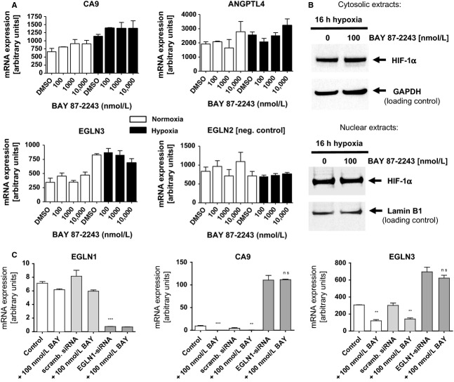
BAY 87-2243 is inactive in RCC4 cells lacking functional VHL protein or in H460 cells silenced for EGLN1.Cancer Med.2013 Oct;2(5):611-24. |
BAY 87-2243 reduces tumor weight, hypoxia-inducible factor (HIF)-1α protein levels, and HIF-1 target gene expression in H460 xenograft tumors. Cancer Med. 2013 Oct;2(5):611-24. |
Products are for research use only; We do not sell to patients
Copyright 2020 InvivoChem LLC | All Rights Reserved


 COA
COA