yingweiwo

Barbadin

Cat No.:V11934 Purity: ≥98%
Barbadin is a selective inhibitor of β-arrestin/β2-adaptin interaction, with IC50s of 19.1 μM and 15.6 μM for β-arrestin1 and β-arrestin2, respectively.
Barbadin
Barbadin Chemical Structure CAS No.: 356568-70-2
Product category: New1
This product is for research use only, not for human use. We do not sell to patients.
Size Price Stock Qty
1mg
5mg
Other Sizes
Official Supplier of:
Alternate Text
Alternate Text
Alternate Text
Alternate Text
Alternate Text
Alternate Text
Alternate Text
Alternate Text
Alternate Text
Alternate Text
Alternate Text
Alternate Text
Alternate Text
Alternate Text
Alternate Text
Alternate Text
Alternate Text
Alternate Text
Alternate Text
Alternate Text
Alternate Text
Alternate Text
Alternate Text
Alternate Text
Alternate Text
Alternate Text
Alternate Text
Alternate Text
Alternate Text
Alternate Text
Alternate Text
Alternate Text


  • Business Relationship with 5000+ Clients Globally
  • Major Universities, Research Institutions, Biotech & Pharma
  • Citations by Top Journals: Nature, Cell, Science, etc.
Top Publications Citing lnvivochem Products
Product Description
Barbadin is a selective inhibitor of β-arrestin/β2-adaptin interaction, with IC50s of 19.1 μM and 15.6 μM for β-arrestin1 and β-arrestin2, respectively. Barbadin-blocking agonists promote endocytosis of β2-adrenergic, V2-vasopressin and angiotensin-II type-1 receptors. Barbadin causes apoptosis.
Biological Activity I Assay Protocols (From Reference)
ln Vitro
Four hours of barbadin administration cause apoptosis and decrease cell viability [2]. Treatment with barbadin for two hours can inhibit G0/G1 phase breast cancer cells [2].
Cell Assay
Apoptosis analysis [2]
Cell Types: MDA MB-231 Cell
Tested Concentrations:
Incubation Duration: 4 hrs (hours)
Experimental Results: Showing the morphological characteristics of apoptosis such as shrinkage, rounding, and shedding, the cell viability percentage diminished to 69.1%, and appeared at 29.9 Apoptosis. Percentage of EBSS (Earle's Balanced Salt Solution) starved cells.

Cell cycle analysis [2]
Cell Types: MDA MB-231 Cell
Tested Concentrations:
Incubation Duration: 2 hrs (hours)
Experimental Results: G0/G1 phase cells were inhibited by 63.7%.
References

[1]. A new inhibitor of the β-arrestin/AP2 endocytic complex reveals interplay between GPCR internalization and signalling. Nat Commun. 2017 Apr 18;8:15054. doi: 10.1038/ncomms15054.

[2]. β-Arrestin inhibition induces autophagy, apoptosis, G0/G1 cell cycle arrest in agonist-activated V2R receptor in breast cancer cells. Med Oncol. 2021 Mar 15;38(4):38.

These protocols are for reference only. InvivoChem does not independently validate these methods.
Physicochemical Properties
Molecular Formula
C19H15N3OS
Molecular Weight
333.406902551651
Exact Mass
333.093
CAS #
356568-70-2
PubChem CID
727640
Appearance
White to light yellow solid powder
LogP
3.7
Hydrogen Bond Donor Count
1
Hydrogen Bond Acceptor Count
4
Rotatable Bond Count
3
Heavy Atom Count
24
Complexity
483
Defined Atom Stereocenter Count
0
SMILES
O=C1N(N)C=NC2SC=C(C1=2)C1C=CC(CC2C=CC=CC=2)=CC=1
InChi Key
OCBXPCSXEQQADU-UHFFFAOYSA-N
InChi Code
InChI=1S/C19H15N3OS/c20-22-12-21-18-17(19(22)23)16(11-24-18)15-8-6-14(7-9-15)10-13-4-2-1-3-5-13/h1-9,11-12H,10,20H2
Chemical Name
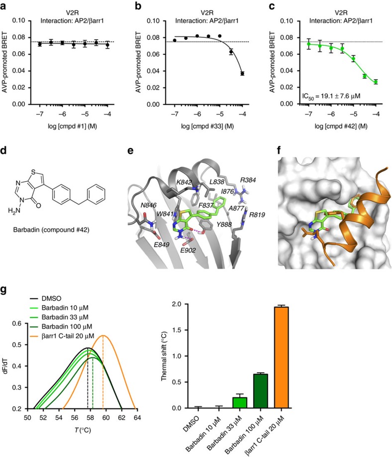
3-amino-5-(4-benzylphenyl)thieno[2,3-d]pyrimidin-4-one
HS Tariff Code
2934.99.9001
Storage

Powder      -20°C    3 years

                     4°C     2 years

In solvent   -80°C    6 months

                  -20°C    1 month

Shipping Condition
Room temperature (This product is stable at ambient temperature for a few days during ordinary shipping and time spent in Customs)
Solubility Data
Solubility (In Vitro)
DMSO : ~50 mg/mL (~149.97 mM)
Solubility (In Vivo)
Solubility in Formulation 1: ≥ 2.5 mg/mL (7.50 mM) (saturation unknown) in 10% DMSO + 40% PEG300 + 5% Tween80 + 45% Saline (add these co-solvents sequentially from left to right, and one by one), clear solution.
For example, if 1 mL of working solution is to be prepared, you can add 100 μL of 25.0 mg/mL clear DMSO stock solution to 400 μL PEG300 and mix evenly; then add 50 μL Tween-80 to the above solution and mix evenly; then add 450 μL normal saline to adjust the volume to 1 mL.
Preparation of saline: Dissolve 0.9 g of sodium chloride in 100 mL ddH₂ O to obtain a clear solution.

Solubility in Formulation 2: ≥ 2.5 mg/mL (7.50 mM) (saturation unknown) in 10% DMSO + 90% Corn Oil (add these co-solvents sequentially from left to right, and one by one), clear solution.
For example, if 1 mL of working solution is to be prepared, you can add 100 μL of 25.0 mg/mL clear DMSO stock solution to 900 μL of corn oil and mix evenly.

 (Please use freshly prepared in vivo formulations for optimal results.)
Preparing Stock Solutions 1 mg 5 mg 10 mg
1 mM 2.9993 mL 14.9966 mL 29.9931 mL
5 mM 0.5999 mL 2.9993 mL 5.9986 mL
10 mM 0.2999 mL 1.4997 mL 2.9993 mL

*Note: Please select an appropriate solvent for the preparation of stock solution based on your experiment needs. For most products, DMSO can be used for preparing stock solutions (e.g. 5 mM, 10 mM, or 20 mM concentration); some products with high aqueous solubility may be dissolved in water directly. Solubility information is available at the above Solubility Data section. Once the stock solution is prepared, aliquot it to routine usage volumes and store at -20°C or -80°C. Avoid repeated freeze and thaw cycles.

Calculator

Molarity Calculator allows you to calculate the mass, volume, and/or concentration required for a solution, as detailed below:

  • Calculate the Mass of a compound required to prepare a solution of known volume and concentration
  • Calculate the Volume of solution required to dissolve a compound of known mass to a desired concentration
  • Calculate the Concentration of a solution resulting from a known mass of compound in a specific volume
An example of molarity calculation using the molarity calculator is shown below:
What is the mass of compound required to make a 10 mM stock solution in 5 ml of DMSO given that the molecular weight of the compound is 350.26 g/mol?
  • Enter 350.26 in the Molecular Weight (MW) box
  • Enter 10 in the Concentration box and choose the correct unit (mM)
  • Enter 5 in the Volume box and choose the correct unit (mL)
  • Click the “Calculate” button
  • The answer of 17.513 mg appears in the Mass box. In a similar way, you may calculate the volume and concentration.

Dilution Calculator allows you to calculate how to dilute a stock solution of known concentrations. For example, you may Enter C1, C2 & V2 to calculate V1, as detailed below:

What volume of a given 10 mM stock solution is required to make 25 ml of a 25 μM solution?
Using the equation C1V1 = C2V2, where C1=10 mM, C2=25 μM, V2=25 ml and V1 is the unknown:
  • Enter 10 into the Concentration (Start) box and choose the correct unit (mM)
  • Enter 25 into the Concentration (End) box and select the correct unit (mM)
  • Enter 25 into the Volume (End) box and choose the correct unit (mL)
  • Click the “Calculate” button
  • The answer of 62.5 μL (0.1 ml) appears in the Volume (Start) box
g/mol

Molecular Weight Calculator allows you to calculate the molar mass and elemental composition of a compound, as detailed below:

Note: Chemical formula is case sensitive: C12H18N3O4  c12h18n3o4
Instructions to calculate molar mass (molecular weight) of a chemical compound:
  • To calculate molar mass of a chemical compound, please enter the chemical/molecular formula and click the “Calculate’ button.
Definitions of molecular mass, molecular weight, molar mass and molar weight:
  • Molecular mass (or molecular weight) is the mass of one molecule of a substance and is expressed in the unified atomic mass units (u). (1 u is equal to 1/12 the mass of one atom of carbon-12)
  • Molar mass (molar weight) is the mass of one mole of a substance and is expressed in g/mol.
/

Reconstitution Calculator allows you to calculate the volume of solvent required to reconstitute your vial.

  • Enter the mass of the reagent and the desired reconstitution concentration as well as the correct units
  • Click the “Calculate” button
  • The answer appears in the Volume (to add to vial) box
In vivo Formulation Calculator (Clear solution)
Step 1: Enter information below (Recommended: An additional animal to make allowance for loss during the experiment)
Step 2: Enter in vivo formulation (This is only a calculator, not the exact formulation for a specific product. Please contact us first if there is no in vivo formulation in the solubility section.)
+
+
+

Calculation results

Working concentration mg/mL;

Method for preparing DMSO stock solution mg drug pre-dissolved in μL DMSO (stock solution concentration mg/mL). Please contact us first if the concentration exceeds the DMSO solubility of the batch of drug.

Method for preparing in vivo formulation:Take μL DMSO stock solution, next add μL PEG300, mix and clarify, next addμL Tween 80, mix and clarify, next add μL ddH2O,mix and clarify.

(1) Please be sure that the solution is clear before the addition of next solvent. Dissolution methods like vortex, ultrasound or warming and heat may be used to aid dissolving.
             (2) Be sure to add the solvent(s) in order.

Biological Data
  • Identification of Barbadin (compound #42) as an inhibitor of the interaction between β2-adaptin and β-arrestin.(a–c) Concentration-response curves of compounds #1, #33 and #42 selected from the BRET-based screen (Fig. 1) using the same β2-adaptin/β-arrestin1 interaction assay. Dotted line represents the level of AVP-promoted BRET upon pre-incubation with DMSO. Data are the mean±s.e.m. of three independent experiments. (d) Chemical structure of Barbadin (IUPAC name: 3-amino-5-(4-benzylphenyl)-3H,4H-thieno[2,3-d]pyrimidin-4-one). (e,f) Docking pose of Barbadin (green sticks) within the groove of β2-adaptin platform subdomain (grey ribbon (e), or surface (f)) that is the site of interaction with β-arrestin (orange ribbon and sticks (f)). β2-adaptin residues known to interact with β-arrestin are labelled and shown as grey stick. Hydrogen-bonds interactions between Barbadin and both Tyr-888 and Glu-902 are depicted as magenta dotted lines (e). Superimposition of Barbadin with the β-arrestin1 C-terminus peptide as in the co-crystal structure (PDB entry 2IV8), where Phe-388, Phe-391 and Arg-395 are the three key residues for β-arrestin binding interaction (shown from top to bottom as orange sticks) (f). (g) Thermal denaturation of β2-adaptin (residues 700–937) and concentration-dependent stabilization effect of Barbadin. The maximum of the first derivatives of fluorescence (dF/dT) data from differential scanning fluorimetry corresponds to the melting temperatures (Tm, indicated with a dotted line) of β2-adaptin in the presence of DMSO, Barbadin or β-arrestin1 C-tail peptide (positive control) at the indicated concentrations.[1].A new inhibitor of the β-arrestin/AP2 endocytic complex reveals interplay between GPCR internalization and signalling. Nat Commun. 2017 Apr 18;8:15054. doi: 10.1038/ncomms15054.
  • Barbadin specifically blocks the interaction between β2-adaptin and β-arrestin.(a–c) BRET-based assay monitoring the interaction between β2-adaptin-YFP and either β-arrestin1-RlucII or β-arrestin2-RlucII. HEK293T cells were pre-incubated with DMSO or Barbadin (100 μM) for 30 min before 45 min receptor stimulation with AVP (100 nM, a), ISO (10 μM, b) or AngII (100 nM, c). Data are the mean±s.e.m. of at least three independent experiments and unpaired t-test were used to assess statistical significance (***P<0.001; **P<0.005). (d,e) BRET-based kinetics monitoring the interaction between β-arrestin1-RlucII and β2-adaptin-YFP (d) or V2R-YFP (e) in HEK293T cells pretreated with DMSO or Barbadin (100 μM) for 30 min before receptor stimulation with AVP (100 nM) for the indicated times. Data are the mean±s.e.m. of three independent experiments. (f) Effect of Barbadin on the co-immunoprecipitation between β-arrestins and AP2. HEK293SL cells expressing V2R and Flag-β-arrestin2 were pretreated for 20 min with DMSO or Barbadin (50 μM) before stimulation by AVP (1 μM) for 2.5 or 5 min. Endogenous AP2 complexes (using the AP1/2 antibody) were immunoprecipitated (IP) from total cell lysates (TCL), and then analysed by western blot using anti-Flag, anti-epsin or anti-adaptin antibodies as described in the Material and Methods. TCLs represent 5% of input used for IP. IP were quantified over three independent experiments and statistical significance was assessed by a two-way ANOVA followed by Bonferroni's post-hoc tests (*P<0.05).[1].A new inhibitor of the β-arrestin/AP2 endocytic complex reveals interplay between GPCR internalization and signalling. Nat Commun. 2017 Apr 18;8:15054. doi: 10.1038/ncomms15054.
  • Barbadin inhibits GPCRs endocytosis.(a,e) Schematic representation of the ebBRET-based assay used to follow agonist-induced receptor loss from the cell surface by monitoring the interaction between receptor-RLucII and rGFP-CAAX (a) or its translocation into endosomes using rGFP-FYVE (e). (b–d,f–h) V2R, β2AR or AT1R interaction with either rGFP-CAAX (b–d, respectively) or rGFP-FYVE (f–h, respectively) was assessed by BRET following HEK293T cells pre-incubation with DMSO or Barbadin (100 μM) for 30 min before AVP (100 nM), ISO (10 μM) or AngII (1 μM) stimulation for the indicated times. Data are the mean±s.e.m. of a least three independent experiments. (i) V2R localization was imaged by BRET. HEK293T cells were transfected with V2R-RlucII and rGFP-CAAX, pretreated with DMSO or Barbadin (100 μM) for 30 min and then stimulated with 100 nM AVP for 30 min. To generate BRET images, the ratio of acceptor photon counts to donor photon counts was calculated for each pixel and expressed as a colour-coded heat map (lowest being black and purple, and highest red and white). Scale bar, 10 μm.[1].A new inhibitor of the β-arrestin/AP2 endocytic complex reveals interplay between GPCR internalization and signalling. Nat Commun. 2017 Apr 18;8:15054. doi: 10.1038/ncomms15054.
Contact Us