| Size | Price | Stock | Qty |
|---|---|---|---|
| 1mg |
|
||
| 5mg |
|
||
| 10mg |
|
||
| 25mg |
|
||
| 50mg |
|
||
| 100mg | |||
| 250mg | |||
| Other Sizes |


| Targets |
Natural product; secondary metabolite
|
|---|---|
| ln Vitro |
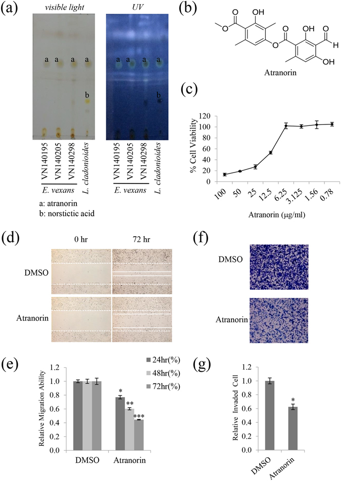
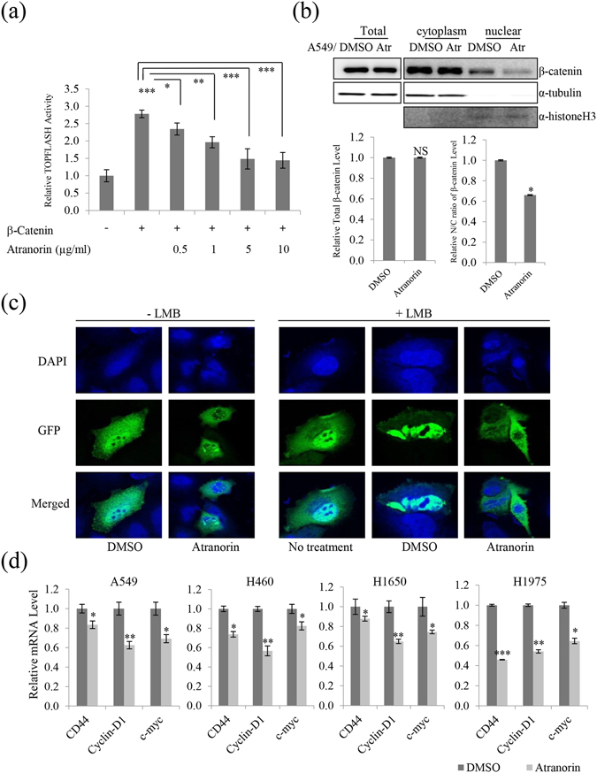
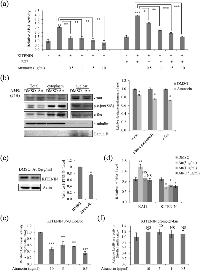
Lichens are symbiotic organisms that produce various secondary metabolites. Here, different lichen extracts were examined to identify secondary metabolites with anti-migratory activity against human lung cancer cells. Everniastrum vexans had the most potent inhibitory activity, and atranorin was identified as an active subcomponent of this extract. Atranorin suppressed β-catenin-mediated TOPFLASH activity by inhibiting the nuclear import of β-catenin and downregulating β-catenin/LEF and c-jun/AP-1 downstream target genes such as CD44, cyclin-D1 and c-myc. Atranorin decreased KAI1 C-terminal interacting tetraspanin (KITENIN)-mediated AP-1 activity and the activity of the KITENIN 3'-untranslated region. The nuclear distribution of the AP-1 transcriptional factor, including c-jun and c-fos, was suppressed in atranorin-treated cells, and atranorin inhibited the activity of Rho GTPases including Rac1, Cdc42, and RhoA, whereas it had no effect on epithelial-mesenchymal transition markers. STAT-luciferase activity and nuclear STAT levels were decreased, whereas total STAT levels were moderately reduced. The human cell motility and lung cancer RT² Profiler PCR Arrays identified additional atranorin target genes. Atranorin significantly inhibited tumorigenesis in vitro [2].
Atranorin was identified as an active secondary metabolite from E. vexans with inhibitory activity against A549 cell motility. Atranorin decreased β-catenin-mediated TOPFLASH activity by suppressing β-catenin nuclear import and downregulated β-catenin/LEF and c-jun/AP-1 downstream target genes. Atranorin affected the expression of KAI1 and KITENIN and downregulated downstream transcriptional factors. Atranorin affected several additional cell motility-related factors in lung cancer cells, including the activity of Rho GTPases, STAT, and the expression of related genes [2]. |
| ln Vivo |
Atranorin inhibited lung cancer invasion and tumor growth in vivo [2]
Results from an in vivo xenograft model further confirmed that atranorin reduced tumor volume, weight, and Ki-67 immunoreactivity (Fig. 6e and f). Consistent with previous results, the main target genes, such as KITENIN, STAT, c-myc, CD44, and/or cyclin-D1 were suppressed in vivo (Fig. 6g and h). Taken together, these results demonstrated that atranorin has antitumorigenic activity in lung cancer models. |
| Cell Assay |
MTT assay [2]
Cells (2 × 10⁴ cells/well) were seeded on a 96-well plate, grown overnight, and then treated with the acetone extracts and atranorin at concentrations of 100 g/mL to 0.78 g/mL for 48 h. After incubation with MTT at 37 °C, cells were lysed with DMSO and absorbance was measured at 570 nm. Wound healing assay [2] A549 cells were plated at a density of 2.5 × 10⁵ cells/well and grown overnight to confluence. Monolayer cells were scratched to create a wound. The cells were then washed twice and incubated in RPMI1640 culture medium supplemented with 2% FBS with 10 μg/mL of the lichen extract or 5 μg/mL atranorin. For the quantitation of relative migration ability, photographs of cells were taken at 0, 24, 48, and 72 h after wounding to measure the width of the wound. The distance migrated by the cells was calculated as the difference between the edges of the wound at time point 1 and at time point 2. For each sample, an average of five wound distances was taken to determine the average rate of migration at a given concentration of lichen extract or atranorin. The migrating rate was determined with the following formula: migrating rate [%] = (width t1 [mm] - width t2 [mm]) / width t1 × 100%. Invasion assay [2] Invasion assays were performed in Transwell chambers with an 8 µm pore polycarbonate membrane coated with 1% gelatin. Invasion assays were performed as previously described. For the quantitation of relative invaded ability, stain the cells adhering to the under-side of the filter and count the number of cells in different five fields of view to get an average sum of cells. Invaded rate [%] = (stained area-sample [mm²] / stained area-control [mm²]) × 100%. Each invasion assay was repeated in three independent experiments. The results are expressed as the mean number of cells migrating per high-power field. Soft agar colony-formation assay [2] Cells (1 × 10⁴) were suspended in soft agar (0.35% agarose), plated onto solidified agar (0.6% agarose) in 6-well plates, and cultured for 3 weeks. Cells were treated twice per week with atranorin (5 g/mL) and DMSO (0.01%). Pixel intensity of the colony area was measured with IMT i-Solution software. To calculate the colony area percentage, the diameter of each colony was quantified as colony size. Data represent the mean of three experiments. |
| Animal Protocol |
Xenograft mouse model [2]
Maintenance of animals and all in vivo experiments were performed according to the Guiding Principles in the Care and Use of Animals (DHEW publication, NIH 80–23). LLC cells (2 × 10⁶ cells) were subcutaneously injected into the flanks of 8-week-old C57BL/6 mice. Drug treatment was initiated 14 days after tumor cell injection, at which time the primary tumor had reached approximately 50 mm3. Animals were treated with 40% DMSO in PBS (vehicle) or 20% atranorin (10 mg/kg) mixed with 20% DMSO every 3 days by intraperitoneal injection for 2 weeks. Tumors were measured every 3 days with an electronic caliper. When primary tumors reached approximately 300 mm3, tumors were surgically removed for further analysis. The tumor volume was determined with the following formula: tumor volume [mm3] = (largest diameter [mm]) × (smallest diameter [mm])² × 0.52. The tumors were histologically examined, and the tissue sections were deparaffinized; rehydrated; rinsed; hybridized with Ki-67 antibodies; and examined as described. |
| References | |
| Additional Infomation |
Atrazoline is a carbonyl compound. It has been reported to exist in Stereocaulon curtatum, Stereocaulon nanodes, and other organisms with relevant data. Background: Atrazoline is a compound with a dihydropyridone structure and is one of the most common secondary metabolites in lichens, a characteristic component of many lichen families, but rarely found in some mosses and higher plants. Various biological properties of atrazoline have been studied over the years. Objective: This review summarizes research on atrazoline, focusing on its diverse biological activities across different fields. Literature describes the anti-inflammatory, analgesic, wound-healing, antibacterial, antifungal, cytotoxic, antioxidant, antiviral, and immunomodulatory activities of dihydropyridone. Furthermore, in vivo animal studies have confirmed the non-toxicity of atrazoline. Conclusion: In summary, atrazoline appears to be an interesting moss substance requiring further investigation to develop its applications, such as in pharmaceutical, medical, or cosmetic fields. Keywords: atrazoline; bioactivity; biosynthesis; lichenin; lichen; secondary metabolites. [1]
Atrazoline significantly inhibited the tumorigenic potential in the mose xenograft model and reduced the level of Ki-67 in the nucleus. Ki-67 is a typical cell cycle-related nuclear protein that is expressed at all stages of the cell cycle. In addition, the molecular mechanism of the anticancer activity of atrazoline proposed in this study has been confirmed in vivo. Combined with the anticancer activity of atrazoline reported in recent literature, our results provide new insights into the anticancer activity of bryophyte species; further research is needed to determine its potential clinical application in the treatment of lung cancer. [2] |
| Molecular Formula |
C19H18O8
|
|---|---|
| Molecular Weight |
374.34142
|
| Exact Mass |
374.1
|
| Elemental Analysis |
C, 60.96; H, 4.85; O, 34.19
|
| CAS # |
479-20-9
|
| PubChem CID |
68066
|
| Appearance |
White to off-white solid powder
|
| Density |
1.4±0.1 g/cm3
|
| Boiling Point |
535.7±50.0 °C at 760 mmHg
|
| Melting Point |
156-158ºC
|
| Flash Point |
189.3±23.6 °C
|
| Vapour Pressure |
0.0±1.5 mmHg at 25°C
|
| Index of Refraction |
1.644
|
| LogP |
6.14
|
| Hydrogen Bond Donor Count |
3
|
| Hydrogen Bond Acceptor Count |
8
|
| Rotatable Bond Count |
6
|
| Heavy Atom Count |
27
|
| Complexity |
564
|
| Defined Atom Stereocenter Count |
0
|
| InChi Key |
YLOYKYXNDHOHHT-UHFFFAOYSA-N
|
| InChi Code |
InChI=1S/C19H18O8/c1-8-5-12(21)11(7-20)17(23)15(8)19(25)27-13-6-9(2)14(18(24)26-4)16(22)10(13)3/h5-7,21-23H,1-4H3
|
| Chemical Name |
(3-hydroxy-4-methoxycarbonyl-2,5-dimethylphenyl) 3-formyl-2,4-dihydroxy-6-methylbenzoate
|
| Synonyms |
Atranorin; 479-20-9; Atranoric acid; Atranorine; Parmelin; Usnarin; Antranoric acid; Parmelin acid;
|
| HS Tariff Code |
2934.99.9001
|
| Storage |
Powder -20°C 3 years 4°C 2 years In solvent -80°C 6 months -20°C 1 month |
| Shipping Condition |
Room temperature (This product is stable at ambient temperature for a few days during ordinary shipping and time spent in Customs)
|
| Solubility (In Vitro) |
DMSO : ~16.67 mg/mL (~44.53 mM)
|
|---|---|
| Solubility (In Vivo) |
Solubility in Formulation 1: 1.67 mg/mL (4.46 mM) in 10% DMSO + 40% PEG300 + 5% Tween80 + 45% Saline (add these co-solvents sequentially from left to right, and one by one), suspension solution; with sonication.
For example, if 1 mL of working solution is to be prepared, you can add 100 μL of 16.7 mg/mL clear DMSO stock solution to 400 μL PEG300 and mix evenly; then add 50 μL Tween-80 to the above solution and mix evenly; then add 450 μL normal saline to adjust the volume to 1 mL. Preparation of saline: Dissolve 0.9 g of sodium chloride in 100 mL ddH₂ O to obtain a clear solution. Solubility in Formulation 2: ≥ 1.67 mg/mL (4.46 mM) (saturation unknown) in 10% DMSO + 90% Corn Oil (add these co-solvents sequentially from left to right, and one by one), clear solution. For example, if 1 mL of working solution is to be prepared, you can add 100 μL of 16.7 mg/mL clear DMSO stock solution to 900 μL of corn oil and mix evenly. (Please use freshly prepared in vivo formulations for optimal results.) |
| Preparing Stock Solutions | 1 mg | 5 mg | 10 mg | |
| 1 mM | 2.6714 mL | 13.3568 mL | 26.7137 mL | |
| 5 mM | 0.5343 mL | 2.6714 mL | 5.3427 mL | |
| 10 mM | 0.2671 mL | 1.3357 mL | 2.6714 mL |
*Note: Please select an appropriate solvent for the preparation of stock solution based on your experiment needs. For most products, DMSO can be used for preparing stock solutions (e.g. 5 mM, 10 mM, or 20 mM concentration); some products with high aqueous solubility may be dissolved in water directly. Solubility information is available at the above Solubility Data section. Once the stock solution is prepared, aliquot it to routine usage volumes and store at -20°C or -80°C. Avoid repeated freeze and thaw cycles.
Calculation results
Working concentration: mg/mL;
Method for preparing DMSO stock solution: mg drug pre-dissolved in μL DMSO (stock solution concentration mg/mL). Please contact us first if the concentration exceeds the DMSO solubility of the batch of drug.
Method for preparing in vivo formulation::Take μL DMSO stock solution, next add μL PEG300, mix and clarify, next addμL Tween 80, mix and clarify, next add μL ddH2O,mix and clarify.
(1) Please be sure that the solution is clear before the addition of next solvent. Dissolution methods like vortex, ultrasound or warming and heat may be used to aid dissolving.
(2) Be sure to add the solvent(s) in order.
|
|
|
Products are for research use only; We do not sell to patients
Copyright 2020 InvivoChem LLC | All Rights Reserved