| Size | Price | Stock | Qty |
|---|---|---|---|
| 5mg |
|
||
| 25mg |
|
||
| 50mg |
|
||
| 100mg |
|
||
| 250mg |
|
||
| 500mg |
|
||
| 1g | |||
| Other Sizes |
Purity: ≥98%
AR-A014418 (AR 0133418; SN4521; GSK-3 inhibitor VIII; AR 014418) is a brand-new, potent, ATP-competitive, and selective GSK3 (Glycogen Synthase Kinase 3) inhibitor with potential for treating neurodegenerative diseases. In cell-free assays, it inhibits GSK3β with an IC50 and Ki of 104 nM and 38 nM, respectively, and has minimal effects on the 26 other kinases that were also tested. By preventing Notch1 expression through inhibition of GSK-3-mediated Notch1 expression, AR-A014418 also demonstrated strong antiproliferative activity in vitro in the treatment of pancreatic cancer cells.
| Targets |
GSK-3β (Ki = 38 nM); GSK-3β (Ki = 38 nM)
- GSK-3β (Glycogen Synthase Kinase-3 Beta) - Selectivity: >100-fold selectivity over GSK-3α and other kinases (e.g., CDK2, ERK2) [1] |
|---|---|
| ln Vitro |
AR-A014418 inhibits tau phosphorylation at a GSK3-specific site (Ser-396) in 3T3 fibroblasts expressing human four-repeat tau protein with IC50 of 2.7 μM, and protects cultured N2A cells from death induced by blocking PI3K/PKB pathway. AR-A014418 prevents neurodegeneration caused by beta-amyloid peptide in hippocampal slices.[1] While in NGP and SH-5Y-SY cells, AR-A014418 lowers neuroendocrine markers and inhibits the growth of neuroblastoma cells.[2]
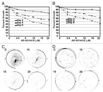
Neuroblastoma cells treated with AR-A014418 had a significant reduction in growth at all doses and time points (P<0.001). A reduction in growth was noted in cell lines on day 6, with 10 μM (NGP-53% vs. 0% and SH-5Y-SY-38% vs. 0%, P<0.001) treatment compared to control, corresponding with a noticeable reduction in tumor marker ASCL1 and CgA expression[2]. Conclusion: Treatment of neuroblastoma cell lines with AR-A014418 reduced the level of GSK-3α phosphorylation at Tyr279 compared to GSK-3β phosphorylation at Tyr216, and attenuated growth via the maintenance of apoptosis. This study supports further investigation to elucidate the mechanism(s) by which GSK-3α inhibition downregulates the expression of NE tumor markers and growth of neuroblastoma[2]. 1. Inhibition of GSK-3β Activity - Experimental Design: Recombinant GSK-3β was incubated with AR-A014418 at concentrations ranging from 0.1–10 μM. Phosphorylation of glycogen synthase (GS) was measured using radiolabeled ATP. - Results: - AR-A014418 potently inhibited GSK-3β activity with an IC₅₀ of ~0.4 μM [1] - The compound competed with ATP binding to the kinase active site [1] 2. Suppression of Neuroblastoma Cell Growth - Cell Line: SH-SY5Y neuroblastoma cells were treated with AR-A014418 (0.1–10 μM) for 72 hours. - Results: - IC₅₀: ~2 μM (MTT assay) [2] - Downregulation of neuroendocrine markers (e.g., chromogranin A, synaptophysin) via GSK-3β-mediated β-catenin stabilization [2] |
| ln Vivo |
AR-A014418 (0–4 mg/kg, i.p.) delays the onset of symptoms, enhances motor function, slows the progression of the disease, and delays the endpoint of the disease in ALS mice with the G93A mutant human SOD1. [3] Additionally, AR-A014418 inhibits mice's acetic acid and formalin-induced nociception by altering the signaling of the NMDA and metabotropic receptors as well as the transmission of TNF- and IL-1 in the spinal cord.[4]
In this study, researchers investigated the antinociceptive effects of AR-A014418, a selective inhibitor of glycogen synthase kinase-3β (GSK-3β) in mice. A 30-minute pretreatment with AR-A014418 (.1 and 1 mg/kg, intraperitoneal [ip]) inhibited nociception induced by an ip injection of acetic acid. AR-A014418 pretreatment (.1 and .3 mg/kg, ip) also decreased the late (inflammatory) phase of formalin-induced licking, without affecting responses of the first (neurogenic) phase. In a different set of experiments, AR-A014418 (.1-10 μg/site) coinjected intraplantarly (ipl) with formalin inhibited the late phase of formalin-induced nociception. Furthermore, AR-A014418 administration (1 and 10 ng/site, intrathecal [it]) inhibited both phases of formalin-induced licking. In addition, AR-A014418 coinjection (10 ng/site, it) inhibited nociception induced by glutamate, N-methyl-D-aspartate (NMDA), (±)-1-aminocyclopentane-trans-1,3-dicarboxylic acid (trans-ACPD), tumor necrosis factor-alpha (TNF-α), and interleukin-1beta (IL-1β) by 47 ± 12%, 48 ± 11%, 31 ± 8%, 46 ± 13%, and 44 ± 11%, respectively. In addition, a 30-minute pretreatment with NP031115 (3 and 10 mg/kg, ip), a different GSK-3 β inhibitor, also attenuated the late phase of formalin-induced nociception. Collectively, these results provide convincing evidence that AR-A014418, given by local, systemic, and central routes, produces antinociception in several mouse models of nociception. The AR-A014418-dependent antinociceptive effects were induced by modulation of the glutamatergic system through metabotropic and ionotropic (NMDA) receptors and the inhibition of the cytokine (TNF-α and IL-1β) signaling. 1. Amelioration of ALS Symptoms in G93A-SOD1 Mice - Animal Model: Transgenic mice expressing G93A-SOD1 (amyotrophic lateral sclerosis model) were treated with AR-A014418 (5 mg/kg/day, intraperitoneal injection) from symptom onset. - Results: - Delayed disease progression (motor function decline) and extended survival by 10–15% [3] - Reduced spinal cord motor neuron loss and glial activation [3] 2. Antinociceptive Effects in Mice - Pain Model: Formalin-induced paw licking assay in male C57BL/6 mice. - Treatment: AR-A014418 (1–10 mg/kg, intraperitoneal) was administered 30 minutes prior to formalin injection. - Results: - Significant reduction in pain-related behaviors during both acute (Phase I) and tonic (Phase II) phases [4] - The effect was reversed by co-administration of a GSK-3β activator [4] |
| Enzyme Assay |
Calcine/propidium iodide uptake is used to determine cell viability. Calcein AM is only taken up by dead cells and becomes orange-red fluorescent when it is cleaved by esterases found within living cells. In a nutshell, N2A cells are cultured in vitro for two days before being exposed to 50 μM LY-294002 in the presence of AR-A014418 or vehicle (DMSO) for 24 hours. Then, 2 μM PI and 1 μM calcein-AM are incubated with N2A cells for 30 min. The cultures are then rinsed three times with Hanks' buffered saline solution containing 2 mM CaCl2, and the cells are observed using fluorescence microscopy with a Zeiss Axiovert 135 microscope. In at least three different experiments, three fields (chosen at random) are analyzed per well (~300 cells/field). The percentage of PI-positive cells compared to all other cells is used to measure cell death. After deducting the quantity of dead cells present in vehicle-treated cultures, specific cell death is calculated in every experiment.
1. GSK-3β Kinase Activity Assay - Reagents: Recombinant GSK-3β, glycogen synthase peptide substrate, ATP (with γ-³²P), and AR-A014418. - Protocol: - GSK-3β (10 nM) was preincubated with AR-A014418 (0.1–10 μM) for 10 minutes at 30°C. - Reaction was initiated by adding ATP (100 μM) and substrate (100 μM), followed by incubation for 30 minutes. - Phosphorylation was quantified by scintillation counting after separating phosphorylated substrate [1] ### Cell Assay 1. Neuroblastoma Cell Viability and Apoptosis Assay - Cell Culture: SH-SY5Y cells were cultured in DMEM/F12 (1:1) with 10% FBS. - Treatment: Cells were exposed to AR-A014418 (0.1–10 μM) for 72 hours. - Assays: - MTT Assay: Cell viability was measured at 570 nm [2] - Annexin V/PI Staining: Apoptosis was analyzed by flow cytometry, showing increased Annexin V-positive cells at ≥5 μM [2] - Western Blot: Downregulation of phosphorylated β-catenin (Ser33/37/Thr41) and upregulation of cleaved caspase-3 [2] |
| Cell Assay |
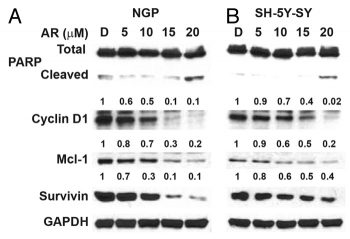
NGP and SH-5Y-SY cells were treated with 0-20 μM of AR-A014418 and cell viability was measured by MTT assay. Expression levels of NE markers CgA and ASCL1, GSK-3 isoforms, and apoptotic markers were analyzed by western blot.[2]
Cell viability is assessed by calcein/propidium iodide uptake. Calcein AM is taken up and cleaved by esterases present within living cells, yielding yellowish-green fluorescence, whereas PI is only taken up by dead cells, which become orange-red fluorescent. In brief, N2A cells are cultured for 2 days in vitro and then treated with 50 μM LY-294002 in the presence of AR-A014418 or vehicle (DMSO) for 24 h. Subsequently, N2A cells are incubated for 30 min with 2 μM PI and 1 μM calcein-AM. The cultures are then rinsed three times with Hanks buffered saline solution containing 2 mM CaCl2, and the cells are visualized by fluorescence microscopy using a Zeiss Axiovert 135 microscope. Three fields (selected at random) are analyzed per well (∼300 cells/field) in at least three different experiments. Cell death is expressed as percentage of PI-positive cells from the total number of cells. In every experiment, specific cell death is obtained after subtracting the number of dead cells present in vehicle-treated cultures. |
| Animal Protocol |
First, 56 Tg mice are split up into four groups to study the effects of GSK-3 inhibition on the clinical symptoms, lifespan, and motor behavior function of ALS. 14 animals per group are given intraperitoneal injections of 0.5 mL of normal saline five times a week starting 60 days after birth, along with 0 μg (control group), 1 μg (group A), 2 μg (group B), or 4 μg (group C) of AR-A014418 per gram of mouse. At the endpoint outlined below, mice are sacrificed.
1. ALS Mouse Model Treatment - Animal: G93A-SOD1 transgenic mice (male, 8–10 weeks old). - Dosing: AR-A014418 (5 mg/kg) dissolved in DMSO:PEG400:saline (1:4:5) was administered intraperitoneally daily until endpoint [3] - Assessment: Motor function was evaluated weekly using rotarod test and grip strength measurements [3] 2. Formalin-Induced Pain Assay - Animal: Male C57BL/6 mice (20–25 g). - Dosing: AR-A014418 (1–10 mg/kg) dissolved in 0.9% saline was injected intraperitoneally 30 minutes before formalin (5% in PBS, 20 μL) was injected subcutaneously into the hind paw [4] - Observation: Paw licking duration was recorded for 60 minutes post-formalin injection [4] |
| ADME/Pharmacokinetics |
Half-life: Approximately 2 hours in mice (intraperitoneal injection) [3]
- Bioavailability: Approximately 15% in rats after oral administration [1] - Tissue distribution: High brain permeability (brain/plasma ratio: 0.8) [1] |
| Toxicity/Toxicokinetics |
- Acute toxicity: - LD₅₀: >100 mg/kg (intraperitoneal injection in mice)[4]
- Subchronic toxicity: - No significant liver or kidney damage was observed in rats after administration at a dose of 10 mg/kg/day for 28 consecutive days[3] - Plasma protein binding rate: approximately 95% in human plasma[1] |
| References |
|
| Additional Infomation |
Glycogen synthase kinase 3 (GSK3) is a serine/threonine kinase closely associated with pathological conditions such as diabetes and Alzheimer's disease. We report the properties of a GSK3 inhibitor, AR-A014418, which inhibits GSK3 in an ATP-competitive manner (IC50 = 104 ± 27 nM, Ki = 38 nM). AR-A014418 showed no significant inhibition of cdk2 or cdk5 (IC50 > 100 μM) and 26 other kinases, indicating its high specificity for GSK3. We report the co-crystallization of AR-A014418 with GSK3β protein and describe the interactions within the ATP-binding pocket, as well as the structural basis for AR-A014418's selectivity. AR-A014418 inhibits tau protein phosphorylation at the GSK3-specific site (Ser-396) in cells stably expressing human tetraduplex tau protein. AR-A014418 can protect N2A neuroblastoma cells from cell death by inhibiting the phosphatidylinositol 3-kinase/protein kinase B survival pathway. In addition, AR-A014418 can also inhibit β-amyloid-mediated neurodegeneration in hippocampal slices. Therefore, AR-A014418 is expected to become an important tool for elucidating the role of GSK3 in cell signaling and Alzheimer's disease. AR-A014418 is the first specific GSK3 inhibitor family of compounds that does not significantly inhibit closely related kinases, such as cdk2 or cdk5. [1] Glycogen synthase kinase (GSK)-3 has recently been considered to be involved in the pathogenesis of neurodegenerative diseases. Although the neuroprotective effect of GSK-3 inhibitors in Alzheimer's disease has been confirmed, their effect on amyotrophic lateral sclerosis (ALS) is still unclear. This study aimed to evaluate the effect of GSK-3 inhibitors in the G93A-SOD1 ALS mouse model. G93A-SOD1 mice were grouped and, at 60 days of age, were intraperitoneally injected with different concentrations of GSK-3 inhibitor VIII (a specific GSK-3 inhibitor that can cross the blood-brain barrier) five days a week. Treatment with GSK-3 inhibitor VIII significantly delayed the onset of symptoms, prolonged the animals' lifespan, and inhibited GSK-3 activity in a concentration-dependent manner. Furthermore, this treatment preserved survival signals and attenuated death and inflammatory signals. These data suggest that GSK-3 plays an important role in the pathogenesis of ALS, and that inhibiting GSK-3 may be a potential therapeutic target for ALS. [2]
1. Mechanism of action: - AR-A014418 binds to the ATP-binding pocket of GSK-3β, preventing phosphorylation of downstream targets (e.g., β-catenin, tau protein) [1] 2. Therapeutic potential: - It has been investigated in the treatment of Alzheimer's disease, cancer, and neurodegenerative diseases [1-3] 3. Synthesis: - This compound is synthesized by the condensation reaction of 4-aminophenylethylamine with a ferulic acid derivative [1] |
| Molecular Formula |
C12H12N4O4S
|
|---|---|
| Molecular Weight |
308.3131
|
| Exact Mass |
308.057
|
| Elemental Analysis |
C, 46.75; H, 3.92; N, 18.17; O, 20.76; S, 10.40
|
| CAS # |
487021-52-3
|
| Related CAS # |
AR-A014418-d3;1216908-63-2
|
| PubChem CID |
448014
|
| Appearance |
Light yellow to yellow solid powder
|
| Density |
1.5±0.1 g/cm3
|
| Melting Point |
208-210?C (dec.)
|
| Index of Refraction |
1.666
|
| LogP |
1.43
|
| Hydrogen Bond Donor Count |
2
|
| Hydrogen Bond Acceptor Count |
6
|
| Rotatable Bond Count |
4
|
| Heavy Atom Count |
21
|
| Complexity |
371
|
| Defined Atom Stereocenter Count |
0
|
| SMILES |
S1C(=C([H])N=C1N([H])C(N([H])C([H])([H])C1C([H])=C([H])C(=C([H])C=1[H])OC([H])([H])[H])=O)[N+](=O)[O-]
|
| InChi Key |
YAEMHJKFIIIULI-UHFFFAOYSA-N
|
| InChi Code |
InChI=1S/C12H12N4O4S/c1-20-9-4-2-8(3-5-9)6-13-11(17)15-12-14-7-10(21-12)16(18)19/h2-5,7H,6H2,1H3,(H2,13,14,15,17)
|
| Chemical Name |
1-[(4-methoxyphenyl)methyl]-3-(5-nitro-1,3-thiazol-2-yl)ure
|
| Synonyms |
GSK-3β Inhibitor VIII; GSK-3beta Inhibitor VIII; SN 4521; SN-4521; SN4521; AR-A014418; AR-A 014418; AR-A-014418; AR-AO-14418; 487021-52-3; 1-(4-methoxybenzyl)-3-(5-nitrothiazol-2-yl)urea; N-(4-METHOXYBENZYL)-N'-(5-NITRO-1,3-THIAZOL-2-YL)UREA; GSK-3beta Inhibitor VIII; 1-[(4-methoxyphenyl)methyl]-3-(5-nitro-1,3-thiazol-2-yl)urea;
|
| HS Tariff Code |
2934.99.9001
|
| Storage |
Powder -20°C 3 years 4°C 2 years In solvent -80°C 6 months -20°C 1 month |
| Shipping Condition |
Room temperature (This product is stable at ambient temperature for a few days during ordinary shipping and time spent in Customs)
|
| Solubility (In Vitro) |
DMSO: ~62 mg/mL (~201.1 mM)
Water: <1 mg/mL (Insoluble) Ethanol: <1 mg/mL |
|---|---|
| Solubility (In Vivo) |
Solubility in Formulation 1: ≥ 2.5 mg/mL (8.11 mM) (saturation unknown) in 10% DMSO + 40% PEG300 +5% Tween-80 + 45% Saline (add these co-solvents sequentially from left to right, and one by one), clear solution.
For example, if 1 mL of working solution is to be prepared, you can add 100 μL of 25.0 mg/mL clear DMSO stock solution to 400 μL PEG300 and mix evenly; then add 50 μL Tween-80 + to the above solution and mix evenly; then add 450 μL normal saline to adjust the volume to 1 mL. Preparation of saline: Dissolve 0.9 g of sodium chloride in 100 mL ddH₂ O to obtain a clear solution. (Please use freshly prepared in vivo formulations for optimal results.) |
| Preparing Stock Solutions | 1 mg | 5 mg | 10 mg | |
| 1 mM | 3.2435 mL | 16.2174 mL | 32.4349 mL | |
| 5 mM | 0.6487 mL | 3.2435 mL | 6.4870 mL | |
| 10 mM | 0.3243 mL | 1.6217 mL | 3.2435 mL |
*Note: Please select an appropriate solvent for the preparation of stock solution based on your experiment needs. For most products, DMSO can be used for preparing stock solutions (e.g. 5 mM, 10 mM, or 20 mM concentration); some products with high aqueous solubility may be dissolved in water directly. Solubility information is available at the above Solubility Data section. Once the stock solution is prepared, aliquot it to routine usage volumes and store at -20°C or -80°C. Avoid repeated freeze and thaw cycles.
Calculation results
Working concentration: mg/mL;
Method for preparing DMSO stock solution: mg drug pre-dissolved in μL DMSO (stock solution concentration mg/mL). Please contact us first if the concentration exceeds the DMSO solubility of the batch of drug.
Method for preparing in vivo formulation::Take μL DMSO stock solution, next add μL PEG300, mix and clarify, next addμL Tween 80, mix and clarify, next add μL ddH2O,mix and clarify.
(1) Please be sure that the solution is clear before the addition of next solvent. Dissolution methods like vortex, ultrasound or warming and heat may be used to aid dissolving.
(2) Be sure to add the solvent(s) in order.
Growth inhibition by GSK-3 inhibitor AR-A014418 in neuroblastoma cells. Cancer Biol Ther. 2014 May;15(5):510-5. |
AR-A014418 attenuation of apoptosis inhibitor expression in NGP and SH-5Y-SY cells. |
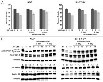
Continuous treatment of AR-A014418 is required for growth suppression of NGP and SH-5Y-SY cells. |
Products are for research use only; We do not sell to patients
Copyright 2020 InvivoChem LLC | All Rights Reserved


 COA
COA