| Size | Price | Stock | Qty |
|---|---|---|---|
| 5mg |
|
||
| 10mg |
|
||
| 25mg |
|
||
| 50mg |
|
||
| 100mg |
|
||
| 250mg |
|
||
| 500mg | |||
| Other Sizes |
Purity: ≥98%
| Targets |
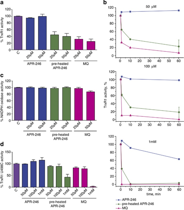
p53 activator; TrxR1 inhibitor
Eprenetapopt (APR246; PRIMA-1MET) targets mutant p53 (mutp53) proteins, reactivating their transcriptional activity with EC50 values ranging from 2.5–10 μM in mutp53-bearing cancer cell lines[2] Eprenetapopt (APR246; PRIMA-1MET) indirectly induces reactive oxygen species (ROS) production, with no direct binding to specific enzymatic targets[1] |
|---|---|
| ln Vitro |
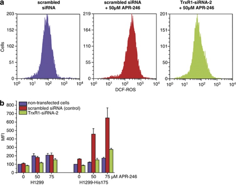
APR-246 (PRIMA-1MET) is the first clinical-stage compound that reactivates mutant p53 and induces apoptosis. APR-246 is a prodrug that is transformed into the active ingredient methylene quinuclidinone (MQ), a Michael acceptor that binds to cysteine residues in mutant p53 and returns it to its wild-type conformation. Drug-resistant ovarian cancer cells with p53 mutations are completely restored to cisplatin and doxorubicin sensitivity by APR-246. Along with reactivating p53, it also dose-dependently lowers intracellular glutathione levels. By increasing ROS and ER stress, inhibiting thioredoxin reductase 1 (TrxR1), and inducing ROS and ER stress, APR-246 can cause apoptosis in a p53-independent manner. Additionally, it was noted that APR-246 alters the GSH/ROS balance in myeloma cells, causing cell death regardless of p53 status[1]. In vitro, PRIMA-1Met/APR-246 effectively stopped the growth of SCLC cell lines expressing mutant p53. It also caused apoptosis, which is characterized by an increase in the proportion of DNA-fragmented cells, caspase-3 activation, PARP cleavage, upregulation of Bax and Noxa, and downregulation of Bcl-2 in the cells[2].
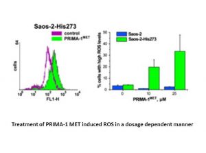
In mutp53-expressing cancer cell lines (SKOV3, OVCAR3, A2780/CP70): Eprenetapopt (APR246; PRIMA-1MET) (5–20 μM) dose-dependently inhibited cell proliferation by 40–70% after 72 hours of incubation, with EC50 values of 4.2 μM (SKOV3), 5.8 μM (OVCAR3), and 7.5 μM (A2780/CP70)[2] - The compound (10 μM) reactivated mutp53, upregulating downstream target genes: p21 (2.5 folds), Bax (2.0 folds), and PUMA (1.8 folds) at the mRNA and protein levels in SKOV3 cells[2] - In HCT116 p53⁻/⁻ cells transfected with mutp53 (R175H, R273H), Eprenetapopt (APR246; PRIMA-1MET) (15 μM) induced apoptosis in 60–70% of cells, as evidenced by annexin V/PI staining and cleavage of caspase-3, -7, and PARP[1] - Eprenetapopt (APR246; PRIMA-1MET) (5–20 μM) increased intracellular ROS levels by 1.5–3.0 folds in mutp53 cancer cells, and ROS scavengers (NAC) abolished its apoptotic effect[1] - In combination with cisplatin (1 μM), Eprenetapopt (APR246; PRIMA-1MET) (5 μM) synergistically inhibited OVCAR3 cell proliferation (inhibition rate increased from 35% to 65%) and enhanced cisplatin-induced apoptosis[2] |
| ln Vivo |
APR-246 demonstrated a favorable safety profile in a Phase I/II clinical dose-finding study on prostate cancer and hematological malignancies, and both clinical and p53-dependent biological responses were noted. APR-246 is well tolerated in studies on animals. When mice with the fast-growing A2780-CP20 tumor xenografts are given just one dose of APR-246, the tumor size is reduced by 21%[1].
In SKOV3 (mutp53) xenograft nude mice: Intraperitoneal administration of Eprenetapopt (APR246; PRIMA-1MET) at 100 mg/kg twice weekly for 4 weeks reduced tumor volume by 60% and tumor weight by 55% compared to vehicle control[2] - Immunohistochemical analysis of tumor tissues showed increased mutp53 nuclear localization, upregulated p21 and Bax expression, and increased TUNEL-positive apoptotic cells (by 45%)[2] - In HCT116 p53⁻/⁻ (R175H mutp53-transfected) xenograft mice: Oral administration of Eprenetapopt (APR246; PRIMA-1MET) at 150 mg/kg daily for 3 weeks prolonged median survival by 30% and reduced tumor growth by 50%[1] - No significant body weight loss or organ toxicity was observed in treated mice, with serum ALT, AST, and creatinine levels within normal ranges[1,2] |
| Enzyme Assay |
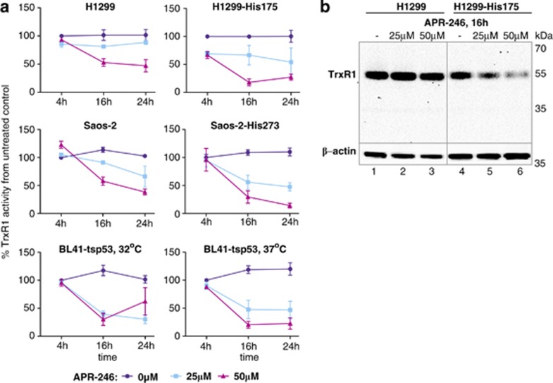
Cells are plated in six-well plates at a density of 15 000 cells per cm2. Next day, cells are treated with different concentrations of APR-246 (0, 25, 50, 75 and 100 μM) and harvested after 4, 12 and 24 h. The cells are lysed, and the clarified supernatants are used for either analysis of TrxR enzymatic activities or western blot. Total protein concentrations are determined with a Bradford reagent kit. Cellular TrxR activity is measured using an adapted Trx-dependent end point insulin reduction assay for microwell plates.
Mutp53 transcriptional activity assay: mutp53-expressing cancer cells (SKOV3) were seeded in 24-well plates and transfected with a p53-responsive luciferase reporter plasmid. After 24 hours, Eprenetapopt (APR246; PRIMA-1MET) (2.5–20 μM) was added, and cells were cultured for another 24 hours. Luciferase activity was measured using a luminometer, and relative transcriptional activity was calculated compared to vehicle control[2] - ROS detection assay: mutp53 cancer cells (HCT116 R175H) were loaded with DCFH-DA probe (10 μM) for 30 minutes, then treated with Eprenetapopt (APR246; PRIMA-1MET) (5–20 μM) for 24 hours. Fluorescence intensity (reflecting ROS levels) was measured by flow cytometry at 488 nm excitation and 525 nm emission[1] |
| Cell Assay |
In 12-well plates, 3 ml of medium and 75 000 OVCAR-3 cells were plated per well. The cells were treated for 20 hours with cisplatin, APR-246, or both the following day after 2.5 ml of the medium had been removed. The following day, cells were collected by trypsinization, twice-washed, and stained with Annexin V and propidium iodine (PI). The samples were stained, and then the LSRII flow cytometer was used to analyze them.
Cell proliferation assay: mutp53-bearing cancer cells (SKOV3, OVCAR3, A2780/CP70) were seeded in 96-well plates (5×10³ cells/well) and incubated overnight. Eprenetapopt (APR246; PRIMA-1MET) (1–40 μM) was added, and cells were cultured for 72 hours. Cell viability was measured by MTT assay, and EC50 values were calculated using GraphPad Prism[2] - Apoptosis assay: HCT116 p53⁻/⁻ cells transfected with mutp53 (R175H, R273H) were seeded in 6-well plates and treated with Eprenetapopt (APR246; PRIMA-1MET) (15 μM) for 24 hours. Cells were stained with annexin V-FITC and PI, then analyzed by flow cytometry. Cleaved caspase-3, -7, and PARP were detected by Western blot[1] - Western blot for p53 target genes: SKOV3 cells were treated with Eprenetapopt (APR246; PRIMA-1MET) (10 μM) for 24 hours. Cells were lysed, and proteins were separated by SDS-PAGE. Blots were probed with antibodies against mutp53, p21, Bax, PUMA, and β-actin. Signals were detected by chemiluminescence[2] - Combination therapy assay: OVCAR3 cells were treated with Eprenetapopt (APR246; PRIMA-1MET) (5 μM) alone, cisplatin (1 μM) alone, or their combination for 72 hours. Cell viability was measured by MTT assay, and synergism was assessed by combination index (CI < 1)[2] |
| Animal Protocol |
Dissolved in PBS; 400 mg/kg/day; i.v. injection
CD-1 Nu/Nu mice SKOV3 xenograft model: Female nude mice (6–8 weeks old) were subcutaneously inoculated with 5×10⁶ SKOV3 cells in Matrigel (1:1). When tumors reached 100–150 mm³, mice were randomly divided into vehicle and treatment groups (n=6/group). Eprenetapopt (APR246; PRIMA-1MET) was dissolved in 5% DMSO + 95% saline and administered intraperitoneally at 100 mg/kg twice weekly for 4 weeks. Tumor volume was measured every 3 days, and mice were euthanized for tumor weight and immunohistochemical analysis[2] - HCT116 R175H xenograft model: Male nude mice (6–8 weeks old) were subcutaneously implanted with 2×10⁶ HCT116 p53⁻/⁻ cells transfected with mutp53 R175H. After tumor establishment (100 mm³), mice received Eprenetapopt (APR246; PRIMA-1MET) (150 mg/kg/day) dissolved in 0.5% CMC via oral gavage for 3 weeks. Survival was monitored daily, and tumor volume was measured twice weekly[1] |
| Toxicity/Toxicokinetics |
In vitro experiments showed that Eprenetapopt (APR246; PRIMA-1MET) had low cytotoxicity to normal human fibroblasts (NHF), with CC50 > 40 μM and a therapeutic index (CC50/EC50) > 8 in SKOV3 cells [2]. In vivo experiments showed that repeated administration of Eprenetapopt (APR246; PRIMA-1MET) (100 mg/kg intraperitoneally twice weekly or 150 mg/kg orally daily) did not cause significant weight loss (<5% change compared to the control group) or significant pathological abnormalities in the liver, kidneys, spleen or heart [1,2]. Serum ALT, AST, creatinine and urea nitrogen levels in the treatment group mice were comparable to those in the solvent control group, indicating no hepatotoxicity or nephrotoxicity [1,2].
|
| References | |
| Additional Infomation |
Eprenetapopt has been used in trials for the treatment of prostate cancer, hematologic malignancies, and platinum-sensitive recurrent high-grade serous ovarian cancer (with p53 mutations). Eprenetapopt is a methylated derivative and structural analog of PRIMA-1 (which reactivates p53 and induces massive apoptosis) with potential antitumor activity. After administration, eprenetapopt covalently modifies the core domain of the cell tumor antigen p53 (p53) mutant via thiol alkylation. These modifications restore the mutant p53 to its wild-type conformation and function, thereby reconstructing endogenous p53 activity, leading to tumor cell cycle arrest and apoptosis. This drug may have synergistic effects with other antitumor drugs. p53 is a tumor suppressor and transcription factor that is normally activated after DNA damage, but it is frequently mutated and overexpressed in cancer cells; it plays a crucial role in DNA repair and apoptosis induction.
Eprenetapopt (APR246; PRIMA-1MET) is a small molecule compound that reactivates mutant p53 proteins by restoring their correct folding and transcriptional activity [1,2]. Its anticancer mechanism involves two key pathways: first, by upregulating p53 target genes (p21, Bax, PUMA) to reactivate mutant p53, thereby inducing cell cycle arrest and apoptosis; second, by inducing intracellular reactive oxygen species (ROS) accumulation, thereby triggering apoptosis signaling pathways [1]. This compound exhibits synergistic anticancer effects with chemotherapeutic drugs (such as cisplatin) in cancer cells expressing mutant p53, enhancing therapeutic efficacy [2]. Currently, it is being developed for the treatment of solid tumors carrying p53 mutations, including ovarian cancer, colorectal cancer, and non-small cell lung cancer [1,2]. |
| Molecular Formula |
C10H17NO3
|
|
|---|---|---|
| Molecular Weight |
199.25
|
|
| Exact Mass |
199.12
|
|
| Elemental Analysis |
C, 60.28; H, 8.60; N, 7.03; O, 24.09
|
|
| CAS # |
5291-32-7
|
|
| Related CAS # |
PRIMA-1;5608-24-2
|
|
| PubChem CID |
52918385
|
|
| Appearance |
White to off-white solid powder
|
|
| Density |
1.2±0.1 g/cm3
|
|
| Boiling Point |
313.8±17.0 °C at 760 mmHg
|
|
| Flash Point |
143.6±20.9 °C
|
|
| Vapour Pressure |
0.0±1.5 mmHg at 25°C
|
|
| Index of Refraction |
1.537
|
|
| LogP |
0.61
|
|
| Hydrogen Bond Donor Count |
1
|
|
| Hydrogen Bond Acceptor Count |
4
|
|
| Rotatable Bond Count |
3
|
|
| Heavy Atom Count |
14
|
|
| Complexity |
236
|
|
| Defined Atom Stereocenter Count |
0
|
|
| SMILES |
O=C1C2([H])C([H])([H])C([H])([H])N(C([H])([H])C2([H])[H])C1(C([H])([H])O[H])C([H])([H])OC([H])([H])[H]
|
|
| InChi Key |
BGBNULCRKBVAKL-UHFFFAOYSA-N
|
|
| InChi Code |
InChI=1S/C10H17NO3/c1-14-7-10(6-12)9(13)8-2-4-11(10)5-3-8/h8,12H,2-7H2,1H3
|
|
| Chemical Name |
2-(hydroxymethyl)-2-(methoxymethyl)-1-azabicyclo[2.2.2]octan-3-one
|
|
| Synonyms |
|
|
| HS Tariff Code |
2934.99.9001
|
|
| Storage |
Powder -20°C 3 years 4°C 2 years In solvent -80°C 6 months -20°C 1 month |
|
| Shipping Condition |
Room temperature (This product is stable at ambient temperature for a few days during ordinary shipping and time spent in Customs)
|
| Solubility (In Vitro) |
DMSO: 10mg/mL
Water: 1 mg/mL (Insoluble) Ethanol: 1 mg/mL |
|---|---|
| Solubility (In Vivo) |
Solubility in Formulation 1: ≥ 2.5 mg/mL (12.55 mM) (saturation unknown) in 10% DMSO + 40% PEG300 + 5% Tween80 + 45% Saline (add these co-solvents sequentially from left to right, and one by one), clear solution.
For example, if 1 mL of working solution is to be prepared, you can add 100 μL of 25.0 mg/mL clear DMSO stock solution to 400 μL PEG300 and mix evenly; then add 50 μL Tween-80 to the above solution and mix evenly; then add 450 μL normal saline to adjust the volume to 1 mL. Preparation of saline: Dissolve 0.9 g of sodium chloride in 100 mL ddH₂ O to obtain a clear solution. Solubility in Formulation 2: ≥ 2.5 mg/mL (12.55 mM) (saturation unknown) in 10% DMSO + 90% (20% SBE-β-CD in Saline) (add these co-solvents sequentially from left to right, and one by one), clear solution. For example, if 1 mL of working solution is to be prepared, you can add 100 μL of 25.0 mg/mL clear DMSO stock solution to 900 μL of 20% SBE-β-CD physiological saline solution and mix evenly. Preparation of 20% SBE-β-CD in Saline (4°C,1 week): Dissolve 2 g SBE-β-CD in 10 mL saline to obtain a clear solution. View More
Solubility in Formulation 3: ≥ 2.5 mg/mL (12.55 mM) (saturation unknown) in 10% DMSO + 90% Corn Oil (add these co-solvents sequentially from left to right, and one by one), clear solution. Solubility in Formulation 4: 100 mg/mL (501.88 mM) in PBS (add these co-solvents sequentially from left to right, and one by one), clear solution; with ultrasonication. |
| Preparing Stock Solutions | 1 mg | 5 mg | 10 mg | |
| 1 mM | 5.0188 mL | 25.0941 mL | 50.1882 mL | |
| 5 mM | 1.0038 mL | 5.0188 mL | 10.0376 mL | |
| 10 mM | 0.5019 mL | 2.5094 mL | 5.0188 mL |
*Note: Please select an appropriate solvent for the preparation of stock solution based on your experiment needs. For most products, DMSO can be used for preparing stock solutions (e.g. 5 mM, 10 mM, or 20 mM concentration); some products with high aqueous solubility may be dissolved in water directly. Solubility information is available at the above Solubility Data section. Once the stock solution is prepared, aliquot it to routine usage volumes and store at -20°C or -80°C. Avoid repeated freeze and thaw cycles.
Calculation results
Working concentration: mg/mL;
Method for preparing DMSO stock solution: mg drug pre-dissolved in μL DMSO (stock solution concentration mg/mL). Please contact us first if the concentration exceeds the DMSO solubility of the batch of drug.
Method for preparing in vivo formulation::Take μL DMSO stock solution, next add μL PEG300, mix and clarify, next addμL Tween 80, mix and clarify, next add μL ddH2O,mix and clarify.
(1) Please be sure that the solution is clear before the addition of next solvent. Dissolution methods like vortex, ultrasound or warming and heat may be used to aid dissolving.
(2) Be sure to add the solvent(s) in order.
| NCT Number | Recruitment | interventions | Conditions | Sponsor/Collaborators | Start Date | Phases |
| NCT04990778 | Withdrawn | Drug: Eprenetapopt Drug: Venetoclax |
Recurrent Mantle Cell Lymphoma Refractory Mantle Cell Lymphoma |
M.D. Anderson Cancer Center | November 30, 2021 | Phase 2 |
Inhibition of TrxR1in vitroby APR-246.Cell Death Dis.2013 Oct 24;4:e881. |
|---|
siRNA knockdown of TrxR1 inhibits APR-246-induced cell death.Cell Death Dis.2013 Oct 24;4:e881. |
siRNA knockdown of TrxR1 inhibits generation of ROS induced by treatment with APR-246.Cell Death Dis.2013 Oct 24;4:e881. |
Inhibition of TrxR1 activity in living cells.Cell Death Dis.2013 Oct 24;4:e881. |
|---|
|
|
Products are for research use only; We do not sell to patients
Copyright 2020 InvivoChem LLC | All Rights Reserved


 COA
COA