yingweiwo

AP20187

Alias: AP20187 AP-20187 AP 20187.
Cat No.:V11419 Purity: ≥98%
AP20187 (B/B Homodimerizer) is a novel cell-permeable andchemical inducer of dimerization, used to dimerize FK506-binding protein (FKBP) fusion proteins and initiate biological signaling cascades and gene expression or disrupt protein-protein interactions.
AP20187
AP20187 Chemical Structure CAS No.: 195514-80-8
Product category: New1
This product is for research use only, not for human use. We do not sell to patients.
Size Price Stock Qty
2mg
5mg
10mg
25mg
50mg
Other Sizes
Official Supplier of:
Alternate Text
Alternate Text
Alternate Text
Alternate Text
Alternate Text
Alternate Text
Alternate Text
Alternate Text
Alternate Text
Alternate Text
Alternate Text
Alternate Text
Alternate Text
Alternate Text
Alternate Text
Alternate Text
Alternate Text
Alternate Text
Alternate Text
Alternate Text
Alternate Text
Alternate Text
Alternate Text
Alternate Text
Alternate Text
Alternate Text
Alternate Text
Alternate Text
Alternate Text
Alternate Text
Alternate Text
Alternate Text


  • Business Relationship with 5000+ Clients Globally
  • Major Universities, Research Institutions, Biotech & Pharma
  • Citations by Top Journals: Nature, Cell, Science, etc.
Top Publications Citing lnvivochem Products
Purity & Quality Control Documentation

Purity: ≥98%

Product Description
AP20187 (B/B Homodimerizer) is a novel cell-permeable and chemical inducer of dimerization, used to dimerize FK506-binding protein (FKBP) fusion proteins and initiate biological signaling cascades and gene expression or disrupt protein-protein interactions. AP20187-mediated activation of a chimeric insulin receptor results in insulin-like actions in skeletal muscle and liver of diabetic mice.
AP20187 (CAS 195514-80-8) is a synthetic, cell-permeable chemical inducer of dimerization (CID) specifically designed to dimerize FK506-binding protein (FKBP) fusion proteins. It is a synthetic analog of FK506 (tacrolimus) that acts as a molecular glue to bring two FKBP domains together, thereby initiating biological signaling cascades, activating gene expression, or disrupting protein-protein interactions. This compound is a powerful research tool for precisely controlling biological processes and studying complex cellular mechanisms.
Biological Activity I Assay Protocols (From Reference)
Targets
AP20187 targets the FK506-binding protein (FKBP) family, specifically engineered fusion proteins containing the FKBPF36V domain. It functions as a homodimerizer, binding to two FKBP domains simultaneously and bringing them into close proximity. This induced proximity can activate chimeric receptors or signaling molecules, such as by dimerizing the intracellular domain of the Fas receptor to induce apoptosis, or by activating a chimeric insulin receptor to trigger insulin-like actions.
ln Vitro
AP20187 (B/B Homodimerizer) (100 nM) treatment of LNCaP cells resulted in a considerable decrease in ro-iCaspase-9 levels and a significant weakening of caspase-9 processing activity [2].
In vitro, AP20187 (100 nM) has been shown to significantly reduce pro-caspase-9 levels and generate the smaller, processed active caspase-9 in LNCaP cells, indicating the induction of apoptosis. It potently inhibits the differentiation of transfected myoblasts in the absence of bFGF, blocks myotube formation and myosin heavy chain expression, and stimulates MAP kinase phosphorylation. AP20187 is also used to induce apoptosis in cells expressing FKBPF36V fusion proteins with the Fas receptor or FADD.
ln Vivo
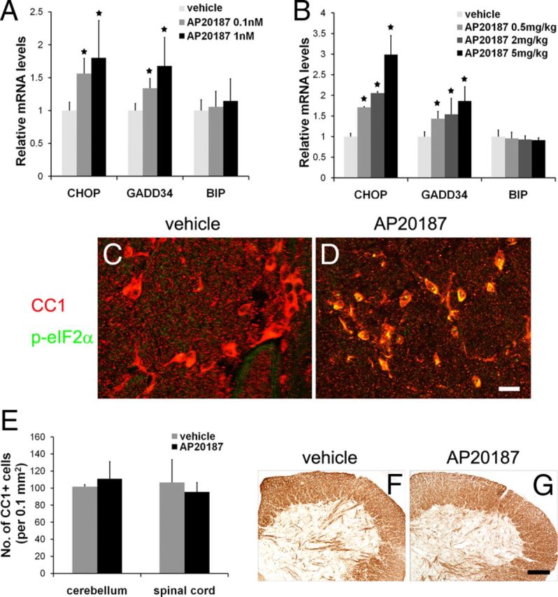
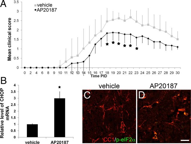
Real-time PCR analysis showed that AP20187 (B/B homodimer) (0.5 mg/kg, 2 mg/kg or 5 mg/kg) treatment significantly enhanced PLP/Fv2E-PERK in PID12 CHOP mRNA levels in the CNS of Fv2E. AP20187 therapy significantly decreased EAE-induced myelin damage during these procedures. AP20187 (B/B Homodimerizer) therapy dramatically reduced the number of degenerated axons and enhanced axonal density in PLP/Fv2E-PERK anatomical lumbar demyelinating lesions [2].
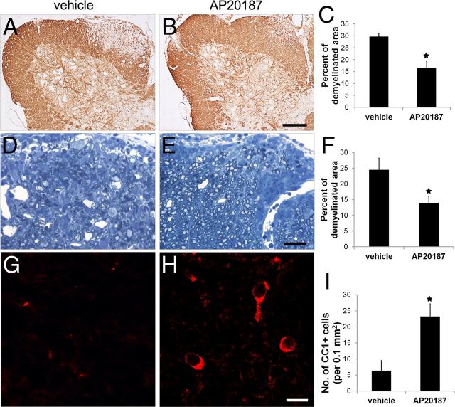
In vivo, AP20187 treatment has demonstrated significant biological effects. In PLP/Fv2E-PERK mice, administration at doses of 0.5, 2, or 5 mg/kg significantly increased CHOP mRNA levels in the central nervous system and alleviated EAE-induced myelin damage. It also significantly reduced the number of degenerating axons and increased axonal density in demyelinating lesions. In diabetic mice, AP20187-mediated activation of a chimeric insulin receptor resulted in insulin-like actions, increasing hepatic glycogen content and muscular glucose uptake.
Enzyme Assay
The in vitro binding assay for AP20187 is typically performed using cell-free systems with purified FKBP domains. The interaction between AP20187 and FKBP can be studied using techniques such as surface plasmon resonance (SPR) or isothermal titration calorimetry (ITC) to determine binding affinity and stoichiometry. Competition binding assays with fluorescently labeled FKBP can also be used to assess the compound's potency. These assays characterize the fundamental molecular interaction that underlies its dimerization activity.
Cell Assay
In vitro cellular assays for AP20187 involve treating cells engineered to express FKBP-fusion proteins with the compound. Common readouts include the activation of specific signaling pathways (e.g., MAP kinase phosphorylation), changes in gene expression (e.g., CHOP mRNA), or the induction of apoptosis (e.g., caspase-9 activation). These assays are used to study signal transduction, protein function, and to validate the specificity of the dimerization system.
Animal Protocol
In vivo animal studies for AP20187 are conducted in transgenic mouse models expressing FKBP-fusion proteins. AP20187 is typically administered systemically via injection. Endpoints include the analysis of target gene expression in tissues, assessment of disease pathology (e.g., myelin damage in EAE models), and evaluation of physiological effects like glucose uptake. These studies demonstrate the compound's utility for in vivo modulation of protein function.
ADME/Pharmacokinetics
AP20187 is a cell-permeable small molecule that is typically administered via injection in vivo. As a synthetic compound, it is designed for research use and exhibits suitable pharmacokinetic properties for systemic administration. Its cell permeability allows it to reach intracellular FKBP fusion proteins. However, detailed pharmacokinetic parameters such as half-life, volume of distribution, and bioavailability are not extensively detailed in the available literature.
Toxicity/Toxicokinetics
Specific toxicity data for AP20187 are not extensively detailed in the available literature. As a research tool, its toxicity profile is evaluated in the context of specific experimental models. The compound is generally well-tolerated at the doses used for inducing dimerization. However, comprehensive toxicological studies are not typically performed for research-use compounds. It is intended for research purposes only and is not for human use.
References

[1]. Photocleavable dimerizer for the rapid reversal of molecular trap antagonists. J Biol Chem. 2014 Feb 21;289(8):4546-52.

[2]. Oligodendrocyte-specific activation of PERK signaling protects mice against experimental autoimmune encephalomyelitis. J Neurosci. 2013 Apr 3;33(14):5980-91.

[3]. The Role of Proprotein Convertase Subtilisin/Kexin Type 9 in Nephrotic Syndrome-Associated Hypercholesterolemia. Circulation. 2016 Jul 5;134(1):61-72.

Additional Infomation
AP20187 is a tertiary amine compound with the structure 2-(aminomethyl)-N,N-dimethylpropane-1,3-diamine, wherein the primary amino groups are acylated by condensation with the carboxyl group of 2-{3-[(1R)-3-(3,4-dimethoxyphenyl)-1-hydroxypropyl]phenoxy}acetic acid, the hydroxyl group of which is esterified by condensation with the carboxyl group of L-piperidine acid, and the nitrogen atom of L-piperidine acid is acylated by condensation with (2S)-2-(3,4,5-trimethoxyphenyl)butyric acid. It is a synthetic, cell-membrane-permeable ligand used to induce homodimerization of fusion proteins containing the DmrB domain. It is an N-acylpiperidine, carboxylic acid ester, aromatic ether, and tertiary amine compound.
AP20187 is a widely used chemical inducer of dimerization (CID) in biomedical research, particularly for studies involving the FKBP system. It is a powerful tool for the conditional regulation of protein activity, allowing researchers to control signaling pathways, gene expression, and apoptosis with temporal precision. Its applications span diverse fields, including signal transduction, cancer research, and neuroscience. It is not approved for clinical use and is strictly a research reagent.
These protocols are for reference only. InvivoChem does not independently validate these methods.
Physicochemical Properties
Molecular Formula
C82H107N5O20
Molecular Weight
1482.7485
Exact Mass
1481.75
CAS #
195514-80-8
PubChem CID
78357784
Appearance
White to yellow solid powder
LogP
11.882
Hydrogen Bond Donor Count
2
Hydrogen Bond Acceptor Count
21
Rotatable Bond Count
42
Heavy Atom Count
107
Complexity
2450
Defined Atom Stereocenter Count
6
SMILES
CC[C@@H](C1=CC(=C(C(=C1)OC)OC)OC)C(=O)N2CCCC[C@H]2C(=O)O[C@H](CCC3=CC(=C(C=C3)OC)OC)C4=CC(=CC=C4)OCC(=O)NCC(CNC(=O)COC5=CC=CC(=C5)[C@@H](CCC6=CC(=C(C=C6)OC)OC)OC(=O)[C@@H]7CCCCN7C(=O)[C@@H](CC)C8=CC(=C(C(=C8)OC)OC)OC)CN(C)C
InChi Key
NSBGUMKAXUXKGI-BPNHAYRBSA-N
InChi Code
InChI=1S/C82H107N5O20/c1-15-61(57-43-71(98-9)77(102-13)72(44-57)99-10)79(90)86-37-19-17-27-63(86)81(92)106-65(33-29-52-31-35-67(94-5)69(39-52)96-7)55-23-21-25-59(41-55)104-50-75(88)83-47-54(49-85(3)4)48-84-76(89)51-105-60-26-22-24-56(42-60)66(34-30-53-32-36-68(95-6)70(40-53)97-8)107-82(93)64-28-18-20-38-87(64)80(91)62(16-2)58-45-73(100-11)78(103-14)74(46-58)101-12/h21-26,31-32,35-36,39-46,54,61-66H,15-20,27-30,33-34,37-38,47-51H2,1-14H3,(H,83,88)(H,84,89)/t61-,62-,63-,64-,65+,66+/m0/s1
Chemical Name
(1R,1'R)-(((((2-((dimethylamino)methyl)propane-1,3-diyl)bis(azanediyl))bis(2-oxoethane-2,1-diyl))bis(oxy))bis(3,1-phenylene))bis(3-(3,4-dimethoxyphenyl)propane-1,1-diyl) (2S,2'S)-bis(1-((S)-2-(3,4,5-trimethoxyphenyl)butanoyl)piperidine-2-carboxylate)
Synonyms
AP20187 AP-20187 AP 20187.
HS Tariff Code
2934.99.9001
Storage

Powder      -20°C    3 years

                     4°C     2 years

In solvent   -80°C    6 months

                  -20°C    1 month

Shipping Condition
Room temperature (This product is stable at ambient temperature for a few days during ordinary shipping and time spent in Customs)
Solubility Data
Solubility (In Vitro)
Ethanol : ~150 mg/mL (~101.16 mM)
DMSO : ≥ 57 mg/mL (~38.44 mM)
Solubility (In Vivo)
Solubility in Formulation 1: 6 mg/mL (4.05 mM) in 10% EtOH + 40% PEG300 + 5% Tween80 + 45% Saline (add these co-solvents sequentially from left to right, and one by one), clear solution; with sonication.
For example, if 1 mL of working solution is to be prepared, you can add 100 μL of 60.0 mg/mL clear EtOH stock solution to 400 μL PEG300 and mix evenly; then add 50 μL Tween-80 to the above solution and mix evenly; then add 450 μL normal saline to adjust the volume to 1 mL.
Preparation of saline: Dissolve 0.9 g of sodium chloride in 100 mL ddH₂ O to obtain a clear solution.

Solubility in Formulation 2: 6 mg/mL (4.05 mM) in 10% EtOH + 90% (20% SBE-β-CD in Saline) (add these co-solvents sequentially from left to right, and one by one), suspension solution; with ultrasonication.
For example, if 1 mL of working solution is to be prepared, you can add 100 μL of 60.0 mg/mL clear EtOH stock solution to 900 μL of 20% SBE-β-CD physiological saline solution and mix evenly.
Preparation of 20% SBE-β-CD in Saline (4°C,1 week): Dissolve 2 g SBE-β-CD in 10 mL saline to obtain a clear solution.

View More

Solubility in Formulation 3: ≥ 2.5 mg/mL (1.69 mM) (saturation unknown) in 10% DMSO + 40% PEG300 + 5% Tween80 + 45% Saline (add these co-solvents sequentially from left to right, and one by one), clear solution.
For example, if 1 mL of working solution is to be prepared, you can add 100 μL of 25.0 mg/mL clear DMSO stock solution to 400 μL PEG300 and mix evenly; then add 50 μL Tween-80 to the above solution and mix evenly; then add 450 μL normal saline to adjust the volume to 1 mL.
Preparation of saline: Dissolve 0.9 g of sodium chloride in 100 mL ddH₂ O to obtain a clear solution.


Solubility in Formulation 4: ≥ 2.5 mg/mL (1.69 mM) (saturation unknown) in 10% DMSO + 90% Corn Oil (add these co-solvents sequentially from left to right, and one by one), clear solution.
For example, if 1 mL of working solution is to be prepared, you can add 100 μL of 25.0 mg/mL clear DMSO stock solution to 900 μL corn oil and mix evenly.

Solubility in Formulation 5: 2.4 mg/mL (1.62 mM) in 4% ethanol 10% PEG-400 2% Tween80 + 84%water. (add these co-solvents sequentially from left to right, and one by one), clear solution; with ultrasonication.

 (Please use freshly prepared in vivo formulations for optimal results.)
Preparing Stock Solutions 1 mg 5 mg 10 mg
1 mM 0.6744 mL 3.3721 mL 6.7442 mL
5 mM 0.1349 mL 0.6744 mL 1.3488 mL
10 mM 0.0674 mL 0.3372 mL 0.6744 mL

*Note: Please select an appropriate solvent for the preparation of stock solution based on your experiment needs. For most products, DMSO can be used for preparing stock solutions (e.g. 5 mM, 10 mM, or 20 mM concentration); some products with high aqueous solubility may be dissolved in water directly. Solubility information is available at the above Solubility Data section. Once the stock solution is prepared, aliquot it to routine usage volumes and store at -20°C or -80°C. Avoid repeated freeze and thaw cycles.

Calculator

Molarity Calculator allows you to calculate the mass, volume, and/or concentration required for a solution, as detailed below:

  • Calculate the Mass of a compound required to prepare a solution of known volume and concentration
  • Calculate the Volume of solution required to dissolve a compound of known mass to a desired concentration
  • Calculate the Concentration of a solution resulting from a known mass of compound in a specific volume
An example of molarity calculation using the molarity calculator is shown below:
What is the mass of compound required to make a 10 mM stock solution in 5 ml of DMSO given that the molecular weight of the compound is 350.26 g/mol?
  • Enter 350.26 in the Molecular Weight (MW) box
  • Enter 10 in the Concentration box and choose the correct unit (mM)
  • Enter 5 in the Volume box and choose the correct unit (mL)
  • Click the “Calculate” button
  • The answer of 17.513 mg appears in the Mass box. In a similar way, you may calculate the volume and concentration.

Dilution Calculator allows you to calculate how to dilute a stock solution of known concentrations. For example, you may Enter C1, C2 & V2 to calculate V1, as detailed below:

What volume of a given 10 mM stock solution is required to make 25 ml of a 25 μM solution?
Using the equation C1V1 = C2V2, where C1=10 mM, C2=25 μM, V2=25 ml and V1 is the unknown:
  • Enter 10 into the Concentration (Start) box and choose the correct unit (mM)
  • Enter 25 into the Concentration (End) box and select the correct unit (mM)
  • Enter 25 into the Volume (End) box and choose the correct unit (mL)
  • Click the “Calculate” button
  • The answer of 62.5 μL (0.1 ml) appears in the Volume (Start) box
g/mol

Molecular Weight Calculator allows you to calculate the molar mass and elemental composition of a compound, as detailed below:

Note: Chemical formula is case sensitive: C12H18N3O4  c12h18n3o4
Instructions to calculate molar mass (molecular weight) of a chemical compound:
  • To calculate molar mass of a chemical compound, please enter the chemical/molecular formula and click the “Calculate’ button.
Definitions of molecular mass, molecular weight, molar mass and molar weight:
  • Molecular mass (or molecular weight) is the mass of one molecule of a substance and is expressed in the unified atomic mass units (u). (1 u is equal to 1/12 the mass of one atom of carbon-12)
  • Molar mass (molar weight) is the mass of one mole of a substance and is expressed in g/mol.
/

Reconstitution Calculator allows you to calculate the volume of solvent required to reconstitute your vial.

  • Enter the mass of the reagent and the desired reconstitution concentration as well as the correct units
  • Click the “Calculate” button
  • The answer appears in the Volume (to add to vial) box
In vivo Formulation Calculator (Clear solution)
Step 1: Enter information below (Recommended: An additional animal to make allowance for loss during the experiment)
Step 2: Enter in vivo formulation (This is only a calculator, not the exact formulation for a specific product. Please contact us first if there is no in vivo formulation in the solubility section.)
+
+
+

Calculation results

Working concentration mg/mL;

Method for preparing DMSO stock solution mg drug pre-dissolved in μL DMSO (stock solution concentration mg/mL). Please contact us first if the concentration exceeds the DMSO solubility of the batch of drug.

Method for preparing in vivo formulation:Take μL DMSO stock solution, next add μL PEG300, mix and clarify, next addμL Tween 80, mix and clarify, next add μL ddH2O,mix and clarify.

(1) Please be sure that the solution is clear before the addition of next solvent. Dissolution methods like vortex, ultrasound or warming and heat may be used to aid dissolving.
             (2) Be sure to add the solvent(s) in order.

Biological Data
  • Persistent activation of PERK signaling specifically in oligodendrocytes was not detrimental to these cells or to myelin in adult animals. A, Real-time PCR analysis showed that AP20187-induced PERK signaling increased the expression of CHOP and GADD34 in the mixed glial cultures generated from neonatal PLP/Fv2E-PERK mice in a dose-dependent manner but did not significantly affect the expression of BIP. B, Real-time PCR analysis showed that AP20187 treatment increased the expression of CHOP and GADD34 in the spinal cords of PLP/Fv2E-PERK mice in a dose-dependent manner but did not significantly affect the expression of BIP. C, D, CC1 and p-eIF2α double immunostaining showed that p-eIF2α was undetectable in oligodendrocytes in the spinal cords of PLP/Fv2E-PERK mice treated with vehicle but became detectable in the oligodendrocytes of mice treated with AP20187. E, Cell counting revealed that the numbers of CC1-positive oligodendrocytes in the white matter of the spinal cords and the cerebellums of PLP/Fv2E-PERK mice treated with AP20187 were comparable with those of mice treated with vehicle. F, G, MBP immunostaining showed that AP20187 treatment did not alter myelin integrity in the spinal cords of adult PLP/Fv2E-PERK mice; N = 3 animals. Error bars indicate SD. ★p < 0.05. Scale bar: C, D, 12 μm; F, G, 200 μm.[2]. Oligodendrocyte-specific activation of PERK signaling protects mice against experimental autoimmune encephalomyelitis. J Neurosci. 2013 Apr 3;33(14):5980-91.
  • Enhancing the PERK-mediated ISR specifically in oligodendrocytes attenuated EAE disease severity. A, Mean clinical score were recorded daily: 0 = healthy; 1 = flaccid tail; 2 = ataxia and/or paresis of hindlimbs; 3 = paralysis of hindlimbs and/or paresis of forelimbs; 4 = tetraparalysis; 5 = moribund or death. N = 12 animals. Error bars indicate SD. ★p < 0.05. B, Real-time PCR analysis showed that AP20187 treatment increased the expression of CHOP in the spinal cords of PLP/Fv2E-PERK mice at PID12; N = 4 animals. Error bars indicate SD. ★p < 0.05. C, D, CC1 and p-eIF2α double immunostaining showed that the level of p-eIF2α was markedly increased in oligodendrocytes in the lumbar spinal cords of PLP/Fv2E-PERK mice treated with AP20187 at PID12; N = 3 animals. Scale bar: C, D, 23 μm.[2]. Oligodendrocyte-specific activation of PERK signaling protects mice against experimental autoimmune encephalomyelitis. J Neurosci. 2013 Apr 3;33(14):5980-91.
  • Enhancing the PERK-mediated ISR specifically in oligodendrocytes ameliorated EAE-induced demyelination and oligodendrocyte loss. A–C, At PID19, MBP immunostaining revealed large demyelinating lesions in the lumbar spinal cords of PLP/Fv2E-PERK mice treated with vehicle. Interestingly, AP20187 treatment significantly reduced the size of demyelinating lesions in PLP/Fv2E-PERK mice; 20 sections per mouse, N = 4 animals. D–F, Toluidine blue staining showed that AP20187 treatment significantly reduced the percentage of the white matter area of the lumbar spinal cord that was demyelinated in PLP/Fv2E-PERK mice at PID19; 5 serial sections per mouse, N = 4 animals. G–I, CC1 immunostaining showed that AP20187 treatment significantly attenuated the oligodendrocyte loss observed at PID19 in the demyelinating lesions in the lumbar spinal cord of PLP/Fv2E-PERK mice; 20 sections per mouse, N = 4 animals. Error bars indicate SD. ★p < 0.05. Scale bar: A, B, 200 μm; D, E, 30 μm; G, H, 15 μm.[2]. Oligodendrocyte-specific activation of PERK signaling protects mice against experimental autoimmune encephalomyelitis. J Neurosci. 2013 Apr 3;33(14):5980-91.
Contact Us