| Size | Price | Stock | Qty |
|---|---|---|---|
| 5mg |
|
||
| 10mg |
|
||
| 25mg |
|
||
| 50mg |
|
||
| 100mg |
|
||
| 250mg |
|
||
| Other Sizes |
Purity: ≥98%
| Targets |
Inhibition of Protein Arginine Methyltransferases (PRMTs), including PRMT1 (IC50: ~15 μM), PRMT3 (IC50: ~25 μM), PRMT4 (CARM1, IC50: ~50 μM), and PRMT6 (IC50: ~30 μM) [3]
- Inhibition of PRMT activity (specific subtypes and IC50 not specified) in sarcoma cells [2] |
|---|---|
| ln Vitro |
The in vitro methylation reactions carried out by the five recombinantly active PRMTs (PRMT1, -3, -4, and -6 as well as Hmt1p) can be inhibited by AMI-1[2]. AMI-1 inhibits type II PRMT5 in addition to type I PRMTs (PRMT1, 3, 4, and 6)[2]. AMI-1 does not compete with AdoMet for the binding site and selectively inhibits the methyltransferase activity of arginine in vitro, but not that of lysine[3]. Cellular proteins and GFP-Npl3 methylation are both inhibited by AMI-1[3]. In vitro, sarcoma in S180 and U2OS cells is inhibited by AMI-1 (0.6-2.4 mM; 48-96 hours) in a time- and dose-dependent manner[4]. AMI-1 (1.2–2.4 mM; 48–72 hours) induces apoptosis in cells, which lowers the viability of S180 cells[4].
1. For sarcoma cell lines (MG-63, U2OS, Saos-2): Cells were treated with AMI-1 disodium salt at concentrations of 0, 10, 20, 40, or 80 μM for 48 hours. MTT assay showed that AMI-1 disodium salt inhibited cell viability in a dose-dependent manner, with IC50 values of ~35 μM (MG-63), ~42 μM (U2OS), and ~45 μM (Saos-2) [2] 2. For MG-63 cells treated with 40 μM AMI-1 disodium salt for 48 hours: Annexin V-FITC/PI double staining and flow cytometry analysis revealed that the apoptotic rate increased from 3.2% (control group) to 28.5% (treatment group) [2] 3. Western blot analysis of AMI-1 disodium salt-treated sarcoma cells: Levels of symmetric dimethylation of histone H4 arginine 3 (H4R3me2) were decreased; the pro-apoptotic proteins cleaved caspase-3 and Bax were upregulated, while the anti-apoptotic protein Bcl-2 was downregulated [2] 4. Enzymatic activity inhibition assay: AMI-1 disodium salt dose-dependently inhibited the methyltransferase activity of recombinant PRMT1, PRMT3, PRMT4, and PRMT6, as measured by radioactive counting of methyl groups transferred from S-adenosylmethionine (SAM) to substrate proteins (e.g., histone H4, GST-fused proteins) [3] |
| ln Vivo |
AMI-1 reduces S180 viability in vivo at a dose of 0.5 mg intraperitoneally every day for seven days[4]. ?In a tumor xenograft model, AMI-1 (0.5 mg; intratumorally; daily; for 7 days) downregulates PRMT5 but does not control PRMT7 expression[4]. ?In a tumor xenograft model, AMI-1 (0.5 mg; intratumorally; daily; for 7 days) reduces the levels of H4R3me2s and H3R8me2s[4].
Nude mouse xenograft model (BALB/c nu/nu, 4-6 weeks old, female): MG-63 sarcoma cells (1×10^7 cells/0.2 mL PBS) were subcutaneously injected into the right back of mice. When tumors reached ~100 mm³, mice were randomly divided into two groups (n=6/group). The treatment group received intraperitoneal injection of AMI-1 disodium salt (50 mg/kg, dissolved in normal saline containing 5% DMSO) every 2 days for 21 days, while the control group received the solvent alone. After treatment, the average tumor volume in the treatment group (~350 mm³) was significantly smaller than that in the control group (~850 mm³), and the average tumor weight in the treatment group (~0.3 g) was significantly lower than that in the control group (~0.7 g). Western blot of tumor tissues showed decreased H4R3me2 levels and increased cleaved caspase-3 expression in the treatment group [2] |
| Enzyme Assay |
1. Recombinant PRMT preparation: Recombinant PRMT1, PRMT3, PRMT4 (CARM1), and PRMT6 proteins were expressed and purified (purification methods not specified in detail). Substrates included histone H4 (for PRMT1, PRMT6) and GST-fused proteins (for PRMT3, PRMT4) [3]
2. Methyltransferase activity assay: The reaction system contained 50 mM Tris-HCl (pH 8.0), 5 mM MgCl2, 1 mM DTT, 0.1 mg/mL BSA, recombinant PRMT enzyme (concentration not specified), substrate protein (concentration not specified), 1 μM [3H]-SAM (radioactive label for methyl groups), and different concentrations of AMI-1 disodium salt (0-100 μM). The system was incubated at 37°C for 1 hour, then terminated by adding 5% trichloroacetic acid (TCA). Precipitated proteins were collected on glass fiber filters, washed with TCA and ethanol, and the radioactivity of the filters was measured using a liquid scintillation counter. The inhibition rate of AMI-1 disodium salt on PRMT activity was calculated, and IC50 values were determined by fitting dose-response curves [3] |
| Cell Assay |
Cell Viability Assay[4]
Cell Types: S180 cells, U2OS cells Tested Concentrations: 0.6 mM, 1.2 mM, 2.4 mM Incubation Duration: 48 hrs (hours), 72 hrs (hours), 96 hrs (hours) Experimental Results: Inhibited the cell viability. Increased the percentages of cells undergoing apoptosis. 1. Cell culture: Sarcoma cell lines (MG-63, U2OS, Saos-2) were cultured in DMEM medium supplemented with 10% fetal bovine serum (FBS) and 1% penicillin-streptomycin, maintained at 37°C in a humidified atmosphere with 5% CO2 [2] 2. MTT cell viability assay: Cells were seeded in 96-well plates at a density of 5×10^3 cells/well and allowed to adhere overnight. Different concentrations of AMI-1 disodium salt (0, 10, 20, 40, 80 μM) were added to each well, and the plates were incubated for 48 hours. After incubation, 20 μL of MTT solution (5 mg/mL) was added to each well, followed by 4 hours of incubation at 37°C. The supernatant was discarded, and 150 μL of DMSO was added to dissolve the formazan crystals. The absorbance at 490 nm was measured using a microplate reader, and cell viability was calculated as (absorbance of treatment group/absorbance of control group) × 100%. IC50 values were derived from dose-response curves [2] 3. Flow cytometry for apoptosis detection: MG-63 cells were seeded in 6-well plates at a density of 2×10^5 cells/well and treated with 40 μM AMI-1 disodium salt for 48 hours. Cells were harvested by trypsinization, washed twice with cold PBS, and resuspended in binding buffer. Then, 5 μL of Annexin V-FITC and 5 μL of propidium iodide (PI) were added to the cell suspension, which was incubated in the dark at room temperature for 15 minutes. Apoptosis was analyzed using a flow cytometer within 1 hour [2] 4. Western blot analysis: Cells treated with AMI-1 disodium salt were harvested, and total proteins were extracted using lysis buffer containing protease inhibitors. Protein concentration was determined by a BCA assay. Equal amounts of proteins (30-50 μg) were separated by SDS-PAGE and transferred to PVDF membranes. Membranes were blocked with 5% non-fat milk in TBST for 1 hour at room temperature, then incubated with primary antibodies (anti-H4R3me2, anti-caspase-3, anti-cleaved caspase-3, anti-Bcl-2, anti-Bax, anti-β-actin) overnight at 4°C. After washing with TBST, membranes were incubated with horseradish peroxidase (HRP)-conjugated secondary antibodies for 1 hour at room temperature. Protein bands were visualized using an enhanced chemiluminescence (ECL) reagent and exposed to X-ray film [2] |
| Animal Protocol |
Animal/Disease Models: 6-7 weeks old male Kunming mice (18-22 g), with S180 cells xenograft[4]
Doses: 0.5 mg Route of Administration: Intratumorally, daily, for 7 days Experimental Results: diminished tumor weight. 1. Xenograft model establishment: Female BALB/c nu/nu nude mice (4-6 weeks old) were acclimated to the laboratory environment for 1 week before the experiment. MG-63 sarcoma cells were harvested in the logarithmic growth phase, washed with PBS, and resuspended in PBS at a concentration of 5×10^7 cells/mL. Each mouse was subcutaneously injected with 0.2 mL of the cell suspension (1×10^7 cells) into the right dorsal region [2] 2. Grouping and drug administration: When the average tumor volume reached ~100 mm³ (usually 7-10 days after cell injection), mice were randomly divided into two groups (n=6/group): control group and AMI-1 disodium salt treatment group. The treatment group received intraperitoneal injection of AMI-1 disodium salt solution (50 mg/kg) every 2 days, and the control group received intraperitoneal injection of the same volume of solvent (normal saline containing 5% DMSO). The administration lasted for 21 days [2] 3. Tumor and body weight monitoring: During the experiment, the body weight of each mouse was measured once a week. The tumor length (L) and width (W) were measured using a vernier caliper once a week, and the tumor volume (V) was calculated using the formula: V = L × W² / 2. At the end of the experiment (21 days after administration), all mice were euthanized by cervical dislocation, and the tumors were dissected and weighed. Part of the tumor tissue was stored at -80°C for subsequent western blot analysis [2] |
| Toxicity/Toxicokinetics |
In the nude mouse xenograft experiment, there was no significant difference in body weight between the AMI-1 disodium salt treatment group (mean weight: approximately 19.5 g) and the control group (mean weight: approximately 20 g) during the 21-day administration period. No obvious clinical symptoms of toxicity (such as somnolence, loss of appetite, and alopecia) were observed in the treatment group. No specific tests were performed on liver and kidney function indicators or plasma protein binding rates. [2]
|
| References |
|
| Additional Infomation |
1. AMI-1 disodium salt is one of the first small molecule inhibitors targeting PRMT synthesized. It exerts its inhibitory effect by binding to the active site of PRMT, interfering with the binding of cofactor SAM to PRMT, thereby reducing the arginine methylation level of substrate proteins[3]. 2. In sarcoma models, AMI-1 disodium salt inhibits tumor growth both in vitro and in vivo by inhibiting PRMT activity, downregulating histone H4R3 methylation, and inducing tumor cell apoptosis through a caspase-dependent pathway (upregulating cleaved caspase-3 and Bax, downregulating Bcl-2)[2].
|
| Molecular Formula |
C21H14N2NA2O9S2
|
|
|---|---|---|
| Molecular Weight |
548.45
|
|
| Exact Mass |
547.993
|
|
| CAS # |
20324-87-2
|
|
| Related CAS # |
AMI-1 free acid;134-47-4
|
|
| PubChem CID |
88489
|
|
| Appearance |
Light brown to brown solid powder
|
|
| LogP |
5.164
|
|
| Hydrogen Bond Donor Count |
4
|
|
| Hydrogen Bond Acceptor Count |
9
|
|
| Rotatable Bond Count |
2
|
|
| Heavy Atom Count |
36
|
|
| Complexity |
859
|
|
| Defined Atom Stereocenter Count |
0
|
|
| SMILES |
O=C(NC1=CC2=CC(S(=O)(O[Na])=O)=CC(O)=C2C=C1)NC3=CC4=CC(S(=O)(O[Na])=O)=CC(O)=C4C=C3
|
|
| InChi Key |
MOUNHKKCIGVIDI-UHFFFAOYSA-L
|
|
| InChi Code |
InChI=1S/C21H16N2O9S2.2Na/c24-19-9-15(33(27,28)29)7-11-5-13(1-3-17(11)19)22-21(26)23-14-2-4-18-12(6-14)8-16(10-20(18)25)34(30,31)32;;/h1-10,24-25H,(H2,22,23,26)(H,27,28,29)(H,30,31,32);;/q;2*+1/p-2
|
|
| Chemical Name |
|
|
| Synonyms |
|
|
| HS Tariff Code |
2934.99.9001
|
|
| Storage |
Powder -20°C 3 years 4°C 2 years In solvent -80°C 6 months -20°C 1 month Note: Please store this product in a sealed and protected environment, avoid exposure to moisture. |
|
| Shipping Condition |
Room temperature (This product is stable at ambient temperature for a few days during ordinary shipping and time spent in Customs)
|
| Solubility (In Vitro) |
|
|||
|---|---|---|---|---|
| Solubility (In Vivo) |
Solubility in Formulation 1: ≥ 2.5 mg/mL (4.56 mM) (saturation unknown) in 10% DMSO + 40% PEG300 + 5% Tween80 + 45% Saline (add these co-solvents sequentially from left to right, and one by one), clear solution.
For example, if 1 mL of working solution is to be prepared, you can add 100 μL of 25.0 mg/mL clear DMSO stock solution to 400 μL PEG300 and mix evenly; then add 50 μL Tween-80 to the above solution and mix evenly; then add 450 μL normal saline to adjust the volume to 1 mL. Preparation of saline: Dissolve 0.9 g of sodium chloride in 100 mL ddH₂ O to obtain a clear solution. Solubility in Formulation 2: ≥ 2.5 mg/mL (4.56 mM) (saturation unknown) in 10% DMSO + 90% (20% SBE-β-CD in Saline) (add these co-solvents sequentially from left to right, and one by one), clear solution. For example, if 1 mL of working solution is to be prepared, you can add 100 μL of 25.0 mg/mL clear DMSO stock solution to 900 μL of 20% SBE-β-CD physiological saline solution and mix evenly. Preparation of 20% SBE-β-CD in Saline (4°C,1 week): Dissolve 2 g SBE-β-CD in 10 mL saline to obtain a clear solution. View More
Solubility in Formulation 3: PBS: 22mg/mL Solubility in Formulation 4: 100 mg/mL (182.33 mM) in PBS (add these co-solvents sequentially from left to right, and one by one), clear solution; with ultrasonication (<60°C). |
| Preparing Stock Solutions | 1 mg | 5 mg | 10 mg | |
| 1 mM | 1.8233 mL | 9.1166 mL | 18.2332 mL | |
| 5 mM | 0.3647 mL | 1.8233 mL | 3.6466 mL | |
| 10 mM | 0.1823 mL | 0.9117 mL | 1.8233 mL |
*Note: Please select an appropriate solvent for the preparation of stock solution based on your experiment needs. For most products, DMSO can be used for preparing stock solutions (e.g. 5 mM, 10 mM, or 20 mM concentration); some products with high aqueous solubility may be dissolved in water directly. Solubility information is available at the above Solubility Data section. Once the stock solution is prepared, aliquot it to routine usage volumes and store at -20°C or -80°C. Avoid repeated freeze and thaw cycles.
Calculation results
Working concentration: mg/mL;
Method for preparing DMSO stock solution: mg drug pre-dissolved in μL DMSO (stock solution concentration mg/mL). Please contact us first if the concentration exceeds the DMSO solubility of the batch of drug.
Method for preparing in vivo formulation::Take μL DMSO stock solution, next add μL PEG300, mix and clarify, next addμL Tween 80, mix and clarify, next add μL ddH2O,mix and clarify.
(1) Please be sure that the solution is clear before the addition of next solvent. Dissolution methods like vortex, ultrasound or warming and heat may be used to aid dissolving.
(2) Be sure to add the solvent(s) in order.
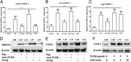
TGF-β is the main component in ISEM that induced PRMT1 upregulation in fibroblasts. ISEM from epithelial cells, TGF-β Ab, and TGF-β Ab were used to stimulate HFL1 cells, and the expressions ofprmt1,cox2, andvegfwere detected by RT-qPCR (A–C). The expressions of PRMT1 and COX2 protein were detected by Western blot (DandE). TGF-β with or without AMI-1 (5 and 10 μM), the pan PRMT enzymatic activity inhibitor, was used to stimulate HFL1, and COX2 expression was detected by Western blot (F).J Immunol.2015 Jul 1;195(1):298-306. |
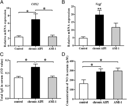
Expressions ofcox2andvegfand concentrations of IgE and NO in serum from chronic AIPI rats with or without AMI-1 administration. The expressions ofcox2(A) andvegf(B) were detected by RT-qPCR in lung tissues from control rats, AIPI rats, and AIPI rats with administration of AMI-1. Total IgE (C) and NO concentrations (D) in serum were detected by ELISA and the Griess method.J Immunol.2015 Jul 1;195(1):298-306. |
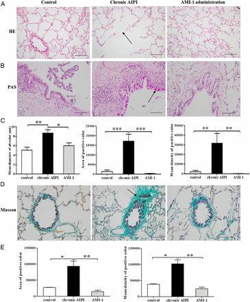
Histopathological changes and remodeling in chronic AIPI rats with and without the administration of AMI-1. Representative images of the histopathological changes by H&E staining (A), PAS staining (B), and Masson staining (D) were from lung sections of E3 rats without Ag challenge, with Ag challenge for 8 wk, and with both Ag challenge and AMI-1 administration for 8 wk, respectively.J Immunol.2015 Jul 1;195(1):298-306. |
Products are for research use only; We do not sell to patients
Copyright 2020 InvivoChem LLC | All Rights Reserved


 COA
COA