| Size | Price | Stock | Qty |
|---|---|---|---|
| 100mg |
|
||
| 250mg |
|
||
| 500mg |
|
||
| Other Sizes |
| Targets |
Sterol regulatory element-binding protein-1c (SREBP-1c) – aloin significantly suppressed the alcohol-dependent induction of SREBP-1c expression (p < 0.01). [3]
AMP-activated protein kinase-α2 (AMPK-α2) – aloin remarkably up-regulated the mRNA levels of AMPK-α2 (p < 0.001). [3] Cytochrome P4502E1 (CYP2E1) – aloin significantly inhibited the alcohol-dependent elevation of CYP2E1 expression (p < 0.05). [3] Toll-like receptor-4 (TLR-4) – aloin suppressed the up-regulation of TLR-4 mRNA expression (p < 0.001). [3] Myeloid differentiation primary response gene 88 (MyD88) – aloin suppressed the up-regulation of MyD88 mRNA expression (p < 0.01). [3] Tumor necrosis factor α (TNF-α) – aloin suppressed the alcohol-induced elevation of hepatic TNF-α (p < 0.05). [3] |
|---|---|
| ln Vitro |
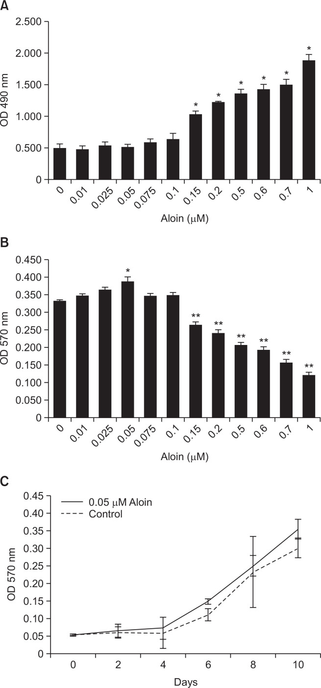
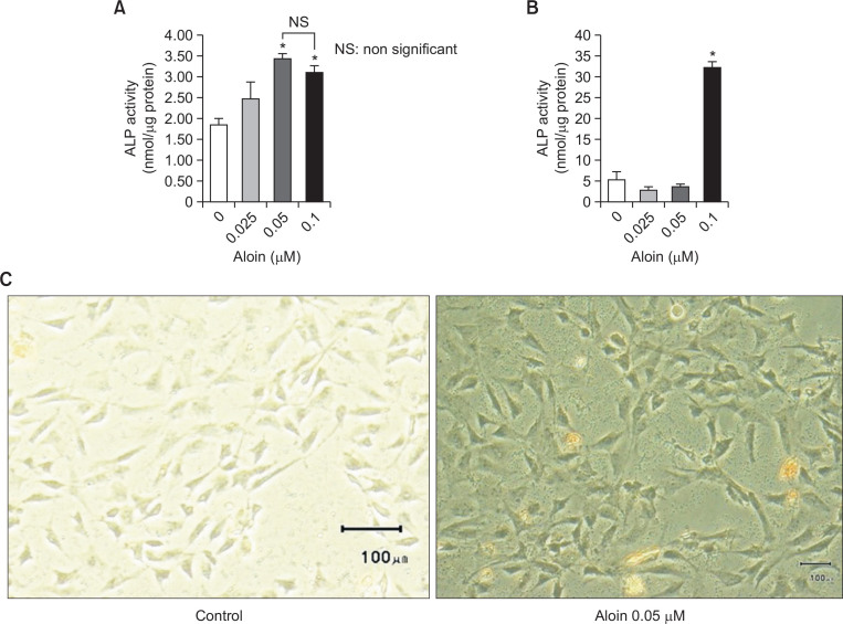
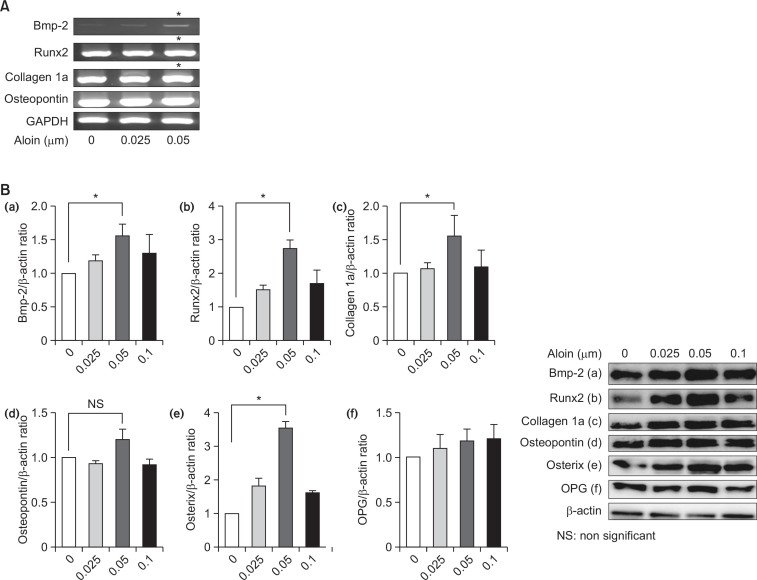
The expression of osteoblast differentiation genes, Bmp-2, Runx2, and collagen 1a is enhanced in a dose-dependent manner by aloin (0.01-1 μM; 10 d) [3]. MC3T3-E1 cells are stimulated to differentiate into osteoblasts by aloin (0.05 μM; 10 d) via the Wnt and Bmp signaling pathways mediated by MAPK [3]. In MC3T3-E1 cells, aloin (0.05 μM; 10 days) boosts ALP activity [3]. Aloe glycoside (10-80 μg/mL; 4.5 hours) attenuates intracellular ROS generation and increases cell survival after biaxial stretch injury (SI), which suppresses alterations in the mitochondrial membrane potential of mouse brain vascular endothelial cells (EC). [4].
In bEnd.3 mouse brain capillary endothelial cells subjected to biaxial stretch injury (SI) to simulate TBI in vitro, aloin treatment at 40 μg/mL (determined as the optimum concentration by LDH release assay) significantly reduced apoptosis. TUNEL assay showed the apoptosis rate was significantly lower in the SI+aloin group compared to the SI+vehicle group at 4 h post-SI (P < 0.01). [5] Aloin reversed the loss of tight junction proteins ZO-1 and occludin after SI. Immunofluorescence and western blot analyses demonstrated that ZO-1 and occludin expression levels were significantly increased in the aloin-treated group compared to the vehicle-treated group at 4 h post-SI (both P < 0.01). [5] Aloin attenuated intracellular reactive oxygen species (ROS) generation after SI. ROS levels were measured using DCFH-DA assay; the highest ROS level occurred at 2 h after SI. Aloin (40 μg/mL) noticeably reduced ROS production at 1 h, 2 h, and 4 h after SI (all P < 0.01). At 2 h and 4 h post-SI, 40 μg/mL aloin had the optimum effect compared with 20 μg/mL (both P < 0.01) and 60 μg/mL (P < 0.01 and P < 0.05, respectively). Flow cytometry also confirmed that aloin significantly decreased DCFH fluorescence intensity (P < 0.01). [5] Aloin protected against changes in mitochondrial membrane potential (ΔΨm) after SI. JC-1 assay revealed that the low ΔΨm (indicated by increased green/red fluorescence ratio) in the vehicle-treated group was remarkably reversed by aloin treatment at 2 h post-SI (P < 0.01), as quantified by fluorescence microscopy and flow cytometry. [5] Aloin regulated MAPK, NF-κB, and apoptosis-associated pathways in vitro. Western blot analyses at 2 h after SI showed that aloin significantly decreased the elevated levels of p-p38/p38 and p-p65/p65 (both P < 0.05), and reduced the increased ratios of Bax/Bcl-2 and cleaved caspase-3/caspase-3 (both P < 0.01) compared to the vehicle group. [5] Cell viability assay (CCK-8) showed that aloin at concentrations of 10, 20, and 40 μg/mL for 4.5 h had no significant effect on bEnd.3 cell viability compared to control (all P > 0.05). At 60 μg/mL and 80 μg/mL, cell viability significantly declined (P < 0.05 for 40 vs 60 μg/mL and 60 vs 80 μg/mL). [5] LDH release assay (an apoptosis-associated index) increased significantly after SI, and 40 μg/mL aloin was determined as the optimum concentration to reduce LDH release at 4 h post-SI. [5] |
| ln Vivo |
Aloin (10-30 mg/kg; single dosage; intraperitoneally given 30 minutes before TBI injury) reduces cerebral edema produced by traumatic brain injury (TBI) via regulating cortical impact injury in rats. Aloe glycoside demonstrates anti-oxidative stress and anti-apoptotic effects in mouse brain capillary endothelial cells [4].
In a chronic alcohol feeding mouse model (alcohol given twice daily by intragastric administration for 11 weeks), co-administration of aloin (10 or 30 mg/kg bw) after each alcohol dose significantly decreased serum alanine aminotransferase (ALT) and aspartate aminotransferase (AST) levels compared to the alcohol group (p < 0.05). [3] Aloin (30 mg/kg bw) significantly suppressed the elevation of serum total cholesterol (TC) (p < 0.05) and aloin (10 and 30 mg/kg bw) significantly suppressed the elevation of serum triglyceride (TG) (p < 0.05 and p < 0.01, respectively) caused by chronic alcohol. Aloin (30 mg/kg bw) also significantly increased serum high density lipoprotein (HDL) level (p < 0.05). No significant decrease was observed in serum low density lipoprotein (LDL) levels. [3] Histopathological examination (H&E staining) showed that aloin co-administration suppressed alcohol-induced steatosis (fat accumulation) and inflammatory injury (Kupffer cell activation), as indicated by diminished Kupffer cells and fatty infiltration of hepatocytes. [3] Aloin (10 and 30 mg/kg bw) significantly attenuated the alcohol-induced elevation of hepatic malondialdehyde (MDA) (p < 0.05 and p < 0.001, respectively) and significantly increased superoxide dismutase (SOD) activity (p < 0.01 and p < 0.001, respectively). Aloin (30 mg/kg bw) significantly inhibited the alcohol-induced over-expression of CYP2E1 mRNA (p < 0.05). No significant effect on alcohol-dependent glutathione (GSH) depletion was observed. [3] Aloin (10 and 30 mg/kg bw) significantly decreased the alcohol-induced elevation of hepatic nitric oxide (NO) (p < 0.05) and hepatic TNF-α (p < 0.05). Aloin (30 mg/kg bw) significantly inhibited the alcohol-induced reduction of anti-inflammatory cytokine IL-10 (p < 0.05). No significant effect on IL-1β was found. [3] Aloin (30 mg/kg bw) significantly decreased the alcohol-induced elevation of serum lipopolysaccharide (LPS) (p < 0.01). [3] Aloin (10 and 30 mg/kg bw) dramatically elevated the mRNA expression of AMPK-α2 compared to the alcohol group (p < 0.001 and p < 0.001, respectively). Aloin (10 and 30 mg/kg bw) markedly inhibited the alcohol-induced over-expression of SREBP-1c mRNA (p < 0.01 and p < 0.001, respectively). [3] Aloin (30 mg/kg bw) significantly reduced the alcohol-induced elevation of TLR-4 mRNA expression (p < 0.001) and significantly decreased MyD88 mRNA expression (p < 0.01). [3] |
| Cell Assay |
bEnd.3 mouse brain capillary endothelial cells were cultured in DMEM containing 10% fetal bovine serum and 1% penicillin/streptomycin in a 37°C humidified incubator with 5% CO₂ and 95% air. Mechanical biaxial stretch injury (SI) was induced using a Cell Injury Controller II system that released a 50-ms burst of nitrogen gas to cause a 7.5-mm downward deformation of the Silastic membrane and adherent cells. Aloin or vehicle was added 30 min before SI. [5]
Cell viability was assessed using CCK-8 assay. Cells were seeded into 96-well plates at 1×10⁴ per well and cultured overnight. Different concentrations of aloin (10, 20, 40, 60, 80 μg/mL) were administered for 4.5 h, then 10 μL of CCK-8 reaction solution was added, incubated for 2 h, and absorbance measured at 450 nm. [5] LDH release was measured using a cytotoxicity detection kit. Supernatant (100 μL) from each group was transferred to 96-well plates, mixed with 100 μL reaction solution, incubated for 30 min in the dark at 25°C, and absorbance measured at 490 nm. [5] Apoptosis was detected by TUNEL assay using an in-situ cell death detection kit. Cells were fixed in 4% paraformaldehyde for 30 min, permeabilized with 0.3% Triton X-100 for 10 min, incubated with TUNEL mixture solution for 1 h at 37°C, then stained with DAPI (1:1000) for 5 min in the dark. Apoptotic cells were observed under a confocal fluorescence microscope and the apoptosis rate was calculated as (apoptotic cells / total cells in a field) × 100%. [5] Immunostaining for tight junction proteins: Cells on Silastic membranes were fixed in cold anhydrous methanol, permeabilized with 0.3% Triton X-100, blocked with 10% bovine serum albumin, then incubated overnight at 4°C with rabbit anti-ZO-1 (1:200) and mouse anti-occludin (1:200) primary antibodies. After washing, cells were incubated with corresponding secondary antibodies (1:400) for 1 h and stained with DAPI (1:1000) for 10 min in the dark at room temperature. Images were captured using a fluorescence microscope. [5] Western blot analysis: Cells were lysed in mixed lysis buffer at the same protein concentration. After denaturation, equal volumes were separated by SDS-PAGE and transferred to PVDF membranes. Membranes were blocked with 5% skimmed milk powder for 1 h, then incubated overnight at 4°C with primary antibodies against ZO-1, occludin, p38, p-p38, p65, p-p65, Bcl-2, Bax, cleaved caspase-3, caspase-3, and β-actin/GAPDH/β-tubulin. After washing, membranes were incubated with HRP-conjugated secondary antibodies (1:5000) for 1 h at room temperature. Protein signals were measured using enhanced chemiluminescence and analyzed with Quantity One software. [5] Intracellular ROS measurement: DCFH-DA assay was used. Cells were incubated with 10 μmol/L DCFH-DA diluent for 20 min in the dark at 37°C, washed three times with DMEM, and fluorescence intensity measured by fluorescence spectrophotometer, fluorescence microscope, and flow cytometry (excitation 488 nm, emission 525 nm). [5] Mitochondrial membrane potential (ΔΨm) measurement: JC-1 assay was used. Cells were incubated with JC-1 reaction solution (1 mL DMEM + 1 mL JC-1 solution) for 20 min in the dark at 37°C, washed twice with JC-1 buffer, and fluorescence images captured under a fluorescence microscope. Red fluorescence indicated healthy cells with normal ΔΨm, green fluorescence indicated apoptotic cells with low ΔΨm. The red/green fluorescence ratio was quantified by flow cytometry. [5] |
| Animal Protocol |
Male Kunming mice (18-22 g body weight) were used. Mice were housed under controlled temperature (23 ± 2°C) and humidity (60 ± 5%) with a 12-h light/dark cycle, fed standard chow and water ad libitum. After 1 week acclimatization, mice were randomly divided into 5 groups (n=12 each): Control (vehicle), Alcohol (alcohol only), Positive control (alcohol + CBT 500 mg/kg bw), Alcohol + low-dose aloin (10 mg/kg bw), Alcohol + high-dose aloin (30 mg/kg bw). [3]
Alcohol (50% v/v in water) was administered intragastrically by gavage twice daily for 11 weeks. The dose was increased gradually: week 1: 4.0 g/kg bw/day (5 mL/kg bw ×2); week 2: 4.7 g/kg bw/day (6 mL/kg bw ×2); week 3: 5.5 g/kg bw/day (7 mL/kg bw ×2); weeks 4-11: 6.3 g/kg bw/day (8 mL/kg bw ×2). Control mice received water. After each alcohol administration, mice received an oral dose of aloin (dissolved in corn oil) or vehicle (corn oil) by gavage. At the end of 11 weeks, mice were anesthetized and sacrificed after 12 h fasting. Blood samples were collected and serum separated by centrifugation (3,000 rpm, 10 min, 4°C). Livers were excised, washed with ice-cold physiological saline (0.9% NaCl), weighed, and sections fixed for pathology; remaining tissues frozen in liquid nitrogen and stored at -80°C. [3] |
| Toxicity/Toxicokinetics |
In this study, no mortality or diarrhea was observed during the entire 11-week treatment period in aloin-treated mice (10 and 30 mg/kg bw). The colon and rectum of aloin-treated mice appeared normal as the control group. Body weight was not affected regardless of treatment. [3]
|
| References |
|
| Additional Infomation |
Aloin A is a C-glycoside compound with the structure β-D-glucopyranose, wherein the terminal hydroxyl group is replaced by the 4,5-dihydroxy-2-(hydroxymethyl)-10-oxo-9,10-dihydroanthracene-9-yl moiety (9S diastereomer). It is a metabolite and a laxative. It is a C-glycoside compound belonging to the anthracene, cyclic ketone, and phenolic classes. Aloin has been reported in Aloe ferox, Aloe africana, and other organisms with relevant data. See also: Aloe leaf (partial); Rhamnus bark (partial); Aloin (note moved to).
Aloin (purity >98%) extracted from Aloe ferox Mill. leaves was used. The study demonstrated that aloin protects against chronic alcoholic liver injury via attenuating lipid accumulation (by regulating AMPK-α2 and SREBP-1c expression), oxidative stress (by reducing MDA and CYP2E1, increasing SOD), and LPS-induced inflammatory response (by suppressing TLR-4 and MyD88, reducing TNF-α and NO, and increasing IL-10). The hepatoprotective effect of aloin was comparable to the conventionally reputed hepatoprotective compound CBT (Compound Biejiaruangan Troche). [3] |
| Molecular Formula |
C21H22O9
|
|---|---|
| Molecular Weight |
418.3940
|
| Exact Mass |
418.126
|
| CAS # |
1415-73-2
|
| Related CAS # |
Aloin B;28371-16-6
|
| PubChem CID |
12305761
|
| Appearance |
Light yellow to yellow solid powder
|
| Density |
1.6±0.1 g/cm3
|
| Boiling Point |
752.6±60.0 °C at 760 mmHg
|
| Melting Point |
148-149ºC
|
| Flash Point |
268.0±26.4 °C
|
| Vapour Pressure |
0.0±2.6 mmHg at 25°C
|
| Index of Refraction |
1.741
|
| LogP |
1.86
|
| Hydrogen Bond Donor Count |
7
|
| Hydrogen Bond Acceptor Count |
9
|
| Rotatable Bond Count |
3
|
| Heavy Atom Count |
30
|
| Complexity |
630
|
| Defined Atom Stereocenter Count |
6
|
| SMILES |
C1=CC2=C(C(=C1)O)C(=O)C3=C([C@H]2[C@H]4[C@@H]([C@H]([C@@H]([C@H](O4)CO)O)O)O)C=C(C=C3O)CO
|
| InChi Key |
AFHJQYHRLPMKHU-OSYMLPPYSA-N
|
| InChi Code |
InChI=1S/C21H22O9/c22-6-8-4-10-14(21-20(29)19(28)17(26)13(7-23)30-21)9-2-1-3-11(24)15(9)18(27)16(10)12(25)5-8/h1-5,13-14,17,19-26,28-29H,6-7H2/t13-,14+,17-,19+,20-,21+/m1/s1
|
| Chemical Name |
(10S)-1,8-dihydroxy-3-(hydroxymethyl)-10-[(2S,3R,4R,5S,6R)-3,4,5-trihydroxy-6-(hydroxymethyl)oxan-2-yl]-10H-anthracen-9-one
|
| HS Tariff Code |
2934.99.9001
|
| Storage |
Powder -20°C 3 years 4°C 2 years In solvent -80°C 6 months -20°C 1 month |
| Shipping Condition |
Room temperature (This product is stable at ambient temperature for a few days during ordinary shipping and time spent in Customs)
|
| Solubility (In Vitro) |
DMSO : ~125 mg/mL (~298.76 mM)
|
|---|---|
| Solubility (In Vivo) |
Solubility in Formulation 1: ≥ 2.08 mg/mL (4.97 mM) (saturation unknown) in 10% DMSO + 40% PEG300 + 5% Tween80 + 45% Saline (add these co-solvents sequentially from left to right, and one by one), clear solution.
For example, if 1 mL of working solution is to be prepared, you can add 100 μL of 20.8 mg/mL clear DMSO stock solution to 400 μL PEG300 and mix evenly; then add 50 μL Tween-80 to the above solution and mix evenly; then add 450 μL normal saline to adjust the volume to 1 mL. Preparation of saline: Dissolve 0.9 g of sodium chloride in 100 mL ddH₂ O to obtain a clear solution. Solubility in Formulation 2: ≥ 2.08 mg/mL (4.97 mM) (saturation unknown) in 10% DMSO + 90% (20% SBE-β-CD in Saline) (add these co-solvents sequentially from left to right, and one by one), clear solution. For example, if 1 mL of working solution is to be prepared, you can add 100 μL of 20.8 mg/mL clear DMSO stock solution to 900 μL of 20% SBE-β-CD physiological saline solution and mix evenly. Preparation of 20% SBE-β-CD in Saline (4°C,1 week): Dissolve 2 g SBE-β-CD in 10 mL saline to obtain a clear solution. (Please use freshly prepared in vivo formulations for optimal results.) |
| Preparing Stock Solutions | 1 mg | 5 mg | 10 mg | |
| 1 mM | 2.3901 mL | 11.9506 mL | 23.9011 mL | |
| 5 mM | 0.4780 mL | 2.3901 mL | 4.7802 mL | |
| 10 mM | 0.2390 mL | 1.1951 mL | 2.3901 mL |
*Note: Please select an appropriate solvent for the preparation of stock solution based on your experiment needs. For most products, DMSO can be used for preparing stock solutions (e.g. 5 mM, 10 mM, or 20 mM concentration); some products with high aqueous solubility may be dissolved in water directly. Solubility information is available at the above Solubility Data section. Once the stock solution is prepared, aliquot it to routine usage volumes and store at -20°C or -80°C. Avoid repeated freeze and thaw cycles.
Calculation results
Working concentration: mg/mL;
Method for preparing DMSO stock solution: mg drug pre-dissolved in μL DMSO (stock solution concentration mg/mL). Please contact us first if the concentration exceeds the DMSO solubility of the batch of drug.
Method for preparing in vivo formulation::Take μL DMSO stock solution, next add μL PEG300, mix and clarify, next addμL Tween 80, mix and clarify, next add μL ddH2O,mix and clarify.
(1) Please be sure that the solution is clear before the addition of next solvent. Dissolution methods like vortex, ultrasound or warming and heat may be used to aid dissolving.
(2) Be sure to add the solvent(s) in order.
|
|
|
Products are for research use only; We do not sell to patients
Copyright 2020 InvivoChem LLC | All Rights Reserved