| Size | Price | |
|---|---|---|
| Other Sizes |


| Targets |
Endogenous Metabolite; Microbial Metabolite
Imidazoline I3 receptor (I3 receptor) [2] |
|---|---|
| ln Vitro |
Well-known for its anti-inflammatory and antioxidant properties, allantoin is a common ingredient in cosmetic products [1]. In a dose-dependent manner, allantoin improved the viability of β-cells generated by STZ while attenuating cytotoxicity and apoptosis. Allantoin enhances the expression of phosphorylated B-cell lymphoma 2 (Bcl-2) and decreases caspase-3. Imidazoline 3 (I3) receptors have been demonstrated to be activated by allantoin [2].
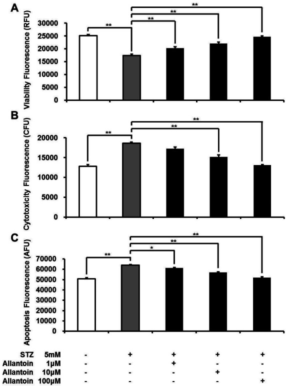
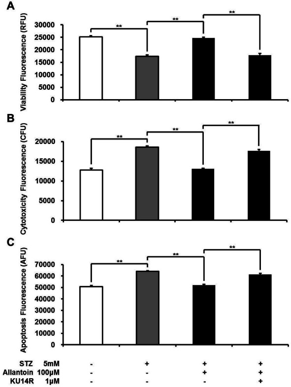
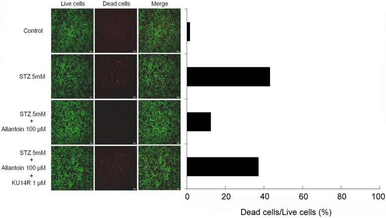
Allantoin (1 µM, 10 µM, and 100 µM) treatment significantly increased cell viability, decreased cytotoxicity and apoptosis induced by 5 mM streptozotocin (STZ) in pancreatic β-cells in a dose-dependent manner, as measured by the ApoTox-Glo triplex assay. [2] In live/dead double staining assay, Allantoin (100 µM) greatly improved the viability of STZ-induced β-cell apoptosis, shown by a marked decrease in red fluorescence emitted by EthD-1 (which binds to DNA of dead cells). Pretreatment with 1 µM KU14R (I3 antagonist) for 30 min reduced the action of allantoin, resulting in increased EthD-1 binding. [2] Flow cytometric analysis using Annexin V/PI staining showed that treatment with 5 mM STZ for 6 h increased the percentage of apoptotic cells to 45.5%. Allantoin (100 µM) reversed this effect and decreased the percentage of apoptotic cells to 32.7%, as shown by movement of cell population from the lower right quadrant to the lower left quadrant. Co-treatment with 1 µM KU14R blocked the action of allantoin and induced apoptosis to 53.9%. [2] Western blotting analysis revealed that in STZ-treated β-cells, the expression level of caspase-3 was significantly increased while Bcl-2 level was significantly decreased. Allantoin (100 µM) significantly suppressed the expression of caspase-3 and significantly increased the expression of Bcl-2. KU14R (1 µM) significantly inhibited these actions of allantoin. [2] The protective effect of Allantoin involved the phospholipase C (PLC) pathway, as the PLC inhibitor U73122 (1 µM) attenuated the protective effect of allantoin in β-cells measured by ApoTox-Glo triplex assay. [2] |
| ln Vivo |
Subchronic treatment of allantoin (1, 3 or 10 mg/kg for 7 days) significantly increased scopolamine-induced cholinergic blockade and latency evaluated in a passive avoidance task in normal young rats. Allantoin therapy (3 or 10 mg/kg for 7 days) also elevated phosphorylated phosphatidylinositol 3-kinase (PI3K), phosphorylated protein kinase B (Akt), and phosphorylated glycogen synthase kinase 3β ( GSK-3β) expression levels. Allantoin dramatically enhances neuronal cell proliferation in immature neurons in the dentate gyrus area of the hippocampus [1]. Daily injection of allantoin for 8 days in STZ-treated rats dramatically lowered plasma glucose and boosted plasma insulin levels [2]. Allantoin decreases SHR blood pressure at 30 minutes, which is the most effective time. Furthermore, SHR treated with allantoin displayed an antihypertensive impact in a dose-dependent manner. Furthermore, in sedated rats, allantoin reduces cardiac contractility and heart rate. In addition, allantoin can considerably improve peripheral blood flow [3].
Subchronic administration of Allantoin (1, 3 or 10 mg/kg, p.o. for 7 days) significantly increased the step-through latency time in the passive avoidance task in both scopolamine-induced cholinergic blockade mice and normal naive mice. In the retention trial of normal naive mice, allantoin at 10 mg/kg significantly increased latency (P < 0.05) compared to vehicle control. In scopolamine-induced memory impaired mice, allantoin at 10 mg/kg significantly ameliorated the reduced step-through latency (P < 0.05). [1] Subchronic administration of Allantoin (1, 3 or 10 mg/kg, p.o. for 7 days) did not show any significant change in spontaneous locomotor activity in the open field test, as measured by total ambulatory distance, compared to vehicle-treated group (One-way ANOVA, F(3,28)=0.998, P > 0.05). [1] Western blot analysis of hippocampal tissue from mice treated with Allantoin (1, 3 or 10 mg/kg, p.o. for 7 days) showed increased expression levels of phosphorylated PI3K (pPI3K), phosphorylated Akt (pAkt), and phosphorylated GSK-3β (pGSK-3β). Allantoin at 10 mg/kg increased pPI3K expression by 27% compared to vehicle control. Allantoin at 3 or 10 mg/kg increased pAkt immunoreactivity by 50% and pGSK-3β immunoreactivity by 125% compared to control. [1] Immunohistochemistry for BrdU incorporation in the subgranular zone (SGZ) of the hippocampal dentate gyrus revealed that subchronic administration of Allantoin (3 or 10 mg/kg, p.o. for 7 days) markedly increased the number of BrdU-incorporated cells (one-way ANOVA, F(3,16)=5.412, P < 0.05). [1] Immunohistochemistry for doublecortin (DCX) in the hippocampal dentate gyrus showed that Allantoin treatment (1 or 3 mg/kg, p.o. for 7 days) significantly increased the number of DCX-immunopositive cells (one-way ANOVA, F(3,16)=5.316, P < 0.05). [1] |
| Enzyme Assay |
ApoTox-Glo triplex assay [2]
The β-cells were seeded into 96-well plates at a total density of 1 × 10⁴ cells per well. Each well contained 200 µl RPMI 1640 medium and the test compound where appropriate. ApoTox-Glo Triplex Assay was used according to the manufacturer’s instructions to measure the β-cells’ viability, cytotoxicity, and apoptosis. After 24 h the Viability/Cytotoxicity reagent, containing both the GF-AFC substrate and the bis-AAF-R110 substrate, was added to all wells and incubated for 30 min. Caspase-Glo 3/7 was added to the wells and mixed briefly for 30 s, then incubated for 30 min at room temperature. Fluorescence was measured at 380EX/510EM to assess viability, 485EX/520EM to assess cytotoxicity, and luminescence was mesured to assess apoptosis. |
| Cell Assay |
The primary cultured cells were devided into 6-well plates. The medium was removed, and the cells were washed once with phosphate-buffered saline (PBS). RPMI 1640 medium containing 25 mM glucose was added to each well with 5 mM STZ and incubated for 6 h to induce cell apoptosis. To know the role of allantoin in the protection of pancreatic β-cells against STZ, allantoin pretreatment at various doses was provided before 30 min prior to the addition of 5 mM STZ and incubated for 6 h. To identify the signaling pathway of allantoin in β-cells, 1 µM KU14R: an I3 binding site antagonist, or 1 µM U73122: the phospholipase C (PLC) inhibitor were provided before 30 min prior to the addition of allantoin as previously described before. All the medium was removed, and the cells were washed three times with PBS prior to processing for the evaluation of morphology[2].
Primary cultured rat pancreatic β-cells were isolated from rat pancreases using collagenase digestion. Cells were cultured in RPMI 1640 medium supplemented with 1% penicillin/streptomycin, 1% amphotericin B and 10% FBS at 37°C with 5% CO2 for 48 h. [2] For ApoTox-Glo triplex assay, β-cells were seeded into 96-well plates at 1×10^4 cells per well in 200 µL RPMI 1640 medium. After 24 h, Viability/Cytotoxicity reagent (containing GF-AFC substrate and bis-AAF-R110 substrate) was added and incubated for 30 min. Then Caspase-Glo 3/7 reagent was added, mixed for 30 s, and incubated for 30 min at room temperature. Fluorescence was measured at 380EX/510EM for viability, 485EX/520EM for cytotoxicity, and luminescence for apoptosis. [2] For live/dead double staining assay, 100 µL of Live/Dead solution was added to the samples and incubated for 15 min at room temperature. The staining solution was removed and samples were viewed under a fluorescence microscope. Living cells were detected at green fluorescence, dead cells at red fluorescence. [2] For Annexin V/PI staining and flow cytometry, β-cells were divided into 12-well plates and treated with reagents for 48 h. Cells were collected, stained with Annexin V-PI, and analyzed using a flow cytometer. [2] For Western blotting analysis, β-cells were pre-cultured with 5 mM STZ for 6 h prior to addition of 100 µM allantoin with or without 1 µM KU14R or vehicle for 30 min. Cells were washed with ice-cold PBS and lysed for 15 min. Protein concentration was measured by BCA assay. Protein samples were separated by SDS-PAGE (10% acrylamide gel) and transferred to PVDF membranes. Membranes were blocked with 5% non-fat milk in TBS-T, then incubated overnight with primary antibodies against caspase-3 and Bcl-2. After incubation with secondary antibodies, antigen-antibody complexes were detected using an ECL kit and band densities were quantified by laser densitometer. [2] |
| Animal Protocol |
Mice: For memory ameliorating study, mice are administered vehicle solution, allantoin (1, 3 or 10 mg/kg, p.o.) or donepezil (5 mg/kg, p.o.) at the same time (10:00-12:00 a.m) and same place for 7 days. For memory enhancing study, mice are administered vehicle solution, allantoin (1, 3 or 10 mg/kg, p.o.) or piracetam (200 mg/kg, i.p.). The final administration of allantoin, donepezil or piracetam is performed 1 h before an acquisition trial in the passive avoidance task[1]
Glucose and insulin levels in STZ-treated rats: The induction of pancreatic cell damage was accomplished by injecting 45 mg/kg STZ dissolved in 10 mM Na-citrated buffer intraperitoneally. STZ-treated rats with blood glucose above 200 mg/dl at 7 days post-injection were included in the group. Total of 24 rats were divided into three groups as follows: Control (STZ) (n = 8), STZ + allantoin (n = 8), STZ + KU14R + allantoin (n = 8). The third group was treated with an intravenous injection of 8 mg/kg/day KU14R; the first and second groups were treated with the same volume of vehicle injected intravenously. After 30 min of KU14R injection, the second and third groups received 10 mg/kg/day of allantoin intravenously. The first group was injected the same volume of vehicle intravenously. The experiments were performed for 8 days. The blood samples were obtained from tail vein everyday. The plasma glucose levels were measured everyday, and the plasma insulin levels were measured on day 0, 4, 6, 8 [2]. Rats: Animals are randomly divided into four groups: (I) the control group treated with the vehicle, saline; (II) the allantoin group treated by intravenous injection of allantoin at 0.5 mg/kg; (III) the allantoin+efaroxan group treated with allantoin at the most effective dose (0.5 mg/kg, i.v.) and efaroxan at effective dose (1.5 mg/kg, i.v.) 30 minutes before injection of allantoin; and (IV) the allantoin treated SHRs group treated by intravenous injection of allantoin at various dose for desired time. After treatment of allantoin, the rats are placed into a holder for the determination of the mean blood pressure[3]. For memory ameliorating study in scopolamine-induced amnestic mice: Allantoin was dissolved in 10% Tween 80 solution. Mice were administered vehicle (10% Tween 80), allantoin (1, 3 or 10 mg/kg, p.o.), or donepezil (5 mg/kg, p.o.) once daily for 7 days. The final administration was performed 1 hour before the acquisition trial of the passive avoidance task. Scopolamine (1 mg/kg, i.p.) or 0.9% saline was administered 30 min before the acquisition trial. [1] For memory enhancing study in normal naive mice: Allantoin (1, 3 or 10 mg/kg, p.o.) or piracetam (200 mg/kg, i.p.) was administered 1 hour before the acquisition trial. Electrical foot shock was 0.25 mA, and latencies were recorded for up to 600 s. [1] For Western blotting: Mice were administered Allantoin (1, 3 or 10 mg/kg, p.o.) or vehicle for 7 days, and sacrificed 1 hour after the final dose. Hippocampal tissue was isolated for analysis. [1] For neurogenesis study: Allantoin was administered (1, 3 or 10 mg/kg, p.o.) for 7 days. On the 6th day, BrdU (50 mg/kg, i.p.) dissolved in 50 mM phosphate-buffered saline was administered 3 times at 2-hour intervals. Mice were sacrificed 1 week after allantoin treatment for tissue preparation. [1] For open field test: Mice were administered Allantoin (1, 3, or 10 mg/kg, p.o.) for 7 days, then placed in the center of a black plexiglas box (45×45×45 cm) and locomotor activity was recorded for 30 min using a video-tracking system. [1] |
| ADME/Pharmacokinetics |
Absorption, Distribution and Excretion
In human studies, allantoin recovery in urine was observed in only two subjects, at 19% and 34%, respectively, and only after administration of high doses. Following intravenous administration, in human models, urinary recovery was nearly quantifiable at doses ranging from 75 to 600 mg. Excretion in human subjects persisted for 72 hours after administration of 240 mg, with similar results observed after subcutaneous injection. Urinary clearance is the primary route of excretion. Some studies suggest that the average renal clearance of allantoin in normal healthy individuals is approximately 123 mL/min. It is generally believed that exogenous allantoin is rapidly excreted. In dogs, after oral administration of allantoin in solid or solution form, urinary excretion ranged from 35% to 92% within 24 hours. In rabbits, allantoin was not detected in urine or feces after oral administration. In humans, after administration of high doses, urinary allantoin recovery in urine was 19% and 34%, respectively, in two subjects. Following intravenous injection, allantoin recovery in urine was nearly quantified in both dogs and humans at doses ranging from 75 to 600 mg. In humans, allantoin excretion continued for 72 hours after a 240 mg injection. Subcutaneous injection yielded similar results. In dogs, intravenously injected uric acid was converted to allantoin within two hours. Metabolism/Metabolites Uricase is the enzyme that converts uric acid to allantoin. Since the human body does not possess endogenous uricase, uric acid is the only final metabolic product in the degradation of purines, while purine nucleotides are metabolic waste products. The presence of allantoin in human urine is a result of a non-enzymatic reaction of uric acid under the influence of reactive oxygen species. Therefore, this non-enzymatic reaction may serve as a potential biomarker for measuring oxidative stress in chronic diseases and aging. Furthermore, because allantoin is an endogenous substance and part of the body's basic metabolic pathways, it does not accumulate. Moreover, allantoin is hardly metabolized in humans and animals. Uric acid is the final stage of purine degradation because humans lack the uricase enzyme to convert uric acid into allantoin. Allantoin is considered a potential toxic substance for Reye's syndrome in the presence of calcium ions. A study has been initiated to find alternative sources of allantoin in humans (lacking uricase). Urate is a strong reducing agent that can non-enzymatically reduce cytochrome c, simultaneously generating CO₂ and H⁺. The stoichiometric ratio of various reactants and products was measured to be 1:2 urate:1... The optimal pH and ionic strength for this reaction are 9.5–10.5 and 10⁻⁵ M, respectively. Based on the results reported in this paper, the following equilibrium equation can be written: uric acid -2 + 2 cytochrome c³⁺ + 2 H₂O → allantoin + 2 cytochrome c²⁺ + H⁺ + HCO₃⁻. The authors propose that allantoin can be generated by the oxidation of uric acid by cytochrome c³⁺, and this may be a potential source of allantoin in human tissues. Uric acid is a major nitrogenous metabolic waste product in birds, but it is also a potent antioxidant. Hominoid primates and birds lack uricase, the enzyme that oxidizes uric acid to allantoin. Therefore, the presence of allantoin in their plasma is due to non-enzymatic oxidation. In most mammals, purine degradation ultimately leads to allantoin formation. Humans lack uricase, the enzyme that catalyzes the conversion of uric acid to allantoin. For more complete data on the metabolism/metabolites of allantoin (11 in total), please visit the HSDB record page. Biological Half-Life In studies of cattle, sheep, and horses, the half-life of allantoin ranged from 1 to 2.5 hours. |
| Toxicity/Toxicokinetics |
Interactions
Twenty or 24 male F344 rats and 20 or 24 female F344 rats were freely fed a diet containing 0.2% allantoin (with or without 0.2% sodium nitrite) for 106 weeks. …Control group rats were fed the untreated diet…Rats in the sodium nitrite treatment group had 0.2% sodium nitrite added to their diet or drinking water. After treatment, all rats were fed the untreated diet…and observed until death. No shortened lifespan was observed in any treatment group. Compared with the untreated control group, the use of allantoin alone or in combination with sodium nitrite did not lead to any increase in tumor incidence… |
| References | |
| Additional Infomation |
Therapeutic Uses
Urea hydantoin is a urea hydantoin found in urine and plants, used in dermatological preparations. Allantoin is a component of comfrey, which stimulates tissue repair and wound healing by promoting cell proliferation. Allantoin also has a significant effect on cell proliferation in degenerated and regenerated peripheral nerves. In humans, the ratio of allantoin to uric acid in plasma increases during oxidative stress, and therefore this ratio is considered an in vivo marker of oxidative stress in humans. A diagnostic marker of oxidative stress during anti-tuberculosis treatment. For more complete data on the therapeutic uses of allantoin (8 types), please visit the HSDB record page. Drug Warnings Skin: For external use only. Eyes: Avoid contact with eyes. Sensitization: Contraindicated in individuals with known hypersensitivity to any of the ingredients in Mederma. /Mederma/ Pharmacodynamics There are currently no adequately controlled and appropriate data to formally confirm the pharmacodynamic properties of allantoin. However, ongoing research indicates that allantoin has moisturizing and keratolytic effects, as well as the ability to increase the water content of the extracellular matrix and promote the shedding of dead skin cells, all of which can promote cell proliferation and wound healing. Allantoin is known to be safe and non-toxic. It is a purine-derived compound with anti-oxidative and anti-inflammatory activities. The study suggests that allantoin has memory-enhancing effects, possibly mediated by activation of the PI3K-Akt-GSK-3β signaling pathway, and may play a role in increasing neuronal cell proliferation in the hippocampal dentate gyrus region. The authors propose that allantoin has therapeutic potential for cognitive dysfunctions observed in Alzheimer's disease. [1] |
| Molecular Formula |
C4H6N4O3
|
|---|---|
| Molecular Weight |
158.1154
|
| Exact Mass |
158.043
|
| Elemental Analysis |
C, 30.39; H, 3.83; N, 35.43; O, 30.36
|
| CAS # |
97-59-6
|
| Related CAS # |
Allantoin-13C2,15N4;1219402-51-3; 97-59-6 (racemic); 7303-80-2 (R-isomer); 3844-67-5 (S-isomer)
|
| PubChem CID |
204
|
| Appearance |
White to off-white solid
|
| Density |
1.7±0.1 g/cm3
|
| Boiling Point |
478ºC
|
| Melting Point |
230 °C (dec.)(lit.)
|
| Flash Point |
230-234°C
|
| Index of Refraction |
1.616
|
| Source |
Microbe
|
| LogP |
-2.89
|
| Hydrogen Bond Donor Count |
4
|
| Hydrogen Bond Acceptor Count |
3
|
| Rotatable Bond Count |
1
|
| Heavy Atom Count |
11
|
| Complexity |
225
|
| Defined Atom Stereocenter Count |
0
|
| SMILES |
O=C(NC1C(=O)NC(=O)N1)N
|
| InChi Key |
POJWUDADGALRAB-UHFFFAOYSA-N
|
| InChi Code |
InChI=1S/C4H6N4O3/c5-3(10)6-1-2(9)8-4(11)7-1/h1H,(H3,5,6,10)(H2,7,8,9,11)
|
| Chemical Name |
1-(2,5-dioxoimidazolidin-4-yl)urea
|
| Synonyms |
5-Ureidohydantoin; SD 101; Allantion; Sebical; Septalan; Allantol; Cordianine; NSC 7606; DL-Allantoin; Glyoxyldiureid; Glyoxyldiureide; Glyoxylic diureide; Psoralon;
|
| HS Tariff Code |
2933.21.0000
|
| Storage |
Powder -20°C 3 years 4°C 2 years In solvent -80°C 6 months -20°C 1 month |
| Shipping Condition |
Room temperature (This product is stable at ambient temperature for a few days during ordinary shipping and time spent in Customs)
|
| Solubility (In Vitro) |
DMSO : ~50 mg/mL (~316.22 mM)
H2O : ~3.85 mg/mL (~24.35 mM) |
|---|---|
| Solubility (In Vivo) |
Solubility in Formulation 1: ≥ 2.5 mg/mL (15.81 mM) (saturation unknown) in 10% DMSO + 40% PEG300 + 5% Tween80 + 45% Saline (add these co-solvents sequentially from left to right, and one by one), clear solution.
For example, if 1 mL of working solution is to be prepared, you can add 100 μL of 25.0 mg/mL clear DMSO stock solution to 400 μL PEG300 and mix evenly; then add 50 μL Tween-80 to the above solution and mix evenly; then add 450 μL normal saline to adjust the volume to 1 mL. Preparation of saline: Dissolve 0.9 g of sodium chloride in 100 mL ddH₂ O to obtain a clear solution. Solubility in Formulation 2: ≥ 2.5 mg/mL (15.81 mM) (saturation unknown) in 10% DMSO + 90% (20% SBE-β-CD in Saline) (add these co-solvents sequentially from left to right, and one by one), clear solution. For example, if 1 mL of working solution is to be prepared, you can add 100 μL of 25.0 mg/mL clear DMSO stock solution to 900 μL of 20% SBE-β-CD physiological saline solution and mix evenly. Preparation of 20% SBE-β-CD in Saline (4°C,1 week): Dissolve 2 g SBE-β-CD in 10 mL saline to obtain a clear solution. View More
Solubility in Formulation 3: ≥ 2.5 mg/mL (15.81 mM) (saturation unknown) in 10% DMSO + 90% Corn Oil (add these co-solvents sequentially from left to right, and one by one), clear solution. Solubility in Formulation 4: 2 mg/mL (12.65 mM) in PBS (add these co-solvents sequentially from left to right, and one by one), clear solution; with ultrasonication (<60°C). |
| Preparing Stock Solutions | 1 mg | 5 mg | 10 mg | |
| 1 mM | 6.3243 mL | 31.6216 mL | 63.2431 mL | |
| 5 mM | 1.2649 mL | 6.3243 mL | 12.6486 mL | |
| 10 mM | 0.6324 mL | 3.1622 mL | 6.3243 mL |
*Note: Please select an appropriate solvent for the preparation of stock solution based on your experiment needs. For most products, DMSO can be used for preparing stock solutions (e.g. 5 mM, 10 mM, or 20 mM concentration); some products with high aqueous solubility may be dissolved in water directly. Solubility information is available at the above Solubility Data section. Once the stock solution is prepared, aliquot it to routine usage volumes and store at -20°C or -80°C. Avoid repeated freeze and thaw cycles.
Calculation results
Working concentration: mg/mL;
Method for preparing DMSO stock solution: mg drug pre-dissolved in μL DMSO (stock solution concentration mg/mL). Please contact us first if the concentration exceeds the DMSO solubility of the batch of drug.
Method for preparing in vivo formulation::Take μL DMSO stock solution, next add μL PEG300, mix and clarify, next addμL Tween 80, mix and clarify, next add μL ddH2O,mix and clarify.
(1) Please be sure that the solution is clear before the addition of next solvent. Dissolution methods like vortex, ultrasound or warming and heat may be used to aid dissolving.
(2) Be sure to add the solvent(s) in order.
| NCT Number | Recruitment | interventions | Conditions | Sponsor/Collaborators | Start Date | Phases |
| NCT05796635 | Completed | Other: Herpecin L | Cold Sores | Focus Consumer Healthcare | 2023-01-04 | Not Applicable |
| NCT04046783 | Completed | Device: patch | Cesarean Section; Dehiscence Scar Keloid Wound Heal |
University of Salerno | 2019-03-02 | |
| NCT05105139 | Completed | Other: Allantoin/ Coal Tar/ Clioquinol
Other: Allantoin/ Coal Tar/ Clioquinol/ Triclosan |
Psoriasis of Scalp Seborrheic Dermatitis |
Laboratorios Silanes S.A. de C.V. | 2021-11-29 | |
| NCT00825565 | Completed | Drug: Alwextin cream | Epidermolysis Bullosa | Northwestern University | 2009-02 | Phase 2 |
| NCT01863407 | Unknown status | Drug: DAM Drug: Normal Saline |
Postoperative Ileus | Beijing Bozhiyin T&S Co., Ltd. | 2013-04 | Phase 3 |
|
|
|
Products are for research use only; We do not sell to patients
Copyright 2020 InvivoChem LLC | All Rights Reserved