| Size | Price | Stock | Qty |
|---|---|---|---|
| 1mg |
|
||
| 5mg |
|
||
| 10mg |
|
||
| 50mg | |||
| Other Sizes |


Purity: ≥98%
AKB-6899 leads to stabilization of HIF-2α which induces sVEGFR-1 production from tumor-associated macrophages and decreases tumor growth in a murine melanoma model. References: Roda JM, Wang Y, Sumner LA, Phillips GS, Marsh CB, Eubank TD. Stabilization of HIF-2α induces sVEGFR-1 production from tumor-associated macrophages and decreases tumor growth in a murine melanoma model. J Immunol. 2012 Sep 15;189(6):3168-77. doi: 10.4049/jimmunol.1103817. Epub 2012 Aug 6. PubMed PMID: 22869907; PubMed Central PMCID: PMC3436995.
| Targets |
PHD3 (prolyl hydroxylase 3) [1]
|
|---|---|
| ln Vitro |
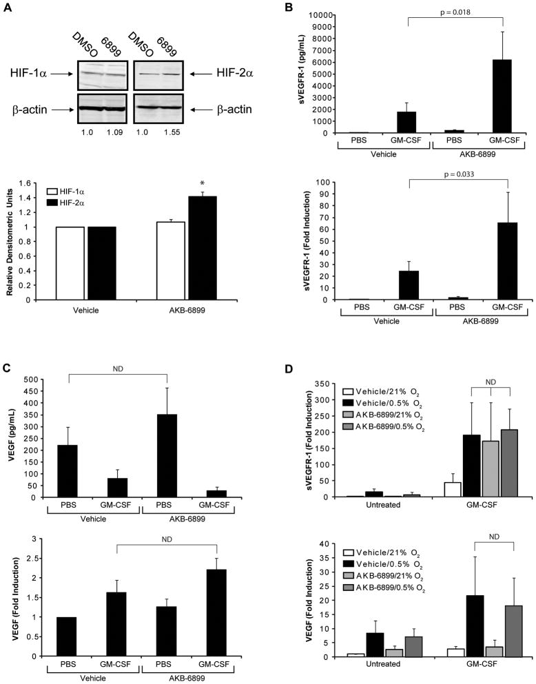
AKB-6899 (10 μM; 24 hours) elevated HIF-2α protein levels without a commensurate increase in HIF-1α. Without altering HIF-1α accumulation or VEGF synthesis, AKB-6899 also boosts the development of soluble forms of VEGF receptor (sVEGFR)-1 by macrophages treated with GM-CSF [1].
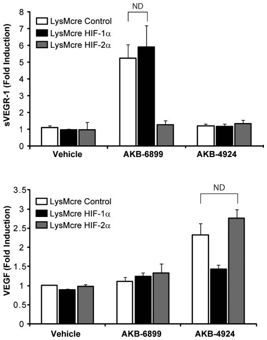
Treatment of murine bone marrow-derived macrophages with 10 μM AKB-6899 for 18 hours increased HIF-2α protein accumulation (p = 0.001) with no corresponding increase in HIF-1α (p = 0.105) as determined by immunoblotting; AKB-6899 did not increase HIF-1α or HIF-2α mRNA levels. [1] Human peripheral blood monocytes stimulated with 100 ng/mL GM-CSF and 10 μM AKB-6899 showed significantly increased sVEGFR-1 production at both protein (p = 0.018) and transcript (p = 0.033) levels compared to GM-CSF alone; VEGF protein (free bioavailable VEGF) was not significantly increased (p = 0.133), and VEGF transcript levels showed no difference between GM-CSF alone and GM-CSF/AKB-6899 (p = 0.558). [1] Stimulation of human monocytes with GM-CSF at 0.5% O2 or with GM-CSF/AKB-6899 at normoxia resulted in equivalent sVEGFR-1 production; AKB-6899 at 0.5% O2 did not further increase sVEGFR-1 compared to normoxia, suggesting maximal HIF-2α stabilization. [1] Using bone marrow-derived macrophages from myeloid-specific HIF-1α or HIF-2α knockout mice, AKB-6899 induced comparable sVEGFR-1 levels in control and HIF-1α-deficient macrophages but failed to induce sVEGFR-1 in HIF-2α-deficient macrophages, confirming that AKB-6899-induced sVEGFR-1 production is dependent on HIF-2α. [1] |
| ln Vivo |
Treatment with AKB-6899 (17.5 mg/kg; intraperitoneal injection; three times per week; for 16 days) effectively reduced tumor growth in a rat melanoma model [1].
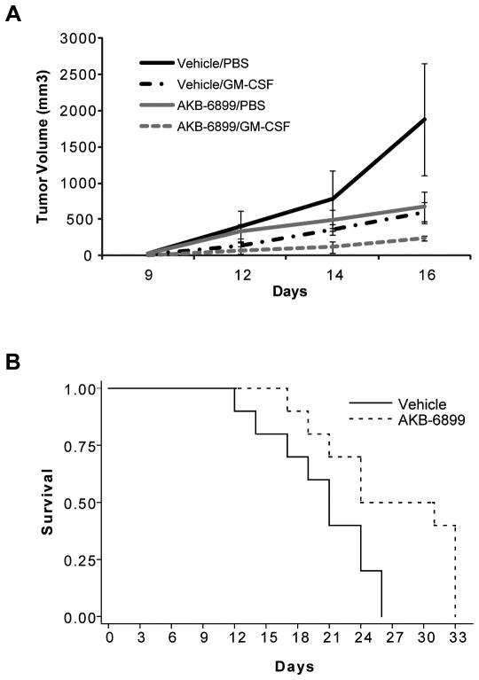
In C57BL/6 mice bearing subcutaneous B16F10 murine melanoma tumors, treatment with intratumoral GM-CSF (100 ng/mouse) plus intraperitoneal AKB-6899 (17.5 mg/kg) three times per week significantly reduced tumor growth compared to either agent alone (p < 0.001 for GM-CSF or AKB-6899 vs. combination at day 16); the combination also significantly prolonged survival (p = 0.001) compared to vehicle control. [1] Mice treated with GM-CSF/AKB-6899 showed increased intratumoral sVEGFR-1 mRNA levels (p = 0.001) but no difference in VEGF mRNA (p = 0.456) compared to controls; tumor vascularity (CD31 staining) was significantly decreased in the combination group (p < 0.001). [1] Lung metastasis, assessed by Pmel17 mRNA, was significantly reduced in mice treated with GM-CSF/AKB-6899 compared to vehicle controls (p < 0.001). [1] The anti-tumor effect of AKB-6899 was abrogated when mice were co-treated with a sVEGFR-1 neutralizing antibody (4 μg/mouse intratumorally) along with AKB-6899; no significant difference in tumor growth was observed between AKB-6899 plus anti-sVEGFR-1 antibody and vehicle controls (p = 0.128). [1] In SCID mice bearing subcutaneous A375 human melanoma xenografts, combination treatment with GM-CSF and AKB-6899 (same regimen) significantly decreased tumor growth (p = 0.05). [1] |
| Enzyme Assay |
Human peripheral blood monocytes were purified from fresh peripheral blood by density gradient centrifugation, then cultured in endotoxin-free medium supplemented with 1% fetal bovine serum, 1% PSA, and 10 μg/mL polymyxin B. Monocytes were treated for 24 hours with 10 ng/mL GM-CSF, 10 μM AKB-6899, or vehicle controls (PBS or DMSO). Cell-free culture supernatants were collected for ELISA (sVEGFR-1 and VEGF), and cells were lysed in Trizol for RNA extraction and real-time PCR. [1]
Bone marrow-derived macrophages from LysMcre control, HIF-1αflox/flox/LysMcre, or HIF-2αflox/flox/LysMcre mice were generated by culturing femoral bone marrow progenitors in medium with 20 ng/mL recombinant murine M-CSF for 5 days. Differentiated macrophages were serum-starved for 2 hours, then treated with 100 ng/mL murine GM-CSF and/or 10 μM AKB-6899 in medium containing 1% FBS, 1% PSA, and 10 μg/mL polymyxin B. After 24 hours, culture supernatants were assayed for VEGF and sVEGFR-1 by ELISA. [1] Human umbilical vein endothelial cells were cultured with GM-CSF and/or AKB-6899; they secreted low basal sVEGFR-1 which did not increase in response to GM-CSF, AKB-6899, or the combination. [1] |
| Cell Assay |
Western Blot Analysis[1]
Cell Types: Mouse bone marrow-derived macrophages Tested Concentrations: 10 μM Incubation Duration: 24 hrs (hours) Experimental Results: HIF was observed to increase -2α protein in cells. |
| Animal Protocol |
Animal/Disease Models: 6-8 weeks old C57BL/6 mice injected with B16F10 murine melanoma cells [1]
Doses: 17.5 mg/kg Route of Administration: intraperitoneal (ip) injection; 3 times a week; continued for 16 days. Experimental Results: Tumor growth demonstrated significant decrease. For the murine melanoma model, 6-8 week old C57BL/6 mice were injected subcutaneously with 1×10^5 B16F10 murine melanoma cells on the left flank. Once tumors became palpable (approximately 5-7 days), mice were randomly allocated to treatment groups. AKB-6899 (17.5 mg/kg) or vehicle control (20% PEG-400 in 5% sucrose) was administered intraperitoneally; GM-CSF (100 ng/mouse) or PBS vehicle was administered intratumorally. Treatments were given three times per week until tumors reached 20 mm in any dimension (approximately 2.5 weeks). Tumor diameters were measured three times per week with calipers, and tumor volumes were calculated as 0.5 × (large diameter) × (small diameter)^2. [1] For survival analysis, mice were inoculated with B16F10 tumors and treated three times weekly with 17.5 mg/kg AKB-6899 or vehicle; survival was defined as time to tumor size of 20 mm in any dimension. [1] For sVEGFR-1 neutralization experiments, mice were treated intraperitoneally three times per week with AKB-6899 or vehicle, and intratumorally with 4 μg of an anti-VEGFR-1 neutralizing antibody or polyclonal goat IgG isotype control in a 50 μL volume. [1] For the human melanoma xenograft model, SCID mice were injected subcutaneously with 1×10^6 A375 human melanoma cells and treated with the same regimen as above. [1] Lung metastases were evaluated by detecting Pmel17 mRNA in lungs via nested real-time PCR. At sacrifice, lungs were excised, flash-frozen, homogenized in liquid nitrogen, and dissolved in Trizol for RNA extraction. [1] |
| Toxicity/Toxicokinetics |
AKB-6899 at concentrations up to 50 μM had no effect on the viability of human peripheral blood monocytes or bone marrow-derived macrophages from LysMcre control, HIF-1αflox/flox/LysMcre, or HIF-2αflox/flox/LysMcre mice, as determined by annexin V and propidium iodide staining after 24 hours of incubation. [1]
|
| References | |
| Additional Infomation |
AKB-6899 was developed for the treatment of chronic anemia, as HIF-2α also controls erythropoietin production; a related compound AKB-6548 is in phase II clinical trials and is well-tolerated, inducing HIF-2α-dependent genes with little effect on HIF-1α-dependent genes. [1]
In contrast to AKB-6899 (PHD3 inhibitor), AKB-4924 is a 4-substituted 3-hydroxy-2-oxo-1,2-dihydropyridine derivative that strongly stabilizes HIF-1α through PHD2 inhibition and increases VEGF production. [1] The anti-tumor and anti-angiogenic effects of AKB-6899 are dependent on sVEGFR-1 production from tumor-associated macrophages, as neutralization of sVEGFR-1 completely reversed the inhibitory effect. [1] Tumor-infiltrating macrophages are the primary source of sVEGFR-1 following AKB-6899/GM-CSF co-treatment, as vascular endothelial cells do not upregulate sVEGFR-1 in response to AKB-6899 or GM-CSF. [1] |
| Molecular Formula |
C14H11FN2O4
|
|---|---|
| Molecular Weight |
290.246546983719
|
| Exact Mass |
290.07
|
| CAS # |
1007377-55-0
|
| Related CAS # |
1007377-55-0
|
| PubChem CID |
49848485
|
| Appearance |
White to yellow solid powder
|
| LogP |
2
|
| Hydrogen Bond Donor Count |
3
|
| Hydrogen Bond Acceptor Count |
6
|
| Rotatable Bond Count |
4
|
| Heavy Atom Count |
21
|
| Complexity |
393
|
| Defined Atom Stereocenter Count |
0
|
| SMILES |
FC1=CC=CC(=C1)C1=CN=C(C(NCC(=O)O)=O)C(=C1)O
|
| InChi Key |
PXWOWORYDKAEJO-UHFFFAOYSA-N
|
| InChi Code |
InChI=1S/C14H11FN2O4/c15-10-3-1-2-8(4-10)9-5-11(18)13(16-6-9)14(21)17-7-12(19)20/h1-6,18H,7H2,(H,17,21)(H,19,20)
|
| Chemical Name |
N-((5-(3-Fluorophenyl)-3-hydroxy-2-pyridinyl)carbonyl)glycine
|
| Synonyms |
AKB-6899 AKB 6899 AKB6899
|
| HS Tariff Code |
2934.99.9001
|
| Storage |
Powder -20°C 3 years 4°C 2 years In solvent -80°C 6 months -20°C 1 month |
| Shipping Condition |
Room temperature (This product is stable at ambient temperature for a few days during ordinary shipping and time spent in Customs)
|
| Solubility (In Vitro) |
DMSO : ~100 mg/mL (~344.53 mM)
|
|---|---|
| Solubility (In Vivo) |
Solubility in Formulation 1: ≥ 2.5 mg/mL (8.61 mM) (saturation unknown) in 10% DMSO + 40% PEG300 + 5% Tween80 + 45% Saline (add these co-solvents sequentially from left to right, and one by one), clear solution.
For example, if 1 mL of working solution is to be prepared, you can add 100 μL of 25.0 mg/mL clear DMSO stock solution to 400 μL PEG300 and mix evenly; then add 50 μL Tween-80 to the above solution and mix evenly; then add 450 μL normal saline to adjust the volume to 1 mL. Preparation of saline: Dissolve 0.9 g of sodium chloride in 100 mL ddH₂ O to obtain a clear solution. Solubility in Formulation 2: ≥ 2.5 mg/mL (8.61 mM) (saturation unknown) in 10% DMSO + 90% (20% SBE-β-CD in Saline) (add these co-solvents sequentially from left to right, and one by one), clear solution. For example, if 1 mL of working solution is to be prepared, you can add 100 μL of 25.0 mg/mL clear DMSO stock solution to 900 μL of 20% SBE-β-CD physiological saline solution and mix evenly. Preparation of 20% SBE-β-CD in Saline (4°C,1 week): Dissolve 2 g SBE-β-CD in 10 mL saline to obtain a clear solution. View More
Solubility in Formulation 3: ≥ 2.5 mg/mL (8.61 mM) (saturation unknown) in 10% DMSO + 90% Corn Oil (add these co-solvents sequentially from left to right, and one by one), clear solution. |
| Preparing Stock Solutions | 1 mg | 5 mg | 10 mg | |
| 1 mM | 3.4453 mL | 17.2265 mL | 34.4531 mL | |
| 5 mM | 0.6891 mL | 3.4453 mL | 6.8906 mL | |
| 10 mM | 0.3445 mL | 1.7227 mL | 3.4453 mL |
*Note: Please select an appropriate solvent for the preparation of stock solution based on your experiment needs. For most products, DMSO can be used for preparing stock solutions (e.g. 5 mM, 10 mM, or 20 mM concentration); some products with high aqueous solubility may be dissolved in water directly. Solubility information is available at the above Solubility Data section. Once the stock solution is prepared, aliquot it to routine usage volumes and store at -20°C or -80°C. Avoid repeated freeze and thaw cycles.
Calculation results
Working concentration: mg/mL;
Method for preparing DMSO stock solution: mg drug pre-dissolved in μL DMSO (stock solution concentration mg/mL). Please contact us first if the concentration exceeds the DMSO solubility of the batch of drug.
Method for preparing in vivo formulation::Take μL DMSO stock solution, next add μL PEG300, mix and clarify, next addμL Tween 80, mix and clarify, next add μL ddH2O,mix and clarify.
(1) Please be sure that the solution is clear before the addition of next solvent. Dissolution methods like vortex, ultrasound or warming and heat may be used to aid dissolving.
(2) Be sure to add the solvent(s) in order.
|
|
|
Products are for research use only; We do not sell to patients
Copyright 2020 InvivoChem LLC | All Rights Reserved


 COA
COA