| Size | Price | Stock | Qty |
|---|---|---|---|
| 10mg |
|
||
| 25mg |
|
||
| 50mg |
|
||
| 100mg |
|
||
| 250mg |
|
||
| 500mg |
|
||
| 1g |
|
||
| Other Sizes |
|


Purity: ≥98%
Emrusolmin (Anle138b) is a novel oligomer modulator for disease-modifying therapy of neurodegenerative diseases such as prion and Parkinson's disease.. Anle138b blocks the activity of conducting Aβ pores without changing the membrane embedded Aβ-oligomer structure, at the mechanistic level. Anle138b blocks interpeptide main chain interactions and impedes the spontaneous formation of ordered β-sheet structures, in particular those with out-of-register antiparallel β-strands.
| Targets |
Oligomeric aggregation
|
|---|---|
| ln Vitro |
It is believed that low aggregates are important neurotoxicants. Emrusolmin strongly inhibits all prion strains examined, mixing BSE derivatives and human prions. Emrusolmin swells the formation of pathogenic aggregates of prion proteins and alpha-synuclein, which form dead deposits in Parkinson's disease and other synucleinopathies (e.g. Louis Emrusolmin). Alpha-synuclein and prion protein pathological oligomers are severely inhibited by emusolmin, which exhibits structural reliance on pathological aggregation [1].
In vitro, Emrusolmin (Anle138b) blocked the formation of pathological aggregates of prion protein (PrP(Sc)) and of α-synuclein (α-syn), which is deposited in PD and other synucleinopathies such as dementia with Lewy bodies (DLB) and multiple system atrophy (MSA). Notably, anle138b strongly inhibited all prion strains tested including BSE-derived and human prions. [1] Emrusolmin (Anle138b)‐doped membranes demonstrated fewer simultaneously active pores and significantly reduced bulk conductance (Fig 5A and B, Appendix Fig S5) compared to membranes lacking the anle138b compound (Fig 5A and B, Appendix Fig S5). Our results indicate that treatment with anle138b alters the pore dynamics, resulting in less stable and shorter lived “open” pores. Decreased pore stability leads to a reduction in the total number of simultaneously conducting pores and significantly decreased conductance across the membrane. AFM data revealed that anle138b treatment did not affect the surface structure of Aβ1‐42 pores (Fig EV2), suggesting that anle138b does not simply prevent Aβ1‐42 from entering lipid bilayer membranes and forming pores. Rather anle138b appears to render conducting Aβ pores to non‐conducting ones—likely through structural change to the membrane embedded region of Aβ1–42—thereby providing one possible mechanism by which anle138b ameliorates LTP and learning deficits in APPPS1ΔE9 mice. Similar effects were observed when the conductance measurements were repeated in oxidized cholesterol [2]. |
| ln Vivo |
Emrusolmin has been demonstrated to bind structurally binding to pathological aggregates and strongly suppresses the production of prion protein and alpha-synapsin nuclear pathological oligomers in vitro and in the chamber [1].
Emrusolmin (Anle138b) showed structure-dependent binding to pathological aggregates and strongly inhibited formation of pathological oligomers in vitro and in vivo both for prion protein and α-synuclein. Both in mouse models of prion disease and in three different PD mouse models, anle138b strongly inhibited oligomer accumulation, neuronal degeneration, and disease progression in vivo. Anle138b had no detectable toxicity at therapeutic doses and an excellent oral bioavailability and blood-brain-barrier penetration. Our findings indicate that oligomer modulators provide a new approach for disease-modifying therapy in these diseases, for which only symptomatic treatment is available so far. Moreover, our findings suggest that pathological oligomers in neurodegenerative diseases share structural features, although the main protein component is disease-specific, indicating that compounds such as anle138b that modulate oligomer formation by targeting structure-dependent epitopes can have a broad spectrum of activity in the treatment of different protein aggregation diseases.[1] Synaptic plasticity and memory function in a mouse model for deposition of amyloid β peptides after oral treatment with Emrusolmin (Anle138b) [2] To initially test the potential of Emrusolmin (Anle138b) as therapeutic strategies to treat amyloid aggregation in Alzheimer's disease, we analyzed its effect in a Drosophila model for amyloid‐induced neurotoxicity. We observed that treatment with anle138b improved survival times when compared to a vehicle‐treated group (Appendix Fig S1). On the basis of these data, we decided to test the efficacy of anle138b in a mouse model for amyloid deposition. We like to state that none of the currently employed animal models for AD fully recapitulate the phenotypes seen in AD patients, and thus, care has to be taken when interpreting such data. In our study, we employed APPPS1Δ9 mice (Jankowsky et al, 2001), a well‐established model for AD‐linked amyloid deposition. Since in the patients therapeutic intervention is normally initiated only after the onset of amyloid plaque formation, we decided to test anle138b in two experimental cohorts. In the “pre‐plaque group,” treatment was initiated before the onset of pathology when mice were 2 months of age, while in the “post‐plaque group” treatment was initiated after the onset of amyloid deposition and memory disturbances in 6‐month‐old mice (Fig EV1; Jankowsky et al, 2004; Lalonde et al, 2005; Reiserer et al, 2007). In both cohorts, anle138b was continuously provided via food pellets. Thus, in the pre‐plaque group, mice were subjected to anle138b or placebo treatment from 2 months of age, and electrophysiological, behavioral, and biochemical analyses were initiated at 6 months of age. A group of wild‐type mice (WT) treated with anle138b served as an additional control. We first measured synaptic plasticity by analyzing hippocampal long‐term potentiation (LTP). While robust hippocampal LTP at the Schaffer collateral synapse was observed in WT control mice treated with anle138b (Fig 1A), LTP was significantly impaired in APPPS1Δ9 mice that received placebo (Fig 1B). Notably, this LTP deficit was completely rescued in APPPS1Δ9 mice treated with anle138b (Fig 1C). These data suggest that oral application of anle138b protects against Aβ‐induced impairment of hippocampal synaptic plasticity. To test whether the effect of anle138b on hippocampal plasticity also improved hippocampus‐dependent memory function, another group of anle138b and placebo‐treated mice were subjected to the Morris water maze test, a well‐established paradigm to assay spatial memory in rodents (Morris, 1984). Anle138b‐treated WT mice displayed robust spatial learning as indicated by decreasing escape latency throughout the 8 days of training (Fig 1D). In contrast, APPPS1Δ9 mice treated with placebo showed a significantly impaired escape latency (Fig 1D). This deficit was partially rescued in APPPS1Δ9 mice that received anle138b. Spatial reference memory was analyzed in a probe test performed after 8 days of training. While WT mice showed a significant preference for the target quadrant, no such effect was observed in placebo‐treated APPPS1Δ9 mice (Fig 1E), confirming memory impairment in APPPS1Δ9 mice. In contrast, anle138b‐treated APPPS1Δ9 mice displayed a significant preference for the target quadrant indicating restored spatial memory (Fig 1E). Swim speed was similar amongst the groups (Fig 1F). We also examined if anle138b would affect basal explorative behavior (Fig 1G) or basal anxiety (Fig 1H). No difference was found amongst the groups suggesting that oral administration of anle138b can protect APPPS1Δ9 mice from deteriorating hippocampal synaptic plasticity and hippocampus‐dependent memory consolidation. Encouraged by these findings, we investigated whether Emrusolmin (Anle138b) could also reinstate synaptic plasticity and memory function when significant amyloid deposition had already occurred employing the post‐plaque group (Fig EV1). To this end, 6‐month‐old APPPS1Δ9 mice were treated with either anle138b or placebo for 4 months. Wild‐type mice treated with anle138b served as an additional control group. Analysis was performed when mice were 10 months of age. In a first cohort, we measured hippocampal LTP. WT mice treated with anle138b showed robust LTP (Fig 2A), while LTP was significantly impaired in placebo‐treated APPPS1Δ9 mice (Fig 2B). Notably, a complete restoration of hippocampal LTP was seen in APPPS1Δ9 mice treated with anle138b (Fig 2C). In conclusion, similar to the pre‐plaque group treatment with anle138b had a marked ameliorating effect on LTP even after the onset of plaque deposition. Objectives: To test the therapeutic potential of Emrusolmin (Anle138b) in a mouse model of MSA. Methods: Two-month-old PLP-hαSyn mice were fed over a period of 4 months with pellets containing Emrusolmin (Anle138b) at two different doses (0.6 and 2 g/kg) and compared to healthy controls and PLP-hαSyn mice fed with placebo pellets. At the end of the treatment, behavioral and histological analyses were performed. Results: We observed a reversal of motor function to healthy control levels when PLP-hαSyn mice were treated with both doses of Emrusolmin (Anle138b). Histological and molecular analyses showed a significant reduction in α-synuclein oligomers and glial cytoplasmic inclusions in animals fed with anle138b compared to nontreated mice. These animals also present preservation of dopaminergic neurons and reduction in microglial activation in SN correlating with the α-synuclein reduction observed. Conclusions: Emrusolmin (Anle138b) reduces α-synuclein accumulation in PLP-hαSyn mice, leading to neuroprotection, reduction of microglial activation, and preservation of motor function supporting the use of anle138b in a future clinical trial for MSA [3]. |
| Enzyme Assay |
Binding studies using intrinsic fluorescence of Emrusolmin (Anle138b) [1]
Based on the molecular structure of Emrusolmin (Anle138b), we considered the possibility that this compound has an intrinsic fluorescence that might be employed for direct molecular binding studies. We found that in aqueous solution with excitation at 300 nm, anle138b has an extinction coefficient of 10,000 (Mcm)−1 but exhibits a very weak intrinsic fluorescence (Fig. 9). The fluorescence properties of anle138b do not change upon addition of monomeric α-syn. However, one observes a strong increase in the fluorescence intensity of anle138b by more than a factor of 30 when pre-aggregated α-syn was added, indicating strong structure-dependent binding of anle138b to aggregated α-syn even at nanomolar concentrations (Fig. 9b). Planar lipid bilayer electrical recording using DOPS and POPE [2] We prepared planar lipid bilayers using the so‐called painted technique (Mueller et al, 1962). Emrusolmin (Anle138b) was mixed with a 1:1 (w/w) solution of DOPS and POPE in chloroform at a concentration of 10 mM with respect to the volume of the lipids. A lipid specific gravity of 0.8 was used for the calculation. This mixture was subsequently dried in a Rotavapor R‐210 (Buchi) and resuspended in decane at a total lipid concentration of 20 mg/ml. Bilayers with embedded anle138b were formed from this solution. Spontaneous membrane formation occurs following the addition of lipid directly over a pore with a diameter of ~250 μm in a Delrin septum (Warner Instruments, Delrin perfusion cup, volume 1 ml). In previous studies, this membrane composition was shown to be stable for long recording times (~4 h; Capone et al, 2012). Control experiments establishing the stability of membranes formed with the addition of anle138b were performed. We used 150 mM KCl, 10 mM HEPES (pH 7.4), and 1 mM MgCl2 as the electrolyte. We observed the following difficulty in the preparation of Emrusolmin (Anle138b) loaded lipids. Anle138b was dissolved in decane along with the lipids prior to membrane painting. Since anle138b is soluble in both the decane and the lipids, the distribution of compound in the lipid membrane that spontaneously forms upon lipid deposition over the aperture can vary. Lipid monolayers bind to either side of the partition and the bilayer membrane forms as the monolayers fuse together at the center, excluding the decane solvent to the perimeter. This solvent annulus acts as a bridge to the Delrin partition and is essential for membrane stability (White, 1972). If a significant proportion of the anle138b is mobile in the decane, the compound could be partitioned to the solvent annulus rather than incorporated into the membrane leading to BLM results that appear similar to that seen with Aβ1‐42 in the absence of compound. This can explain why anle138b modulated the activity of the pores in only 50% of the cases. Lipid bilayer preparation for AFM imaging [2] For liposome preparation, DOPS and POPE lipids were used in a 1:1 ratio. Liposomes were prepared by mixing 20 μl of each lipid (5 mg/ml) dissolved in chloroform, and Emrusolmin (Anle138b), also in chloroform, was added to a 1,000:1 lipid to anle138b molar ratio. Then, liposomes were allowed to dry overnight in vacuum. The dried lipid film (and anle138b) was hydrated with peptide solution (1:60 peptide to lipid molar ratio) to facilitate peptide incorporation in the lipid bilayer, resulting in proteoliposome formation. For controls, the dried lipid film (and anle138b) was hydrated with 200 μl of HEPES buffer and vortexed occasionally for an hour. |
| Cell Assay |
CyQUANT [2]
Primary neuronal cultures were produced from E17.5 CD1 Swiss embryos. On DIV, 10 cultures were treated conditioned medium supplemented with Emrusolmin (Anle138b) to a final concentration of 1 μM in 0.05% DMSO (Roth, A994.2) or 0.05% DMSO as vehicle. After 24 h Aβ1‐40 oligomers, monomers or buffer (n = 4 each) was applied at 10 μM and incubated for 48 h. CyQUANT® Direct Cell Proliferation Assay was used according to manufacturer's protocol to determine membrane integrity. After 30‐min incubation, fluorescence was measured with a Tecan infinite 200. Statistical analysis was performed in GraphPad Prism. MTT assay [2] Cell viability was measured using the MTT assay with the same sample preparation as for the CyQUANT assay. Briefly, after Emrusolmin (Anle138b) and Aβ1‐40 treatment, the cell culture medium was supplemented with MTT to a final concentration of 0.5 mg/ml and incubated for 1 h at 37°C in a standard cell culture incubator. Subsequently, medium was removed and metabolites suspended in 500 μl DMSO. Absorption at 800 nm was measured using a Tecan infinite 200. |
| Animal Protocol |
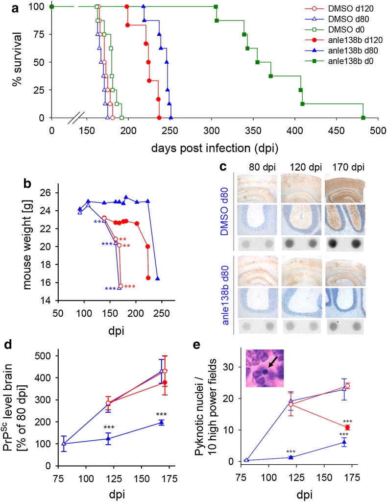
Animal/Disease Models: Two-month-old PLP-hαSyn mice [3]
Doses: 0.6 and 2 g/kg Route of Administration: Oral Experimental Results: Prevention of motor deficits and neurodegeneration PLP-hαSyn mice. For long-term survival experiments, Emrusolmin (Anle138b) was administered orally in DMSO/peanut butter as described above. In a first set of experiments, 5 mg anle138b were given once daily starting either at day 80 or day 120 post i.c. infection. Animals of each treatment group were monitored daily for signs of disease by trained animal caretakers from day 80 post infection. The animals were sacrificed, when they had reached the terminal stage of the disease based on clinical signs (ataxia, tremor, difficulty in righting up from a position lying on its back and tail stiffness). Typically the disease progress through the terminal stage will lead to the death of the animal within 1 or 2 days. In addition, groups of four mice per experimental group were sacrificed at predefined time points as indicated in Fig. 3c–e. From all animals, one brain hemisphere and one half of the spleen were freshly frozen at −80 °C for biochemical analysis. The other hemisphere and the remaining half of the spleen as well as all inner organs were fixed in 4 % formaldehyde solution for histological analysis. In a further experiment, treatment with anle138b was started on the day of i.c. infection with a dose of 5 mg anle138b twice daily. [1] Oral rotenone in vivo mouse model of Parkinson’s disease [1] One-year old C57Bl/6J mice were maintained in a constant light/dark cycle (12 h/12 h). Water and food were given ad libitum. Mice were divided into 4 groups (“no rotenone”, “rotenone/normal feed”, “rotenone/placebo feed”, “rotenone/Emrusolmin (Anle138b)”). A further group of mice (“rotenone/hemivagotomy”) that was used for comparison was hemivagotomized previous to rotenone treatment as previously described. This procedure partially prevents the spread of pathology from the enteric nervous system to the CNS resulting in a decreased dopaminergic cell death. Oral rotenone treatment was performed as previously described. Briefly, mice were treated with 0.01 ml/g body weight of solutions containing either only vehicle [4 % carboxymethylcellulose and 1.25 % chloroform] or rotenone (0.625 mg/ml rotenone, 4 % carboxymethylcellulose and 1.25 % chloroform) once a day, 6 days/week for 4 months. Treatment was administered orally with the help of a 1.2 × 60-mm gavage. During treatment, two groups of rotenone-treated mice were supplied with the same batch of food pellets that either contained the compound (“rotenone/anle138b” group) or were prepared without anle138b (“rotenone/placebo feed” group). The other two groups (“no rotenone” and “rotenone/normal feed”) were fed with standard food pellets. The locomotor abilities of the mice were tested using an acceleration protocol of the rotarod test, as previously described. The test was repeated once a month, four times a day on each animal over three consecutive days. In addition, a 1-h stool collection test was performed on 4 consecutive days once a month as previously described to assess gut motility and function of the enteric nervous system. Transgenic α-synuclein mouse model [1] The in vivo effect of Emrusolmin (Anle138b) was tested in a well-established murine α-synucleinopathy model {(Thy1)-h[A30P]α-syn} on a genetic background of C57/Bl6 mice. Anle138b treatment was tested against placebo treatment with the vehicle (DMSO/peanut butter). Furthermore, untreated non-transgenic C57/Bl6 mice were examined as age-matched controls. The animals were evaluated clinically regarding survival time, motor performance and body weight. In addition, we conducted histological post mortem evaluation of α-syn deposition. Treatment with anle138b and placebo, respectively, was initiated at the age of 8 weeks. Transgenic animals in the treatment and placebo group were matched both in regard to litter and sex. Anle138b was solved in DMSO and mixed with peanut butter. Five days prior to the first dose of anle138b, initial doses of 200 μl peanut butter were given to the mice once daily. During the first 2 weeks of treatment, 2 mg of anle138b dissolved in 10 μl DMSO mixed with 200 μl peanut butter were given. After 2 weeks of treatment, the dose was increased to 5 mg in 10 μl DMSO/200 μl peanut butter. At the age of 33 weeks, the dose was increased to 2 × 5 mg per day. All mice were monitored daily for signs of disease. Detailed clinical assessment including behaviour and movement was performed once a week. Every 2 weeks, rotarod performance and body weight was monitored. In order to investigate the prophylactic effect of Emrusolmin (Anle138b), we treated healthy, plaque‐free, adult, APPPS1Δ9 mice with placebo‐ or anle138b‐containing dry food pellets for 4 months from 2 to 6 months of age (pre‐plaque group; Fig EV1). Age‐ and sex‐matched wild‐type littermates were also treated and served as controls. Similarly, in order to investigate the therapeutic effect of anle138b, we treated symptomatic APPPS1ΔE9 mice and treated them for 4 months from 6 to 10 months of age (post‐plaque group; Fig EV1). Controls were age‐ and sex‐matched wild‐type littermates treated with anle138b or placebo. Anle138b was administered orally. Dry food pellets were prepared containing 2g anle138b per kg food (SSNIFF). This amounted to an estimated daily dose of 500 mg/kg (at an approx. 6‐g daily food consumption and a 25‐g average body weight). Based on pharmacokinetic studies, 40–70 μM anle138b reached the brain during most of the wake phase (Wagner et al, 2015). Placebo food was prepared from the same batch but without anle138b (SSNIFF). Of note, our previous PK studies in mice have shown that after a single bolus the half‐life of anle138b in the brain is approximately 4 h. [2] Two‐month‐old male transgenic mice were randomized in three different groups: one fed with placebo food pellets, another fed with pellets containing Emrusolmin (Anle138b) at 0.6 g/kg of food), and a last group fed with pellets containing 2 g of anle138b per kg of food. The dose of 2 g of anle138b per kg of food was used in previous experiments in mice43 and establishes during the wake phase a concentration of 60 μM in the brain. Two‐month‐old C57/BL6 healthy nontransgenic animals (WT) fed with placebo pellets were used as a healthy control (n = 10). Food pellets were provided to the animals throughout the whole experiment. After 4 months of treatment, behavioral analyses were performed followed by sacrifice of the animals and brain extraction.[3] |
| ADME/Pharmacokinetics |
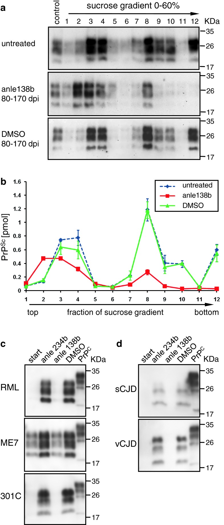
Pharmacokinetic (PK) analysis of the different application protocols used in mice modelling CJD as well as Parkinson’s disease (vide infra) revealed that Emrusolmin (Anle138b) has an excellent oral bioavailability and excellent blood–brain-barrier penetration, and that concentrations shown to be active in the PMCA assay were obtained in the brain (Fig. 5). Key findings in regard to PK are that (i) oral application of Emrusolmin (Anle138b) consistently results in approximately 50 % of the AUC (area under the curve) values compared to i.p. application, which indicates very good oral bioavailability, (ii) application in peanut butter results in slower resorption and prolonged availability of Emrusolmin (Anle138b), (iii) a higher dose results in a proportional increase in serum and brain levels indicating linear PK without saturation of clearance, and (iv) at all time points in all experimental groups anle138b reaches ~3-fold higher levels in the brain than in the serum, which corroborates excellent blood–brain-barrier penetration. Thus, anle138b has a better bioavailability compared to several potential anti-aggregation drugs investigated previously, for which concentrations in the brain were found to be low [56]. Dose–response analysis for the effect of anle138b in vivo (Suppl. Fig. 9) is compatible with pharmacokinetic data and the dose–response curves obtained in vitro. In some experiments, even a reduction of PrPSc levels could be obtained with anle138b (Suppl. Fig. 10). At the same time, investigation of the brain extracts with HPLC in combination with high resolution mass spectroscopy did not identify metabolites of anle138b (Suppl. Fig. 11) in the brain, indicating that anle138b is most probably the active oligomer-modulating molecule. [1]
|
| References |
|
| Additional Infomation |
Emrusolmin restores hippocampal synaptic and transcriptional plasticity as well as spatial memory in a mouse model for Alzheimer's disease, when given orally before or after the onset of pathology.
Drug Indication Treatment of multiple system atrophy Mechanism of Action There is evidence that anle138b blocks the activity of conducting Aβ pores without changing the membrane embedded Aβ-oligomer structure. Alzheimer's disease is a devastating neurodegenerative disease eventually leading to dementia. An effective treatment does not yet exist. Here we show that oral application of the compound anle138b restores hippocampal synaptic and transcriptional plasticity as well as spatial memory in a mouse model for Alzheimer's disease, when given orally before or after the onset of pathology. At the mechanistic level, we provide evidence that anle138b blocks the activity of conducting Aβ pores without changing the membrane embedded Aβ-oligomer structure. In conclusion, our data suggest that anle138b is a novel and promising compound to treat AD-related pathology that should be investigated further. In conclusion, our data show that anle138b can reinstate synaptic plasticity and memory function in a mouse model for amyloid pathology via mechanisms that—at least in part—appear to involve the blockage of Aβ‐induced pores in membranes. Careful analysis of this activity indicates that the oligomers are still in the membrane, but pores have a changed conductivity profile, mainly staying open for shorter time and lacking the possibility of building up large currents as seen when anle138b was absent. Taking into account that anle138b was also shown to ameliorate disease phenotypes in a mouse model for Tau pathology, thus being to the best of our knowledge one of the first compounds that seems to causatively interfere with both of the two major hallmarks of AD, we suggest that anle138b to further be validated in clinical trials for its potential to treat AD and perhaps other aggregopathies.[2] MSA is a fatal neurodegenerative disease characterized by autonomic failure and severe motor impairment. Its main pathological hallmark is the accumulation of α-synuclein in oligodendrocytes, leading to glial and neuronal dysfunction and neurodegeneration. These features are recapitulated in the PLP-hαSyn mouse model expressing human α-synuclein in oligodendrocytes. At present, there is no effective disease-modifying therapy. Previous experiments have shown that the aggregation inhibitor, anle138b, reduces neurodegeneration and behavioral deficits in mouse models of other proteinopathies. [3] |
| Molecular Formula |
C16H11BRN2O2
|
|---|---|
| Molecular Weight |
343.18
|
| Exact Mass |
342
|
| Elemental Analysis |
C, 56.00; H, 3.23; Br, 23.28; N, 8.16; O, 9.32
|
| CAS # |
882697-00-9
|
| Related CAS # |
882697-00-9
|
| PubChem CID |
44608289
|
| Appearance |
Off-white to light yellow solid powder
|
| Density |
1.6±0.1 g/cm3
|
| Boiling Point |
527.3±50.0 °C at 760 mmHg
|
| Flash Point |
272.7±30.1 °C
|
| Vapour Pressure |
0.0±1.3 mmHg at 25°C
|
| Index of Refraction |
1.670
|
| LogP |
4.96
|
| Hydrogen Bond Donor Count |
1
|
| Hydrogen Bond Acceptor Count |
3
|
| Rotatable Bond Count |
2
|
| Heavy Atom Count |
21
|
| Complexity |
370
|
| Defined Atom Stereocenter Count |
0
|
| SMILES |
BrC1C=C(C2NN=C(C3C=C4C(OCO4)=CC=3)C=2)C=CC=1
|
| InChi Key |
RCQIIBJSUWYYFU-UHFFFAOYSA-N
|
| InChi Code |
InChI=1S/C16H11BrN2O2/c17-12-3-1-2-10(6-12)13-8-14(19-18-13)11-4-5-15-16(7-11)21-9-20-15/h1-8H,9H2,(H,18,19)
|
| Chemical Name |
5-(1,3-Benzodioxol-5-yl)-3-(3-bromophenyl)-1H-pyrazole
|
| Synonyms |
Anle 138b; Anle138b; 882697-00-9; Emrusolmin; ANLE-138-b; 3-(1,3-benzodioxol-5-yl)-5-(3-bromophenyl)-1h-pyrazole; Emrusolmin [INN]; E7WRA77JET; 3-(2H-1,3-BENZODIOXOL-5-YL)-5-(3-BROMOPHENYL)-1H-PYRAZOLE; Anle-138b; Anle138b
|
| HS Tariff Code |
2934.99.03.00
|
| Storage |
Powder -20°C 3 years 4°C 2 years In solvent -80°C 6 months -20°C 1 month |
| Shipping Condition |
Room temperature (This product is stable at ambient temperature for a few days during ordinary shipping and time spent in Customs)
|
| Solubility (In Vitro) |
DMSO : ≥ 50 mg/mL (~145.70 mM)
|
|---|---|
| Solubility (In Vivo) |
Solubility in Formulation 1: ≥ 2.5 mg/mL (7.29 mM) (saturation unknown) in 10% DMSO + 40% PEG300 + 5% Tween80 + 45% Saline (add these co-solvents sequentially from left to right, and one by one), clear solution.
For example, if 1 mL of working solution is to be prepared, you can add 100 μL of 25.0 mg/mL clear DMSO stock solution to 400 μL PEG300 and mix evenly; then add 50 μL Tween-80 to the above solution and mix evenly; then add 450 μL normal saline to adjust the volume to 1 mL. Preparation of saline: Dissolve 0.9 g of sodium chloride in 100 mL ddH₂ O to obtain a clear solution. Solubility in Formulation 2: ≥ 2.5 mg/mL (7.29 mM) (saturation unknown) in 10% DMSO + 90% Corn Oil (add these co-solvents sequentially from left to right, and one by one), clear solution. For example, if 1 mL of working solution is to be prepared, you can add 100 μL of 25.0 mg/mL clear DMSO stock solution to 900 μL of corn oil and mix evenly. (Please use freshly prepared in vivo formulations for optimal results.) |
| Preparing Stock Solutions | 1 mg | 5 mg | 10 mg | |
| 1 mM | 2.9139 mL | 14.5696 mL | 29.1392 mL | |
| 5 mM | 0.5828 mL | 2.9139 mL | 5.8278 mL | |
| 10 mM | 0.2914 mL | 1.4570 mL | 2.9139 mL |
*Note: Please select an appropriate solvent for the preparation of stock solution based on your experiment needs. For most products, DMSO can be used for preparing stock solutions (e.g. 5 mM, 10 mM, or 20 mM concentration); some products with high aqueous solubility may be dissolved in water directly. Solubility information is available at the above Solubility Data section. Once the stock solution is prepared, aliquot it to routine usage volumes and store at -20°C or -80°C. Avoid repeated freeze and thaw cycles.
Calculation results
Working concentration: mg/mL;
Method for preparing DMSO stock solution: mg drug pre-dissolved in μL DMSO (stock solution concentration mg/mL). Please contact us first if the concentration exceeds the DMSO solubility of the batch of drug.
Method for preparing in vivo formulation::Take μL DMSO stock solution, next add μL PEG300, mix and clarify, next addμL Tween 80, mix and clarify, next add μL ddH2O,mix and clarify.
(1) Please be sure that the solution is clear before the addition of next solvent. Dissolution methods like vortex, ultrasound or warming and heat may be used to aid dissolving.
(2) Be sure to add the solvent(s) in order.
| NCT Number | Recruitment | interventions | Conditions | Sponsor/Collaborators | Start Date | Phases |
| NCT04685265 | Completed | Drug: anle138b Drug: Placebo |
Parkinson Disease | MODAG GmbH | 2020-12-22 | Phase 1 |
| NCT04208152 | Completed | Drug: anle138b Drug: Placebo |
Healthy Volunteers | MODAG GmbH | 2019-12-06 | Phase 1 |
| NCT06568237 | Recruiting | Drug: TEV-56286 Drug: Placebo |
Multiple System Atrophy | Teva Branded Pharmaceutical Products R&D, Inc. | 2024-10-02 | Phase 2 |
| NCT05532358 | Completed | Drug: anle138b (TEV-56286) Drug: Fluvoxamine 100 mg QD for 5 days |
Healthy Volunteers | MODAG GmbH | 2022-09-12 | Phase 1 |
|
|
Products are for research use only; We do not sell to patients
Copyright 2020 InvivoChem LLC | All Rights Reserved


 COA
COA